ORIGINAL RESEARCH
Pituicytoma, Spindle Cell Oncocytoma, and
Granular Cell Tumor: Clarification and
Meta-Analysis of the World Literature since 1893
M.F. Covington S.S. Chin A.G. Osborn
BACKGROUND AND PURPOSE: Pituicytoma, SCO, and GCT are poorly understood entities with con-fusing nomenclature and undetermined imaging characteristics. Our purpose was to confirm published cases of pituicytoma, SCO, and GCT with the newest 2007 World Health Organization criteria and elucidate imaging findings that distinguish these tumors from common entities such as pituitary adenoma.
MATERIALS AND METHODS:A literature search identified 145 published cases (81 GCTs, 48 pituicy-tomas, and 16 SCOs). Case diagnoses were blindly reviewed by a neuropathologist according to the latest WHO criteria, resulting in 112 pathologically documented cases (64 GCTs, 35 pituicytomas, and 13 SCOs). Imaging illustrations from proved cases were reviewed to determine location, configuration, attenuation and signal intensity, and enhancement characteristics.
RESULTS: Only pituicytomas presented as purely intrasellar lesions (7/33). Most GCTs were purely suprasellar (28/45). All SCOs were both intra- and suprasellar (13/13). Twenty-five percent of pituicy-tomas (6/22) and GCTs (7/30) appeared separate from the pituitary gland. All SCOs were infiltrating. nine percent of entities appeared isointense to brain on T1-weighted image (34/43). Seventy-four percent of pituicytomas enhanced homogeneously (14/19). Twelve of 23 GCTs and 5/7 SCOs enhanced heterogeneously. Most GCTs were hyperattenuated to brain on CT (18/20). Eleven of 13 cases enhanced homogeneously. Visual disturbances were common symptoms for all entities (67/ 112). Diabetes insipidus was rare (4/112).
CONCLUSIONS:Pituicytoma may be considered for purely intrasellar masses that are clearly separate from the pituitary gland. GCT should receive consideration for purely suprasellar lesions that are hyperattenuated to brain on CT. SCO should be considered for infiltrating pituitary masses with a mixed intra- and suprasellar location. A history of diabetes insipidus helps to exclude these tumors.
ABBREVIATIONS:DI ⫽ diabetes insipidus; GCT ⫽ granular cell tumor; SCO ⫽ spindle cell oncocytoma
P
rimary nonadenomatous pituitary gland tumors are rare, poorly understood entities with confusing nomenclature. The 2007WHO Classification of Tumors of the Central Nervous Systemclarified and redefined criteria for pituicytoma, codi-fying it as a separate diagnostic entity distinct from GCT of the neurohypophysis.1The 2007 update also added a new entity— SCO—to the spectrum of nonadenomatous sellar neoplasms.2 The purpose of our study was to apply the new World Health Organization (WHO) criteria to published cases and elucidate imaging findings that might distinguish these rare neoplasms from each other as well as more common lesions such as pitu-itary adenoma and lymphocytic hypophysitis.Materials and Methods
A comprehensive literature search of English and non-English studies by using PubMed and Google Scholar was completed between Sep-tember and October 2010 to identify all previously published cases of
pituicytoma, SCO, and neurohypophyseal GCT. To include all names assigned to these tumors over past decades, the following keywords were used in the search: pituicytoma, spindle cell oncocytoma, gran-ular cell tumor, infundibuloma, “choristoma”, grangran-ular cell myoblas-toma, Abriksossoff tumor, and pilocytic astrocytoma.
Search results were screened by the primary author to include only those tumors involving the sellar and suprasellar region (eg, most GCTs involve extracranial sites such as the tongue and esophagus3;
pilocytic astrocytoma—previously used synonymously for what is now termed pituicytoma—typically does not involve the infundibu-lum or pituitary gland4). This search identified 145 potential cases
(81 GCTs, 48 pituicytomas, and 16 SCOs).
Full-text articles of each report were obtained. The radiologic and histologic images from each article were compiled into electronic databases by the primary author. Captions or other identifying text accompanying each image were removed to facilitate blinded case review.
The histologic image data base was reviewed by a board-certified neuropathologist (S.S.C.) who was blinded as to tumor entity and instructed to classify each case, if possible, as pituicytoma, GCT, or SCO in accordance with the latest 2007 WHO criteria. For cases in which provided histologic images were insufficient to meet WHO criteria, all available written descriptions of the study’s pathologic diagnoses were provided. This included descriptions of histology, immunohistochemistry, and ultrastructure. After review of this infor-mation, the neuropathologist was again asked whether sufficient
Received February 10, 2011; accepted after revision April 28.
From the Departments of Pathology (S.S.C.), and Radiology (A.G.O.), University of Utah School of Medicine (M.F.C.), Salt Lake City, Utah.
Paper previously presented in part at: Annual Meeting of the American Society of Neuroradiology, June 6 –9, 2011; Seattle, Washington.
Please address correspondence to Anne Osborn, MD, Department of Radiology, University of Utah Medical Center, 30 N 1900 E #1A71, Salt Lake City, UT 84132; e-mail: anne.osborn@hsc.utah.edu
http://dx.doi.org/10.3174/ajnr.A2717
BRAIN
ORIGINAL
WHO criteria were met to classify the case definitively. Any cases that could not be assigned to 1 of our 3 entities based on histology, written reports, or both were recorded as unclassifiable and eliminated from further consideration.
The review process resulted in 112 pathologically documented cases in total from 65 articles (64 GCTs,3,5-4435 pituicytomas4,45-63
and 13 SCOs64-70). Histologic images were of too poor quality to
analyze in 7 cases that were received through interlibrary loan (3 GCTs, 2 pituicytomas, and 2 SCOs), and these were marked as un-classifiable along with 33 additional cases. In no case was a tumor reassigned from 1 entity (eg, pituicytoma) to another (eg, SCO or GCT). Most articles were published in pathology, neurosurgery, or other clinically oriented journals. Only 5 articles were published in radiology journals.
Of the 112 pathologically documented cases, only 58 (30 GCTs, 22 pituicytomas, and 6 SCOs) contained CT images, MR images, or both. Original MR images were available for 19 cases of pituicytoma, 7 cases of SCO, and 24 cases of GCT. T1WI was available for 15 pituicytomas, 4 SCOs, and 24 GCTs. T2WI was available for 8 pituicy-tomas and 17 GCTs. Results of postcontrast scans were available for 19 pituicytomas, 7 SCOs, and 23 GCTs.
Information regarding CT signal intensity characteristics were available for only GCTs (n⫽20). Results of CT enhancement pat-terns were available in 13 of these cases.
Original CT and MR images were compiled into a database and reviewed by a senior neuroradiologist (A.G.O.). Findings including location (suprasellar/infundibulum, intrasellar/pituitary gland, or both), configuration (round or infiltrating and separation from the pituitary gland), attenuation and signal intensity, and enhancement characteristics were tabulated.
Finally, M.F.C. extrapolated the age, sex, and clinical symptoms at presentation from each pathologically documented case.
Results
Location
Pituicytoma was the only tumor that ever presented as a purely intrasellar lesion (7/33) (Table 1). Most pituicytomas were either suprasellar (13/33) or combined intra- and suprasellar lesions (13/33). GCTs were either suprasellar (28/45) or both intra- and suprasellar (17/45). No SCOs were purely intra- or suprasellar; all SCOs were both intra- and suprasellar (13/13).
Separation from Pituitary Gland
Only 25% each of pituicytomas (6/22) and GCTs (7/30) could be clearly separated from the pituitary gland. Seventy-five per-cent of all tumor entities were infiltrating and could not be separated from the underlying pituitary gland (39/52).
Imaging Findings
Signal intensity characteristics and enhancement patterns from CT imaging were available for only the GCTs. Eighteen of 20 were hyperattenuated compared with brain on noncon-trast CT. Postconnoncon-trast scans were available in 13/20 cases. Eighty-four percent (11/13) enhanced homogeneously. One tumor (8%) enhanced inhomogeneously and 1 tumor showed no enhancement after contrast administration.
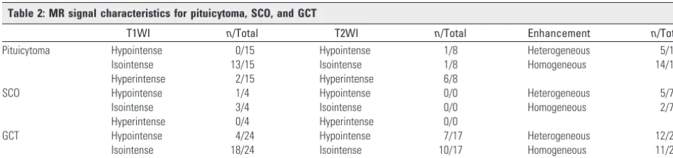
Forty-three cases had 1 or more MR images illustrated (Table 2). T1WI was available in all 43 cases. Seventy-nine percent of all entities, regardless of tumor type, appeared isointense compared with cortex on T1WI (34/43). T2WI was available in only 25 cases. Pituicytomas were generally hyper-intense on T2WI compared with gray matter (6/8), whereas GCTs were predominately isointense (10/17). No confirmed cases of SCO had published images of T2-weighted scans.
Contrast-enhanced T1WI was available for review in 49 cases. Seventy-four percent of pituicytomas enhanced homogeneously (14/19); the remainder showed heteroge-neous enhancement (5/19). Enhancement patterns for the other 2 entities were mixed (12/23 GCTs and 5/7 SCOs dem-onstrated heterogeneous enhancement, whereas the remain-der enhanced homogeneously).
Clinical Findings
Visual disturbances (eg, bitemporal hemianopsia, decreased visual acuity) were the most common presenting symptom for all entities (67/112; Table 3). With pituicytomas, the next most common presenting symptoms were headache (10/35), hypopituitarism (9/35), fatigue (8/35), and decreased libido (7/35). For SCO, hypopituitarism was the next most common symptom (6/13) followed by headache (4/13). For GCT, the next most common symptoms were headache (21/64), amen-orrhea (12/64), fatigue (8/64), and memory loss (7/64).
[image:2.594.53.283.56.104.2]DI was especially uncommon with these tumors. Only 1 pathologically documented case of pituicytoma56and 3 cases of GCT36,39,44had DI. Similarly, increased levels of prolactin were reported for only 1 case of pituicytoma,55 2 cases of SCO,64,70and 4 cases of GCT.11,17,22,31Galactorrhea was re-ported only in 1 case of GCT.11
Table 2: MR signal characteristics for pituicytoma, SCO, and GCT
T1WI n/Total T2WI n/Total Enhancement n/Total
Pituicytoma Hypointense 0/15 Hypointense 1/8 Heterogeneous 5/19
Isointense 13/15 Isointense 1/8 Homogeneous 14/19
Hyperintense 2/15 Hyperintense 6/8
SCO Hypointense 1/4 Hypointense 0/0 Heterogeneous 5/7
Isointense 3/4 Isointense 0/0 Homogeneous 2/7
Hyperintense 0/4 Hyperintense 0/0
GCT Hypointense 4/24 Hypointense 7/17 Heterogeneous 12/23
Isointense 18/24 Isointense 10/17 Homogeneous 11/23
[image:2.594.52.534.616.729.2]Hyperintense 2/24 Hyperintense 0/17
Table 1: Anatomic location for cases of pituicytoma, SCO, and GCT
Location Pituicytoma SCO GCT
Sellar 7/33 0/13 0/45
Sellar/suprasellar 13/33 13/13 17/45
Sex
The male:female ratio was 18:17 (51.4% male) for pituicy-toma, 5:8 (61.5% female) for SCO, and 22:42 (65.5% female) for GCT.
Age
The average age at diagnosis was 50.3 years for pituicytoma, 59.4 years for SCO, and 49.2 years for GCT.
Discussion
Pituicytoma, SCO, and GCT of the neurohypophysis are rare, poorly understood neoplasms of the sellar region. Pituicytoma has an especially notable history of frequently changing no-menclature and shifting diagnostic criteria.62Synonyms for pituicytoma over past decades have included choristoma, granular cell myoblastoma, infundibuloma, pilocytic astrocy-toma, and even granular cell tumor. Not until the 2007 edition of theWHO Classification of Tumors of the Central Nervous Systemwas pituicytoma designated as a distinct diagnostic en-tity, separate from GCT. The 2007 edition also added SCO as a new distinct entity in the differential diagnosis of sellar neoplasms.
Given their striking similarities on imaging to other much more common lesions such as pituitary adenoma, the pre-operative diagnosis of pituicytoma, SCO, and GCT has been problematic. The inability to distinguish these lesions from entities such as pituitary adenoma is important because these tumors, unlike pituitary adenomas, tend to be very vascular and are prone to heavy bleeding during surgical resec-tion.5,52,65 This has often resulted in the need to stabilize the patient, abort the surgery, and consider reoperation at a later date, potentially after embolization of tumor vascula-ture.62If reoperation does not occur, symptomatic recurrence is common.8
Detecting clues that might establish the preoperative diag-nosis of these entities has been problematic due to the
excep-tional rarity of these tumors. No single institution is likely to see more than a handful of these tumors over the course of several decades. The literature consists mostly of single case reports and a few very small case series. Therefore, only a ret-rospective meta-analysis of all published cases could poten-tially detect meaningful differential diagnostic information.
In addition, considering these entities’ history of frequently changing and often overlapping nomenclature, confirmation of published case diagnoses by a board-certified neuropathol-ogist by using the new 2007 WHO criteria was a necessary prerequisite to imaging analysis. Confirming the precise his-tologic diagnosis in each case has allowed us to determine that all cases included in our meta-analysis do indeed represent true cases of pituicytoma, SCO, or GCT.
Major limitations of our study include dependence on the often-limited information provided for the figures, images, and text as included in each original case report or case series. Less than 10% of cases were published in the radiology litera-ture. Precise delineation of imaging parameters were univer-sally absent from nonradiology journals and were present in only 2 of the 5 radiology manuscripts used in our imaging analysis. We did not have access to any of the original patient scans for review or comparison to published reports.
Pituicytoma
The definitive histologic description of pituicytoma46and the subsequent acceptance of pituicytoma as a distinct tumor en-tity were only recently established. Given the lack of known clinical and imaging findings specific to pituicytoma, these tumors are typically diagnosed preoperatively as pituitary ad-enoma.47Attempted resection of these presumed adenomas often results in unexpected heavy intraoperative bleeding, subtotal resection, and a high risk of symptomatic tumor recurrence.62
Our study identifies specific imaging findings (Fig 1) that could permit a neuroradiologist to make the preoperative di-Fig 1.Pituicytoma. Sagittal T1WI image (A) and coronal T1WI postcontrast scan (B) show a rounded suprasellar mass that is clearly separate from the pituitary gland. (From Gibbs WN, Monuki ES, Linskey ME et al. Pituicytoma: diagnostic fea-tures on selective carotid angiography and MR imaging. AJNR Am J Neuroradiol 2006:27:1639 – 42. Used with permission.)
Table 3: Presenting symptoms for pituicytoma, SCO, and GCT
Pituicytoma (n⫽35) No. Cases SCO (n⫽13) No. Cases GCT (n⫽64) No. Cases
Visual disturbance 18 Visual disturbance 8 Visual disturbance 41
Headache 16 Panhypopituitarism 5 Headache 21
Fatigue 8 Headache 4 Amenorrhea 12
Decreased libido 7 Fatigue 2 Fatigue 8
Hypopituitarism 6 Weight loss 2 Memory loss 7
[image:3.594.51.528.57.306.2]agnosis of pituicytoma in selected cases. Although a minority of pituicytomas will present in this manner, the diagnosis might be suggested if imaging shows a mass that is purely intrasellar and clearly separate from the pituitary gland. We found no similar presentation in any of our pathologically documented cases of SCO or GCT. Such a presentation also would be rare for pituitary adenoma, lymphocytic hypophysi-tis, or physiologic pituitary hyperplasia.71
Patients with pituicytoma almost never present with diabe-tes insipidus, galactorrhea, or prolactinemia. When these symptoms are present, more common diagnoses such as pitu-itary adenoma or lymphocytic hypophysitis are likely. Instead, to support the diagnosis of pituicytoma, the radiologist should look for a classic history of visual disturbance, headache, or both.
Spindle Cell Oncocytoma
SCO (Fig 2) was first described in 2002 by Roncaroli et al69 in their series of 5 cases. Only 16 total cases have been reported in the literature. Of these, we were able to confirm the histo-pathology of 13 cases. The remaining 3 cases were marked as unclassifiable due to the poor quality of images received through interlibrary loan.
Our analysis of pathologically documented cases of SCO was limited by the small number of imaging studies available in the published literature. However, all 13 cases of the patho-logically documented SCO presented as combined intra- and
suprasellar lesions. Thus it is unlikely that a lesion presenting as a purely intra- or suprasellar mass on imaging is a SCO.
SCOs arise from the adenohypophysis whereas pituicy-toma and GCT derive from the neurohypophysis.69,70 There-fore, if imaging localizes a tumor to the neurohypophysis, the diagnosis of SCO may be excluded. Moreover, all cases of SCO were infiltrating and none could be seen separately from the pituitary gland itself. It is therefore not possible to distin-guish SCOs from more common lesions such as pituitary ad-enoma or lymphocytic hypophysitis on the basis of imaging features alone.
SCO typically presents with visual disturbance, panhypo-pituitarism, and headache. From our analysis, the incidence of panhypopituitarism seems to be more common with SCO than either pituicytoma or GCT. This discrepancy may possi-bly be explained by SCO’s exclusive derivation from the ade-nohypophysis. There are no reported cases of SCO presenting with DI.
We could identify no specific imaging or clinical findings that allowed us to suspect the preoperative diagnosis of SCO. In contrast, there are imaging and clinical clues that help ex-clude SCO when present. These inex-clude a mass that clearly arises from the neurohypophysis or masses that are exclusively either intra- or suprasellar on imaging studies. Individuals presenting with a pituitary mass and concomitant diabetes insipidus are unlikely to have a SCO.
Fig 2.Spindle cell oncocytoma. Coronal T1WI (A) demonstrates a mixed intra- and suprasellar infiltrating pituitary lesion. Coronal T1-postcontrast scan (B) reveals a heterogeneous pattern of enhancement. Sagittal T1WI (C) shows enlargement of the anterior pituitary by the infiltrating mass and displacement of the unaffected neurohypophysis. (From Vajtai I, Sahli R, Kappeler A. Spindle cell oncocytoma of the adenohypophysis: report of a case with a 16-year follow-up.Pathol Res Pract2006:202:745–50. Used with permission.)
[image:4.594.56.532.44.202.2] [image:4.594.52.524.252.411.2]Granular Cell Tumor
Boyce and Beadles72first described GCT (Fig 3) as a distinct neurohypophyseal tumor in 1893. Many GCTs are asymp-tomatic and consist of small nests of tumor cells that do not have any space-occupying effects.7 Such tumor cell nests are common in the general population. Necropsy of 200 pitu-itary glands found GCT nests in 5.7% of cases,12and analysis of 1364 pituitary glands identified GCT nests in 6.45% of cases.73
As first reported in 1951,42these tumor cell nests may en-large over time and become clinically symptomatic, usually in middle-aged or older adults.22To date, symptomatic GCT of the neurohypophysis has been described in 81 cases, 64 of which we could confirm on histopathologic review.
Our analysis shows that GCT is almost always hyperattenu-ated to brain on CT and usually demonstrates a homogeneous pattern of enhancement. Most GCTs are entirely suprasellar in location. None of our 64 pathologically documented cases presented as a purely intrasellar mass. Therefore, GCT should be excluded from the differential diagnosis of purely intra-sellar lesions.
Clinical findings for GCT are nonspecific and most com-monly include visual disturbance, headache, and amenorrhea. Similar to our other entities, DI, prolactinemia, and galactor-rhea are uncommon presenting features of GCT.
The diagnosis of GCT should be considered for purely su-prasellar masses that are hyperattenuated compared with brain on CT and enhance homogeneously. MR imaging offers few clues that raise preoperative suspicion for GCT. On MR imaging, GCT tends to be isointense on both T1WI and T2WI. The pattern of enhancement for GCTs is equally heter-ogeneous or homheter-ogeneous after gadolinium administration.
Conclusions
Pituicytoma, SCO, and GCT represent rare but important differential diagnostic considerations of sellar and supra-sellar lesions. The preoperative diagnosis of these tumors has been difficult owing to a lack of specific imaging and clinical findings. From our analysis, we have identified im-aging and clinical clues that make the potential preopera-tive diagnosis of these entities possible (Fig 4). A diagnosis of pituicytoma may be considered for masses that are purely intrasellar and clearly separate from the pituitary gland on imaging. GCT should receive diagnostic consideration for lesions that are hyperattenuated compared with brain on non-contrast CT and of a purely suprasellar location. SCO should be considered only for infiltrating pituitary masses with a mixed intra- and suprasellar location. Neither pituicytoma, SCO, nor GCT should be highly considered for patients who present with DI, prolactinemia, or galactorrhea. Instead, these rare tumors most often present with visual disturbance and headache.
Disclosures: Anne G. Osborn.Ownership Interest:Amirsys, Amirsys Publishing, Details: shareholder;Other Financial Relationships:Amirsys Publishing, Details: CEO.
References
1. Louis DN, Ohgaki H, Weistler OD, et al.WHO Classification of Tumours of the
Central Nervous System, 4th ed. Lyon, France: IARC Press; 2007:309
2. Fuller GN, Scheithauer BW.The 2007 revised World Health Organization
(WHO) classification of tumours of the central nervous system: newly codi-fied entities.Brain Pathol2007;17:304 – 07
3. Kobayashi TK, Bamba M, Oka H.Granular cell tumour of the
neurohypoph-ysis on cytological squash preparations.Cytopathology2006;17:153–54
4. Katsuta T, Inoue T, Nakagaki H, et al.Distinctions between pituicytoma and
ordinary pilocytic astrocytoma: case report.J Neurosurg2003;98:404 – 06
5. Aquilina K, Kamel M, Kalimuthu SG, et al.Granular cell tumour of the
[image:5.594.56.343.42.347.2]hypophysis: a rare sellar tumour with specific radiological and operative fea-tures.Br J Neurosurg2006;20:51–54
6. Becker DH, Wilson CB.Symptomatic parasellar granular cell tumors.
Neuro-surgery1981;8:173– 80
7. Boucher-Schwarz HG, Fries G, Bornemann A, et al.Suprasellar granular cell
tumor.Neurosurgery1992;31:751–54
8. Buhl R, Hugo HH, Hempelmann RG, et al.Granular-cell tumour: a rare
su-prasellar mass.Neuroradiology2001;43:309 –12
9. Burston J, John R, Spencer H.“Myoblastoma” of the neurohypophysis.
J Pathol Bacteriol1962;83:455– 61
10. Cohen-Gadol AA, Pichelmann MA, Link MJ, et al.Granular cell tumor of the
sellar and suprasellar region: clinicopathologic study of 11 cases and litera-ture review.Mayo Clin Proc2003;78:567–73
11. Cusick JF, Ho KC, Hagen TC, et al.Granular-cell pituicytoma associated with
multiple endocrine neoplasia type 2.J Neurosurg1982;56:594 –96
12. Doron Y, Behar A, Beller AJ.Granular-cell “myoblastoma” of the
neurohy-pophysis.J Neurosurg1965;22:95–99
13. Benites Filho PR, Sakamoto D, Machuca TN, et al.Granular cell tumor of the
neurohypophysis: report of a case with unusual age presentation.Virchows
Arch2005;447:649 –52
14. Glazer N, Hauser H, Slade H.Granular cell tumor of the neurohypophysis.
Am J Roentgenol Radium Ther Nucl Med1956;76:324 –26
15. Graziani N, Dufour H, Figarella-Branger D, et al.Suprasellar granular-cell
tumour, presenting with intraventricular haemorrhage.Br J Neurosurg1995; 9:97–102
16. Harland WA.Granular-cell myoblastoma of the hypophyseal stalk.Cancer
1953;6:1134 –38
17. Higuchi M, Tsuji M, Ikeda H.Symptomatic hypophyseal granular cell tumour:
endocrinological and clinicopathological analysis.Br J Neurosurg1997;11: 582– 86
18. Jenevein EP.A neurohypophysial tumor originating from pituicytes.Am J
Clin Pathol1964;41:522–26
19. Ji CH, Teng MM, Chang T.Granular cell tumour of the neurohypophysis.
Neuroradiology1995;37:451–52
20. Kasashima S, Oda Y, Nozaki J, et al.A case of atypical granular cell tumor of the
neurohypophysis.Pathol Int2000;50:568 –73
21. Lafitte C, Aesch B, Henry-Lebras F.Granular cell tumor of the pituitary stalk.
Case report.J Neurosurg1994;80:1103– 07
22. Lee CC, Liu CH, Wei CP, et al.Symptomatic granular cell tumor of the
neuro-hypophysis.J Formos Med Assoc2004;103:58 – 62
23. Lima A, Antunes L, Tome F.Pituicytoma or granular-cell myoblastoma of the
pituitary gland? Report of a case.J Neurosurg1960;17:778 – 82
24. Liwnicz BH, Liwnicz RG, Huff JS.Giant granular cell tumor of the suprasellar
area: immunocytochemical and electron microscopic studies.Neurosurgery 1984;15:246 –51
25. Losa M, Saeger W, Mortini P.Acromegaly associated with a granular cell
tu-mor of the neurohypophysis: a clinical and histological study. Case report.
J Neurosurg2000;93:121–26
26. Moriyama E, Matsumoto Y, Meguro T, et al.Suprasellar granular cell tumor.
Neurol Med Chir (Tokyo)1996;36:237– 40
27. Mumert ML, Walsh MT, Chin SS.Cystic granular cell tumor mimicking
Rathke cleft cyst.J Neurosurg2011;114:325–28
28. Nishio S, Takeshita I, Yoshimoto K, et al.Granular cell tumor of the pituitary
stalk.Clin Neurol Neurosurg1998;100:144 – 47
29. Nishioka H, IiK, Llena JF, et al.Immunohistochemical study of granular cell
tumors of the neurohypophysis.Virchows Arch B Cell Pathol Incl Mol Pathol 1991;60:413–17
30. Policarpio-Nicolas ML, Le BH, Mandell JW, et al.Granular cell tumor of the
neurohypophysis: report of a case with intraoperative cytologic diagnosis.
Diagn Cytopathol2008;36:58 – 63
31. Popovic V, Pekic S, Skender-Gazibara M, et al.A large sellar granular cell
tu-mor in a 21-year-old woman.Endocr Pathol2007;18:91–94
32. Radhakrishnan W, Misra BK, Rout D.Granular cell tumour (choristoma) of
the neurohypophysis—a report of two cases.Indian J Pathol Microbiol1997; 40:71–74
33. Rhee JS, Wackym PA, Hague K, et al.Granular cell tumor of the pituitary fossa.
Ann Otol Rhinol Laryngol2002;111:754 –58
34. Satyamurti S, Huntington HW.Granular cell myoblastoma of the pituitary.
Case report.J Neurosurg1972;37:483– 86
35. Schaller B, Kirsch E, Tolnay M, et al.Symptomatic granular cell tumor of the
pituitary gland: case report and review of the literature.Neurosurgery1998;42: 166 –71
36. Schlachter LB, Tindall GT, Pearl GS.Granular cell tumor of the pituitary gland
associated with diabetes insipidus.Neurosurgery1980;6:418 –21
37. Shuangshoti S, Chantra K, Navalitloha Y, et al.Atypical granular cell tumor of
the neurohypophysis: a case report with review of the literature.J Med Assoc
Thai1998;81:641– 46
38. Symon L, Burston J, Gonz JC.Granular cell myoblastoma of the
neuro-hypophysis: report of two cases.J Neurosurg1971;35:82– 89
39. Talerman A, Dawson-Butterworth K.Granular-cell myoblastoma of the
pitu-itary.Postgrad Med J1966;42:216 –18
40. Ulrich J, Landolt A, Benini A.Granular-cell tumor of the third ventricle of the
brain. Clinical findings, light and electron microscopy.Acta Neuropathol1974; 26:215–23
41. Vaquero J, Leunda G, Cabezudo JM.Granular pituicytomas of the pituitary
stalk.Acta Neurochir (Wien)1981;59:209 –15
42. Luthy F, Klingler M.The tumoret tumor of the posterior pituitary lobe.
Schweiz Z Pathol Bakteriol1951;14:721–29
43. Waller RR, Riley FC, Sundt TM.A rare cause of the chiasmal syndrome.Arch
Ophthalmol1972;88:269 –72
44. Wilkinson MD, Fulham MJ, Besser M.Neuroimaging findings in a suprasellar
granular cell tumor.J Comput Assist Tomogr2003;27:26 –29
45. Benveniste RJ, Purohit D, Byun H.Pituicyotma presenting with spontaneous
hemorrhage.Pituitary2006;9:53–58
46. Brat DJ, Scheithauer BW, Staugaitis SM, et al.Pituicytoma: a distinctive
low-grade glioma of the neurohypophysis.Am J Surg Pathol2000;24:362– 68
47. Hammoud DA, Munter FM, Brat DJ, et al.Magnetic resonance imaging
fea-tures of pituicytomas: analysis of 10 cases.J Comput Assist Tomogr2010;34: 757– 61
48. Cenacchi G, Giovenali P, Castrioto C, et al.Pituicytoma: ultrastructural
evi-dence of a possible origin from folliculo-stellate cells of the adenohypophysis.
Ultrastruct Pathol2001;25:309 –12
49. Chen KT.Crush cytology of pituicytoma.Diagn Cytopathol2005;33:255–57
50. Figarella-Branger D, Dufour H, Fernandez C, et al.Pituicytomas, a
misdiag-nosed benign tumor of the neurohypophysis: report of three cases.Acta Neu-ropathol2002;104:313–19
51. Furtado SV, Ghosal N, Venkatesh PK. Et al.Diagnostic and clinical
implica-tions of pituicytoma.J Clin Neurosci2010;17:938 – 43
52. Gibbs WN, Monuki ES, Linskey ME, et al.Pituicytoma: diagnostic features on
selective carotid angiography and MR imaging.AJNR Am J Neuroradiol2006; 27:1639 – 42
53. Hurley TR, D’Angelo CM, Clasen RA, et al.Magnetic resonance imaging and
pathological analysis of a pituicytoma: case report.Neurosurgery1994;35: 314 –17
54. Kowalski RJ, Prayson RA, Mayberg MR.Pituicytoma.Ann Diagn Pathol2004;
8:290 –94
55. Nakasu Y, Nakasu S, Saito A.Pituicytoma: two case reports.Neurol Med Chir
(Tokyo)2006;46:152–56
56. Phillips JJ, Misra A, Feuerstein BG, et al.Pituicytoma: characterization of a
unique neoplasm by histology, immunohistochemistry, ultrastructure, and array-based comparative genomic hybridization.Arch Pathol Lab Med2010; 134:1063– 69
57. Schultz AB, Brat DJ, Oyesiku NM, et al.Intrasellar pituicytoma in a patient
with other endocrine neoplasms.Arch Pathol Lab Med2001;125:527–30
58. Shah B, Lipper MH, Laws ER, et al.Posterior pituitary astrocytoma: a rare
tumor of the neurohypophysis: a case report.AJNR Am J Neuroradiol2005;26: 1858 – 61
59. Takei H, Goodman JC, Tanaka S, et al.Pituicytoma incidentally found at
au-topsy.Pathol Int2005;55:745– 49
60. Thiryayi WA, Gnanalingham KK, Reid H, et al.Pituicytoma: a misdiagnosed
benign tumour of the posterior pituitary.Br J Neurosurg2007;21:47– 48
61. Ulm AJ, Yachnis AT, Brat DJ, et al.Pituicytoma: report of two cases and clues
regarding histogenesis.Neurosurgery2004;54:753–57
62. Wolfe SQ, Bruce J, Morcos JJ.Pituicytoma: case report.Neurosurgery2008;63:
E173–74
63. Zhi L, Yang L, Quan H, et al.Pituicytoma presenting with atypical histological
features.Pathology2009;41:505– 09
64. Borges MR, Lillehei KO, Kleinschmidt-Demasters BK.Spindle cell oncocytoma
with late recurrence and unique neuroimaging characteristics due to recur-rent subclinical intratumoral bleeding.J Neurooncol2011;101:145–54
65. Borota OC, Schethauer BW, Fougner SL, et al.Spindle cell oncocytoma of the
adenohypophysis: report of a case with marked cellular atypia and recurrence despite adjuvant treatment.Clin Neuropathol2009;28:91–95
66. Coire CI, Horvath E, Smyth HS, et al.Rapidly recurring folliculostellate cell
tumor of the adenohypophysis with the morphology of a spindle cell oncocytoma: case report with electron microscopic studies.Clin Neuropathol 2009;28:303– 08
67. Dahiya S, Sarkar C, Hedley-Whyte ET, et al.Spindle cell oncocytoma of the
adenohypophysis: report of two cases.Acta Neuropathol2005;110:97–99
68. Kloub O, Perry A, Tu PH, et al.Spindle cell oncocytoma of the
adeno-hypophysis: report of two recurrent cases.Am J Surg Pathol2005;29:247–53
69. Roncaroli F, Scheithauer BW, Cenacchi G, et al.‘Spindle cell oncocytoma’ of
the adenohypophysis: a tumor of folliculostellate cells?Am J Surg Pathol 2002;26:1048 –55
70. Vajtai I, Sahli R, Kappeler A.Spindle cell oncocytoma of the adenohypophysis:
report of a case with a 16-year follow-up.Pathol Res Pract2006;202:745–50
71. Osborn AG, Salzman KL, Barkovich AJ.Diagnostic Imaging. Brain, 2nd ed. Salt
Lake City: Amirsys Publishing; 2010: II:24 –27, II:42– 45
72. Boyce R, Beadles CF.A further contribution to the study of the pathology of
the hypophysis cerebri.J Pathol Bacteriol1893;1:359 – 83
73. Luse SA, Kernohan JW.Granular-cell tumors of the stalk and posterior lobe of