ORIGINAL RESEARCH
Quantitative Cerebrovascular Reserve Measured
by Acetazolamide-Challenged Dynamic CT
Perfusion in Ischemic Adult Moyamoya Disease:
Initial Experience with Angiographic Correlation
K.H. Kang H.S. Kim S.Y. Kim
BACKGROUND AND PURPOSE: CT perfusion is a much more readily accessible imaging method to assess cerebral hemodynamic status than single-photon emission CT. We prospectively assessed quantitative cerebrovascular reserve by using acetazolamide (ACZ)-challenged CT perfusion for eval-uating hemodynamic impairment in ischemic adult Moyamoya disease and compared it with angio-graphic findings.
MATERIALS AND METHODS: Sixteen adult patients with ischemic Moyamoya disease and 12 age-matched normal control subjects underwent both ACZ-challenged CT perfusion and digital subtraction angiography. Normalized baseline hemodynamic parameters and their percent changes (PCs) were calculated in 56 hemispheres. We classified the degrees of distal carotid artery stenosis according to modified Suzuki stage and determined the presence of basal Moyamoya vessels (BMVs). The values of normalized parameters and their PCs were compared with angiographic findings.
RESULTS: Normalized baseline mean transit time (MTT) and PC of normalized cerebral blood flow (CBF) were significantly correlated with angiographic stages in all of the vascular territories; however, the correlation coefficient of the normalized baseline MTT was lower than that of the PC of CBF. In the external borderzone and the middle cerebral arterial territory, the hemispheres with extensive BMVs exhibited significantly lower PC values of CBF and significantly higher normalized baseline MTT values than those in hemispheres with diminished BMVs and in normal control subjects.
CONCLUSION:Among the hemodynamic parameters measured by ACZ-challenged CT perfusion, the PC of CBF correlated highly significantly with angiographic stage; however, the normalized baseline CT perfusion parameters showed weak or no significant correlation.
M
oyamoya disease is an idiopathic progressive occlusive cerebrovascular disorder of unknown etiology. This dis-ease has angiographically been characterized by numerous en-larged lenticulostriated and thalamoperforating arteries, as well as dural, leptomeningeal, and pial collateral vessels.1-4 The most common clinical presentation is recurrent transient ischemic attacks or strokes, resulting in significant morbidity. In Moyamoya disease, as in other chronic steno-occlusive ce-rebrovascular diseases, the steno-occlusive change in the main cerebral arteries decreases the cerebral perfusion pressure, and collateral circulation maintains cerebral blood flow (CBF).4 According to a few earlier reports, patients with Moyamoya diseases have decreased CBF, increased cerebral blood volume (CBV), and delayed mean transit time (MTT).3-6These hemo-dynamic changes in Moyamoya disease have been explained by decreases in the cerebral perfusion pressure and associated vasodilation.3A few studies investigated the relationship be-tween cerebral angiographic findings and hemodynamic sta-tus in Moyamoya disease.5-7Most patients show extensive de-velopment of basal Moyamoya vessels (BMV) and present ischemic events.Acetazolamide (ACZ), a carbonic anhydrase inhibitor, in-directly dilates the cerebrovasculature by increasing carbon dioxide levels in the blood stream.8,9As a result, ACZ-chal-lenged single-photon emission CT (SPECT) increases the con-trast in radioactivity levels observed between regions of ade-quate vascular reserve and those of inadeade-quate reserve. Cerebrovascular reserve (CVR) obtained by using SPECT pro-vides important information on hemodynamic status in pa-tients with chronic cerebrovascular occlusive diseases.10 How-ever, SPECT usually has to be performed in a 2-day setting due to tracer kinetics. Moreover, it provides less morphologic in-formation than CT or MR imaging. The dynamic CT perfu-sion (CTP) is a much more readily accessible imaging method to assess cerebral hemodynamic status in patients with cere-bral steno-occlusive arterial disease than SPECT, and its per-fusion maps can be generated in a short time at a workstation.11
The aim of the present study was to quantitatively evaluate CVR in ischemic adult Moyamoya disease by using ACZ-chal-lenged dynamic CTP (ACZ-CTP) and to compare it with an-giographic findings.
Methods
Study Population
Our institutional review board approved this prospective study, and written informed consent was obtained from every participant in ac-cordance with the guidelines of the institutional review board at our institution. Between June 2005 and May 2007, 45 patients with sus-pected Moyamoya presenting with transient ischemic attack were
re-Received December 11, 2007; accepted after revision March 17, 2008.
From the Department of Diagnostic Radiology, Ajou University, School of Medicine, Gyeonggi-do, South Korea.
This study was supported by a 2007 grant from Ajou University School of Medicine.
Please address correspondence to Ho Sung Kim, Department of Diagnostic Radiology, Ajou University, School of Medicine, Mt 5, Woncheon-dong, Yeongtong-gu, Suwon-si, Gyeonggi-do, 442-749, South Korea; e-mail: J978005@lycos.co.kr
DOI 10.3174/ajnr.A1129
BRAIN
ORIGINAL
ferred to our institute through neurologic clinics for evaluation of cerebral vascular and perfusion status via digital subtraction angiog-raphy (DSA) and CTP. Two patients were excluded because of history of renal disease or allergic reaction to contrast material, and 7 patients were excluded because we did not acquire their informed consent. Five pediatric patients were also excluded due to radiation hazard. Thirty-one patients underwent both DSA and ACZ-CTP, including precontrast CT scan. Both DSA and ACZ-CTP were performed within 3 days of each other. During the period, patients were selected for prospective study if steno-occlusive diseases of the bilateral distal internal carotid arteries (ICAs) around ICA bifurcation were detected on DSA. Stenotic arteries were considered to be diseased according to the modified Suzuki classification.1Other criteria for inclusion in this
study included the normal finding of posterior circulation on DSA. Ten patients who had concomitant proximal posterior cerebral artery stenosis were excluded. MR study including diffusion-weighted im-aging was also performed in all of the referred patients to exclude cerebral infarct. Among 21 patients, 5 additional adult patients who had definite territorial infarct on MR images were excluded from the study. Finally, 16 adult patients with ischemic Moyamoya diseases were enrolled in this study. ACZ-CTP was also performed in 12 age-matched normal control subjects who were volunteers for the evalu-ation of neurologic symptoms such as dizziness. Normal values of baseline hemodynamic parameters, including CBV, CBF, MTT, and percent changes (PCs) of each parameter, were determined based on age-matched normal control subjects. Among 16 study patients, there were 5 men (age range, 32– 44 years; mean age, 38 years) and 11 women (age range, 31– 66 years; mean age, 45 years). All of the study patients experienced transient ischemic attacks, and 7 patients expe-rienced headache. The clinical features and demographic data of the study patients are summarized in Table 1.
CTP Imaging Protocol
CT scans were performed in the transverse plane by using a 64-chan-nel multidetector CT scanner (Brilliance 64 Chan64-chan-nel CT; Philips Medical Systems, Cleveland, Ohio). The patient’s head was fixed in place with a head holder and was positioned by using light beams.
CTP consisted of a 60-second series with 30 gantry rotations per-formed in cine mode during the intravenous administration of iodin-ated contrast material. Images were acquired and reconstructed at a temporal sampling rate of 1 image per 2 seconds, resulting in a series of 30 images for each assessed section. After unenhanced CT of the whole brain, 8 adjacent 5-mm-thick sections were selected by starting at the level of the basal ganglia. A 50-mL bolus of nonionic contrast media (Omnipaque, iodine 300 mg/mL; Amersham Health, Prince-ton, NJ) was administered into an antecubital vein by using a power injector at an injection rate of 4.5 mL/s. At 2 seconds after initiation of the contrast injection, a cine (continuous) scan was initiated with parameters of 80 kVp and 150 mAs. The gantry angle was parallel to and above the orbital roof to avoid radiation exposure to the lens. CTP was repeatedly performed, approximately 20 minutes after in-travenous bolus injection of 1000 mg of ACZ (Diamox; Wyeth, Mari-etta, Pa). All of the rest of the CTP and ACZ-CTP were obtained during the same session with patients remaining in supine position.
Data Processing
CTP data were analyzed using brain perfusion software (Extended Brilliance Workstation v 3.0, Philips Medical Systems). The software relies on the central volume principle to calculate perfusion parame-ters from the time-concentration curve. It has been reported that this principle is the most accurate for low injection rates of iodinated contrast agent.12The software first performs a motion correction and
then noise reduction was done by using an anisotropic, edge-preserv-ing spatial filter. The software applies curve fittedge-preserv-ing by a least-mean-squares method to obtain mathematical descriptions of the time-at-tenuation curves, and the MTT map was calculated by a closed-form (noniterative) deconvolution operation from the time-concentration curve of a particular voxel and the arterial input function (AIF).12An
AIF was selected by placing a small circular region of interest (ROI) within the earliest appearing and most densely enhancing artery (usu-ally one of MCA ipsilateral to a less affected hemisphere). A venous function was selected by placing a circular ROI within a superior sagittal sinus. For each voxel, the CBV map was calculated from the areas under the time-concentration curves. The CBF map for each voxel was finally calculated according to the following equation com-bining the CBV and MTT value: CBF⫽CBV/MTT.13
Data Analysis
Two experienced neuroradiologists independently and blindly evalu-ated the angiographic findings of 56 hemispheres in 16 study patients and 12 normal control subjects who underwent ACZ-CTP. The ICA lesions were evaluated by a 4-stage angiographic classification scale that was modified from that proposed by Suzuki and Takaku1(Table
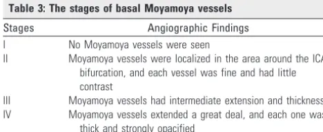
2). The development of BMVs was also graded into 4 stages as shown by a previous report (Table 3).3Our study patients were classified into
2 groups according to the extent of BMV development (poor BMV, stage 1 and 2; extensive BMV, stage 3 and 4). After the blinded study, discrepancies were resolved by consensus. Finally, the consensus data of the angiographic findings were compared with CTP parameters.
For quantification of the hemodynamic parameters, 2 experi-enced neuroradiologists independently drew 8 standardized elliptical mirrored ROIs manually on the basal ganglia (BG) section level (around foramen of Monro) of the reference CT image (Fig 1) over the cortical gray matter of the anterior cerebral artery (ACA) distri-bution, the middle cerebral artery (MCA) distridistri-bution, the anterior external borderzone (EBZ), and the putamen in each cerebral hemi-sphere. The ROI placements in EBZ were performed based on
previ-Table 1: Clinical data of 16 adult patients with ischemic Moyamoya diseases
Patient
No. Age/Sex
Clinical Symptoms
Duration of TIA
Bypass Surgery
1 39/F TIA 12 months EIAB
2 44/M Headache, TIA 7 months Negative*
3 33/F Headache, TIA 8 months Negative*
4 64/F Headache, TIA 4 months Negative*
5 66/F Headache, TIA 2 months EIAB
6 32/M Headache, TIA 24 months Negative*
7 36/M TIA 24 months Negative*
8 45/F TIA 1 month Negative*
9 42/F TIA 7 months Negative*
10 39/F TIA 36 months Negative*
11 56/F Headache, TIA 1 month Negative*
12 31/F TIA 24 months EIAB
13 35/F Headache, TIA 12 months Negative*
14 37/M TIA 8 months EIAB
15 42/M TIA 11 months Negative*
16 48/F TIA 1 month Negative*
Note:—TIA indicates transient ischemic attack; EIAB, external internal arterial bypass; F, female; M, male.
[image:2.594.52.285.66.252.2]ous reports.14,15The observer also drew mirrored ROIs in the bilateral
cerebellar hemispheres for normalization of the hemodynamic pa-rameters. Large cortical vessels were automatically excluded via brain perfusion software. From each ROI, the absolute values of CBF, CBV, and MTT were calculated. Then, for normalization of each baseline parameter, the ratio of the values of baseline parameters obtained from the ROIs on each cerebral hemisphere to those obtained from the ROIs on the ipsilateral cerebellar hemisphere was calculated.5
These normalized values of baseline parameters were used for quan-titative analysis. For the ACZ-CTP study, a section at the same level as the one selected for the baseline study was selected. For the evaluation of vascular reserve capacity, PC was calculated as follows: PC (%)⫽ (NVACZ⫺NVBaseline) ⫼NVBaseline⫻100, where NVBaseline and NVACZrepresented normalized values of the hemodynamic parame-ters before and after intravenous injection of ACZ, respectively. In-terobserver variability was calculated for all of the quantitative analyses.
Modified Suzuki angiographic stage was compared with the nor-malized baseline hemodynamic parameter and the PCs. In addition, the patients were dichotomized into BMV stages 1 and 2 (poor BMV group) versus 3 and 4 (extensive BMV group), with additional 1-way analysis of variance (ANOVA) tests to compare differences of nor-malized hemodynamic values among the poor BMV group, extensive BMV group, and normal control subjects. Finally, by using 12 normal control subjects, a range of normal values was developed, and patients were categorized as being abnormal if baseline values and their PCs were 2 SDs beyond the mean.
Statistical Analysis
Interobserver variability for quantitative CTP analysis was evaluated by intraclass correlation coefficients. The correlation coefficient be-tween the values of the hemodynamic parameters and the modified Suzuki angiographic stages was calculated by using the Pearson cor-relation coefficient. The ANOVA, including the Tukey honestly sig-nificant difference test and the Duncan method for pairwise multiple comparison procedures, was used to determine differences among quantitative hemodynamic values in the extensive BMV group, poor BMV group, and normal control subjects. APvalue less than .05 was considered to be statistically significant.
Results
The angiographic stages ipsilateral to ICA occlusive lesions of the 32 hemispheres in 16 adult Moyamoya patients were as follows: stage 1, 7 hemispheres; stage 2, 8 hemispheres; stage 3, 11 hemispheres; and stage 4, 6 hemispheres. For all of the vascular territories (BG, ACA, MCA, and EBZ), normal values (%) of the PCs of absolute CBV, CBF, and MTT in the 24 cerebral hemispheres of normal control subjects were 19.22⫾ 17.53, 25.51⫾12.74, and⫺15.45⫾9.59, respectively. Nor-mal values (%) of the PCs of norNor-malized CBV, CBF, and MTT in the same hemispheres were 1.42⫾2.31,⫺2.12⫾3.25, and
⫺2.24⫾3.57, respectively. The PCs of absolute CTP values for normal control subjects are absolutely positive (eg, 10% and 20%). However, the PCs of normalized CTP values for normal control subjects are usually close to 0%, because ACZ increases CBF of both cerebral and cerebellar hemispheres. The intraclass correlation coefficients were 0.79 (95% confi-dence intervals, 0.67– 0.82), 0.84 (95% conficonfi-dence intervals, 0.79 – 0.87), 0.82 (95% confidence intervals, 0.77– 0.89), and 0.87 (95% confidence intervals, 0.83– 0.93) for the normalized CBV, CBF, and MTT, and PC of CBF ROIs, respectively.
Among normalized baseline and ACZ-challenged CTP pa-rameters, the PC of CBF showed significant good or excellent correlation with the angiographic stage in the ROIs of BG, MCA, and EBZ. The correlation in the ACA territory was sig-nificant but fair (Table 4 and Fig 2). The normalized baseline MTT also showed significant correlation with the angio-graphic stage in the ROIs of all of the vascular territories; how-ever, the correlation coefficients were lower than those of the PCs of CBF (Table 4 and Fig 3). The normalized baseline CBV displayed significant correlation with the angiographic stage in the EBZ and also the normalized baseline CBF with the angiographic stage in the MCA territory. The PC of MTT showed significant pair correlation with the angiographic stage in the EBZ (Table 4).
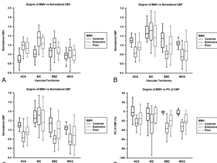
[image:3.594.52.285.58.225.2]The presence of BMV development in 32 hemispheres con-sisted of 19 extensive BMV hemispheres and 13 poor BMV hemispheres. In the ROIs of the EBZ and the MCA territory, the PCs of CBF in the extensive BMV hemispheres were sig-nificantly lower than those in diminished BMV hemispheres and normal control subjects (Table 5 and Fig 4). In the ROIs of the MCA and the ACA territories, the normalized baseline MTT values in the hemispheres with extensive BMV were sig-nificantly higher than those in the hemispheres of diminished BMV and normal control subjects (Table 5 and Fig 4). The values of all of the normalized baseline hemodynamic param-eters and the PC of CBF were significantly different between
Table 2: Modified Suzuki angiographic stages
Stages Angiographic Findings
I Mild-to-moderate stenosis around carotid bifurcation with absent or slightly developed ICA Moyamoya:* almost all of both ACA and MCA branches are opacified in antegrade fashion
II Severe stenosis around carotid bifurcation or occlusion of either of proximal ACA or MCA with well-developed ICA Moyamoya:* either ACA or MCA branches or both are clearly defective, but at least several of ACA or MCA branches remain opacified in antegrade fashion
III Occlusion of both proximal ACA and MCA with well-developed ICA Moyamoya:* only a few of either ACA or MCA branches or both are faintly opacified in antegrade fashion through the meshwork of ICA Moyamoya
IV Complete occlusion of both proximal ACA and MCA with absent or small amount of ICA Moyamoya:* without opacification of either ACA or MCA branches in antegrade fashion
Note:—Data were modified from the staging system proposed by Suzuki and Takaku.1 When the proximal ACA was hypoplastic, stage was determined by evaluating the proximal MCA involvement, opacification of MCA branches, and degree of development of ICA Moyamoya. ICA indicates internal carotid artery; ACA, anterior cerebral artery; MCA, middle cerebral artery.
* ICA Moyamoya indicates Moyamoya vessels at or around the terminal part of the ICA.
Table 3: The stages of basal Moyamoya vessels
Stages Angiographic Findings
I No Moyamoya vessels were seen
II Moyamoya vessels were localized in the area around the ICA bifurcation, and each vessel was fine and had little contrast
III Moyamoya vessels had intermediate extension and thickness IV Moyamoya vessels extended a great deal, and each one was
thick and strongly opacified
[image:3.594.52.286.285.381.2]the extensive BMV and poor BMV groups in the EBZ (Table 5).
Discussion
Our present study of ACZ-CTP in patients with ischemic adult Moyamoya diseases showed 2 major findings. First, the CTP is suitable for quantitative evaluation of CVR after ACZ chal-lenge and certainly provides information similar to that de-scribed previously with positron-emission tomography (PET) or SPECT.16,17Second, the PC of CBF is more correlated with angiographic stages than the baseline MTT, which has been shown previously to have positive correlation with the degrees of arterial stenosis in patients with Moyamoya disease.3In our study, there was no case presenting any major or minor com-plications associated with ACZ administration. SPECT usually requires 2-day protocol to evaluate cerebral perfusion, whereas ACZ-CTP study in our imaging protocol was per-formed within 30 minutes. Moreover, SPECT or PET does not routinely provide a quantitative CVR parameter. On the other hand, quantitative analysis using ACZ-CTP could be made
immediately after image processing. Nevertheless, radiation exposure is still a drawback of CTP imaging (ACZ-CTP: 0.2 mSv⫻2⫽0.4 mSv versus Diamox SPECT: 0.011 mSv⫻2⫽ 0.022 mSv); therefore, we excluded pediatric patients from this study and simplified our imaging protocol, including pre-contrast CT and ACZ-CTP. Our second major finding indi-cates that, among the resting and stress hemodynamic param-eters, the PC of CBF was correlated most significantly with angiographic stages, suggesting that the PC of CBF is a reliable parameter of cerebral hemodynamics in patients with Moya-moya disease. Previous report showed that MTT correlates positively with angiographic stages.3MTT can be measured by using CTP or MR perfusion imaging. By using dynamic sus-ceptibility contrast-enhanced MR imaging, Kikuchi et al18 showed that CVR impairment could be evaluated with MTT. However, our present study showed weak or fair correlation between baseline MTT value and angiographic stage. Baseline CBF value is used mostly in clinical practice; however, the regional CBF value is an insensitive indicator for the severity of occlusive cerebrovascular disease, because the value does not vary with a small change of cerebral perfusion pressure.19In our study, the baseline CBF did not significantly correlate with angiographic stages. Previous studies showed increased base-line CBV value distal to stenotic or occluded cerebral arter-ies20,21; however, we found that this parameter was variable compared with angiographic stage and clinical symptoms.
[image:4.594.134.456.41.211.2]As shown previously with PET and SPECT,4the hemody-namic status of the hemispheres with extensive BMV is differ-ent from that of hemispheres with diminished BMV. Patidiffer-ents with hemispheres with extensive BMV exhibited impaired perfusion in the cortices, whereas patients with hemispheres with diminished BMV did not, suggesting that intracerebral anastomoses may not provide an adequate blood supply to the cerebral cortices, even if they are fully developed. Piao et al4 reported that an extensive development of BMV is a sign of severe hemodynamic impairment in adult patients with isch-emic Moyamoya disease. Moyamoya vessels would develop to compensate for the misery cerebral perfusion. In our study, hemispheres with extensive BMV exhibited significantly lower PC of CBF and significantly higher baseline MTT than those in hemispheres with diminished BMV and normal control, indi-Fig 1.ROIs drawn in a reference CT image (A) and vessel-removed MTT map (B). The ROIs were placed on cortical regions in the MCA territory, EBZ, ACA territory, and putamen in the section of BG level around the foramen of Monro.
Table 4: Correlation coefficient between normalized baseline- and ACZ-challenged CT perfusion parameters and angiographic stages in each vascular territory
Variable BG* ACA* MCA* EBZ*
Observer 1
CBV vs AS 0.39† 0.29 0.29 0.69†
CBF vs AS ⫺0.16 ⫺0.24 ⫺0.50† ⫺0.33
MTT vs AS 0.47† 0.39† 0.52† 0.68†
PC of CBV vs AS ⫺0.36 0.11 ⫺0.29 ⫺0.19
PC of CBF vs AS ⫺0.65† ⫺0.40† ⫺0.81† ⫺0.82†
PC of MTT vs AS 0.25 0.32 0.33 0.48†
Observer 2
CBV vs AS 0.37 0.26 0.31 0.67†
CBF vs AS ⫺0.14 ⫺0.27 ⫺0.47† ⫺0.31
MTT vs AS 0.46† 0.38† 0.53† 0.64†
PC of CBV vs AS ⫺0.33 0.14 ⫺0.29 ⫺0.19
PC of CBF vs AS ⫺0.69† ⫺0.45† ⫺0.83† ⫺0.79†
PC of MTT vs AS 0.28 0.34 0.31 0.49†
Note:—CBV indicates normalized cerebral blood volume; CBF, normalized cerebral blood flow; MTT, normalized mean transit time; PC, percent change; AS, angiographic stage; BG, basal ganglia; ACA, anterior cerebral artery; MCA, middle cerebral artery; EBZ, anterior external border zone.
[image:4.594.53.285.281.437.2]rectly suggesting that the CVR parameters obtained by CTP are comparable with those obtained by SPECT or PET.
Previous study found that the values of hemodynamic parameters obtained from CTP before and after the ACZ challenge are correlated with those obtained from PET.16 However, there are different underlying physiologic mech-anisms between CTP and PET imaging. Moreover, both
methods have certain restrictions: CTP measurements de-pend on injection rate and cardiac output, whereas PET measurements offer low spatial resolution and assume that the time-activity curve of radiotracer in the radial artery is same as in the intracranial arteries, which may not hold true in patients with carotid stenosis or occlusion and collateral flows. Consequently, the data obtained with the 2 methods Fig 2.A 48-year-old woman with transient ischemic attack (case 16).A, Anteroposterior view of right internal carotid arteriogram shows severe stenosis in the proximal M1 portion of MCA with developed BMVs (modified Suzuki stage II).B, Anteroposterior view of left internal carotid arteriogram shows occlusion of distal ICA without antegrade flow (modified Suzuki stage IV). CBF maps before (C) and after (D) ACZ administration show decreased CVR in the left anterior EBZ (white arrow) and MCA territory (black arrow) ipsilateral to the hemisphere with higher modified Suzuki stage compared with contralateral hemisphere with lower modified Suzuki stage. Corresponding SPECT before (E) and after (F) ACZ administration show decreased CVR in the same areas.
[image:5.594.56.530.44.357.2] [image:5.594.56.532.425.605.2]may not exactly be interchangeable. Previous studies with ACZ-CTP lack normal data from healthy subjects, thus providing no reference values to determine abnormal find-ings. On the other hand, we determined normal values of baseline hemodynamic parameters and PCs of the parame-ters based on the data of 12 normal control subjects in our institution. Therefore, these normal values of hemody-namic parameters were the references in our study.
There are several limitations regarding the quantification of CTP. First, in the normal clinical setting, CTP commonly
[image:6.594.53.532.57.227.2]leads to the selection of major cerebral vessels for the AIF. These vessels lie over the circle of Willis and, therefore, are influenced by collateral recruitment. It is reasonable to expect that an AIF on the affected hemisphere will be subjected to more dispersion than that on the unaffected or slightly af-fected hemisphere because of increase of length of the collat-eral pathways supplying this region. However, the problem with using a single unaffected artery as a reference artery for the whole brain is that CBF values on the affected side may be underestimated, whereas MTT values may be overestimated.
Table 5: Differences of the mean values of normalized hemodynamic parameters according to the presence of BMV
Variable
BG ACA MCA EBZ
BMV(⫹) BMV(⫺) BMV(⫹) BMV(⫺) BMV(⫹) BMV(⫺) BMV(⫹) BMV(⫺)
Observer 1
CBV 1.34 1.22 1.21 1.14 1.06 0.98 1.24* 1.07*
CBF 1.27 1.21 0.85 0.97 0.75 0.89 0.78* 0.99*
MTT 1.10 1.04 1.45* 1.21* 1.46 1.20 1.76* 1.20*
PC of CBV ⫺2% 5% ⫺5% ⫺6% ⫺3% 4% ⫺11% ⫺5%
PC of CBF ⫺29% ⫺15% ⫺24% ⫺17% ⫺33%* ⫺18%* ⫺39%* ⫺21%*
PC of MTT 25% 20% 31% 22% 30% 22% 37% 26%
Observer 2
CBV 1.33 1.20 1.22 1.17 1.09 1.01 1.27* 1.10*
CBF 1.29 1.19 0.92 0.95 0.77 0.89 0.82* 0.96*
MTT 1.11 1.01 1.45* 1.24* 1.42 1.22 1.71* 1.22*
PC of CBV ⫺4% 5% ⫺7% ⫺9% ⫺6% 1% ⫺9% ⫺4%
PC of CBF ⫺32% ⫺16% ⫺21% ⫺13% ⫺32%* ⫺19%* ⫺33%* ⫺19%*
PC of MTT 27% 24% 35% 27% 27% 19% 34% 27%
Note:—CBV indicates normalized cerebral blood volume; CBF, normalized cerebral blood flow; MTT, normalized mean transit time; PC, percent change; BG, basal ganglia; ACA, anterior cerebral artery; MCA, middle cerebral artery; EBZ, anterior external border zone; BMV, basal Moyamoya vessel.
* Values are statistically significant (P⬍.05) by using analysis of variance and the multiple comparison test.
[image:6.594.81.506.260.579.2]In Moyamoya disease, the presence of steno-occlusion of the main cerebral arteries and collateral vessels always leads to the delay and dispersion of the bolus of the contrast agent. We used the AIF obtained from an MCA branch ipsilateral to the less affected hemisphere at the level of the BG, which is fine and narrow in patients with Moyamoya disease, and, there-fore, would be susceptible to partial volume averaging. A new method that can compensate for or resolve this problem is desirable. Second, the above limitations may result in a wide range of absolute measurements, making the detection of ab-normal values difficult. Intrasubject ab-normalization of data has been used frequently to deal with these problems. The use of hemispheric ratios takes advantage of the basic symmetry of the brain to improve sensitivity for identifying localized dis-ease. Moreover, intraobserver and interobserver variability for absolute hemodynamic parameter values can significantly be improved if the values are normalized. We used the baseline parameters and their PCs obtained from the hemispheric ratio to cerebellum, and a previous report indicates that the calcu-lation of ratios eliminates variations caused by the choice of AIF, and it may also reduce the variability in the proportion of gray and white matter that is included in the regional analy-sis.22Finally, there are no standardized guidelines for placing ROIs. Larger ROIs may result in greater volume averaging of gray and white matters, thus lowering quantitative values for CBF compared with results obtained by using smaller ROIs centered in the cortex. In the present study, an observer man-ually drew ROIs over the cortical gray matter of the expected territory of ACA, MCA, EBZ, and BG, with care not to involve substantial parts of the cerebral white matter. Large cortical blood vessels were automatically excluded from the ROIs.
Conclusion
The CTP is feasible for the evaluation of quantitative CVR and certainly provides information that is similar to that described previously with PET or SPECT. Moreover, the CTP is a much more readily accessible imaging method than PET or SPECT, and the quantitative analysis using CTP could be made imme-diately after image processing. The quantitative CVR (PC of CBF), measured by ACZ-CTP, is more significantly correlated with angiographic stage than normalized baseline CTP pa-rameters in adult ischemic Moyamoya disease. Therefore, CTP evaluation for the CVR in patients with Moyamoya dis-ease requires ACZ challenge. Future evaluation of this tech-nique in prebypass and postbypass patients is warranted to determine the potential clinical use.
Acknowledgments
We acknowledge Scott Pohlman of the Philips Medical Sys-tems for his cooperation and valuable advice.
References
1. Suzuki J, Takaku A.Cerebrovascular Moyamoya disease: disease showing ab-normal net-like vessels in base of brain.Arch Neurol1969;20:288 –99 2. Calamante F, Ganesa V, Kirkham FJ, et al.MR perfusion imaging in Moyamoya
syndrome: potential implications for clinical evaluation of occlusive cerebro-vascular disease.Stroke2001;32:2810 –16
3. Togao O, Mihara F, Yoshiura T, et al.Cerebral hemodynamics in Moyamoya disease: correlation between perfusion-weighted MR imaging and cerebral angiography.AJNR Am J Neuroradiol2006;27:391–97
4. Piao R, Oku N, Kitagawa K, et al.Cerebral hemodynamics and metabolism in adult Moyamoya disease: comparison of angiographic collateral circulation.
Ann Nucl Med2004;18:115–21
5. Yamada I, Himeno Y, Suzuki S.SPECT and MRI evaluations of the posterior circulation in Moyamoya disease.J Nucl Med1996;37:1613–17
6. Mugikura S, Rakahashi S, Higano S.The relationship between cerebral infarc-tion and angiographic characteristics in childhood Moyamoya disease.AJNR Am J Neuroradiol1999;20:336 – 43
7. Yamada I, Himeno Y, Nagaoka T, et al.Moyamoya disease: evaluation with diffusion-weighted and perfusion echo-planar MR imaging.Radiology1999; 212:340 – 47
8. Vorstrup S, Henriksen L, Paulson OB.Effect of acetazolamide on cerebral blood flow and cerebral metabolic rate for oxygen.J Clin Invest1984;74: 1634 –39
9. Sullivan HG, Kingsbury TB 4th, Morgan ME, et al.The rCBF response to Dia-mox in normal subjects and cerebrovascular disease patients.J Neurosurg
1987;67:525–34
10. Yamamoto S, Watanabe M, Uematsu T, et al.Correlation of angiographic cir-culation time and cerebrovascular reserve by acetazolamide-challenged sin-gle photon emission CT.AJNR Am J Neuroradiol2004;25:242– 47
11. Hoeffner EG, Case I, Jain R, et al.Cerebral perfusion CT: technique and clinical applications.Radiology2004;231:632– 44
12. Wintermark M, Maeder P, Thiran JP, et al.Quantitative assessment of regional cerebral blood flows by perfusion CT studies at low injection rates: a critical review of the underlying theoretical models.Eur Radiol
2001;11:1220 –30
13. Axel L.Tissue mean transit time from dynamic computed tomography by a simple deconvolution technique.Invest Radiol1983;8:94 –99
14. Van der Zwan A, Hillen B, Tulleken CAF, et al.Variability of the territories of the major cerebral arteries.J Neurosurg1992;77:927– 40
15. Hupperts RMM, Lodder J, Heuts-van Raak, et al.Borderzone brain infarcts on CT taking into account the variability in vascular supply areas.Cerebrovasc Dis
1996;6:294 –300
16. Bisdas S, Nemitz O, Berding G, et al.Correlative assessment of cerebral blood flow obtained with perfusion CT and positron emission tomography in symp-tomatic stenotic carotid disease.Eur Radiol2006;16:2220 –28
17. Honda M, Ezaki Y, Kitagawa N, et al.Quantification of the regional cerebral blood flow and vascular reserve in Moyamoya disease using split-dose iodo-amphetamine I 123 single-photon emission computed tomography.Surg Neurol2006;66:155–59
18. Kikuchi K, Murase K, Miki H, et al.Quantitative evaluation of mean transit times obtained with dynamic susceptibility contrast-enhanced MR imaging and with 133 Xe SPECT in occlusive cerebrovascular disease.AJR Am J Roent-genol2002;179:229 –35
19. Powers WJ.Cerebral hemodynamics in ischemic cerebrovascular disease.Ann Neurol1991;29:231– 40
20. Chen A, Shyr MH, Chen TY, et al.Dynamic CT perfusion imaging with acet-azolamide challenge for evaluation of patients with unilateral cerebrovascu-lar steno-occlusive disease.AJNR Am J Neuroradiol2006;27:1876 – 81 21. Derdeyn CP, Videen TO, Yundt KD, et al.Variability of cerebral blood volume
and oxygen extraction: stages of cerebral haemodynamic impairment revis-ited.Brain2002;125:595– 607