MOLECULAR CHARACTERIZATION AND CYTOPATHOLOGICAL STUDIES OF OLIVE LATENT VIRUS
1 (OLV-1) AND OLIVE LATENT VIRUS 2
1
Om-Hashem M. EL-Banna,
1
Plant Pathol,
2
Virus and Phytoplasma Res. Dept., Plant Pathol
ARTICLE INFO ABSTRACT
In a preliminary study for the assessment of the sanitary status of olive trees in Egypt during April and May in the two successive season 2014
symptomatic and asymptomatic ten olive cultivars from three diff
Marsa Matrouh and Beheira. RNA was extracted and used directly for one step RT
primer for each virus. Electron microscopy of sap extracts from olive infected leaves with the two viruses most commonly
showed isometric particles consistent to Oleavirus
Chenopodium quinoa
of chloroplast membrane, deformation of chloroplast, tubular like structural, crossing the all wall and deformation and lysis of the nucleus. Nucleotide sequenc
from the coat protein gene of OLV
OLV-2 showed homology ranged from 98% to 94 % with Italy and Poland Isolates for OLV % to 94 % with USA and
under study were identified as an isolates of OLV
Copyright©2017, Om-Hashem M. EL-Banna et al., This
unrestricted use, distribution, and reproduction in any medium, provided the original work is properly cited.
INTRODUCTION
Olive (Olea europaea L) belongs to Family:
Olea is one of the most extensively cultivated fruit crops worldwide (FAO, 2015). Olive is one of the crops of worldwide importance. Olive leaves are important for their secondary metabolites such as the phenolic compounds. Olive fruits are very high in vitamin E, vitamin k and other powerful antioxidants benefit, it prevents heart diseases; diabetes, lowers blood pressure and lowers bad cholesterol (Pereira 2007). It is a very important crop in Egypt and widely cultivated for oil production. The estim
cultivated olive trees in Egypt is around 203.000 Fadden’s of which the total production is 500.000 tons per year (Anomynous, 2015). Egypt represents about 18% of the total olive production and considered the second largest producers in the world (FAO, 2016). Olive trees are hosts to number of diseases caused by viruses, phytoplasmas, bacteria, fungi, and agents of diseases of unknown etiology.
*Corresponding author: Sahar A. Youssef,
Virus and Phytoplasma Res. Dept., Plant Pathol, Res. Res. Centre, Giza, Egypt.
ISSN: 0975-833X
Article History:
Received 01st June, 2017
Received in revised form 21st July, 2017
Accepted 22nd August, 2017
Published online 30th September, 2017
Citation: Om-Hashem M. EL-Banna, Engy M. Maher, Sahar A.Youssef and A. A. Shalaby Studies of Olive Latent Virus 1 (OLV-1) and Olive Latent Virus 2 (OLV
9, (09), 57910-57916.
Key words:
OLV-1, OLV-2, RT-PCR, Nigative stain,
Ultrastructural Changes, Sequence Analysis.
RESEARCH ARTICLE
MOLECULAR CHARACTERIZATION AND CYTOPATHOLOGICAL STUDIES OF OLIVE LATENT VIRUS
AND OLIVE LATENT VIRUS 2 (OLV-2) ISOLATED FROM OLIVE TREES IN EGYPT
Banna,
2Engy M. Maher,
2,*Sahar A. Youssef and
Plant Pathol, Dept., Fac. Agric., Cairo Univ., Egypt
Res. Dept., Plant Pathol, Res. Inst., Agric. Res. Centre, Giza, Egypt
ABSTRACT
In a preliminary study for the assessment of the sanitary status of olive trees in Egypt during April and May in the two successive season 2014-2015. Three hundred shoots were collected from symptomatic and asymptomatic ten olive cultivars from three diff
Marsa Matrouh and Beheira. RNA was extracted and used directly for one step RT
primer for each virus. Electron microscopy of sap extracts from olive infected leaves with the two viruses most commonly found Olive Latent Virus-1 (OLV-1) and Olive Latent Virus
showed isometric particles consistent to Necrovirus (OLV-1) and bacilliform particles consistent to Oleavirus (OLV-2). Ultrastructure changes in inoculated Chenopodium amaranticolor
enopodium quinoa showed necrolization and vaculation of the cytoplasm, layside and degenerated of chloroplast membrane, deformation of chloroplast, tubular like structural, crossing the all wall and deformation and lysis of the nucleus. Nucleotide sequencing analysis for 230bp amplified fragment from the coat protein gene of OLV-1 and 222bp amplified fragment from the coat protein gene of
2 showed homology ranged from 98% to 94 % with Italy and Poland Isolates for OLV
% to 94 % with USA and Italy for OLV-2. Based on molecular studies and cytopathological, viruses under study were identified as an isolates of OLV-1 and OLV-2.
This is an open access article distributed under the Creative Commons use, distribution, and reproduction in any medium, provided the original work is properly cited.
) belongs to Family: Oleaceae, genus: is one of the most extensively cultivated fruit crops worldwide (FAO, 2015). Olive is one of the crops of worldwide importance. Olive leaves are important for their secondary metabolites such as the phenolic compounds. Olive in E, vitamin k and other powerful antioxidants benefit, it prevents heart diseases; diabetes, lowers blood pressure and lowers bad cholesterol (Pereira et al.,
2007). It is a very important crop in Egypt and widely cultivated for oil production. The estimated acreage of cultivated olive trees in Egypt is around 203.000 Fadden’s of which the total production is 500.000 tons per year (Anomynous, 2015). Egypt represents about 18% of the total olive production and considered the second largest producers Olive trees are hosts to number of diseases caused by viruses, phytoplasmas, bacteria, fungi, and
Virus and Phytoplasma Res. Dept., Plant Pathol, Res. Inst., Agric.
Viruses were found to be the most serious disease causing problem in olive in different countries. Currently, fifteen different virus species belonging to 9 different genera have been identified in olive trees (M
2002). A number of viruses have been reported and mostly detected in symptomless olive trees such as: Olive latent virus -1 (OLV-1) which belongs to Family:
Necrovirus, positive-sense ssRNA (Serce
latent virus -2 (OLV-2) that belongs to Family: Genus: Oleavirus, positive sense ssRNA (Martelli
and Bjelis et al., 2007). The difficulty of recognizing and diagnosing virus-infected olive trees during field s
due to a lack of disease symptoms, the absence of differential woody indicator plants for bioassays and the unreliability of serological tests (ELISA). All these factors have made olive tree virus diagnosis very problematic (Alabdullah
However, Molecular methods based on polymerase chain reaction (PCR) amplification of the pathogen nucleic acid enable greater sensitivity especially when the target is in low concentration, or the pathogen has uneven distribution as in asymptomatic hosts (Olmos et al.,
and Hadidi and Candresse, 2001).
International Journal of Current Research
Vol. 9, Issue, 09, pp.57910-57916, September, 2017
Banna, Engy M. Maher, Sahar A.Youssef and A. A. Shalaby, 2017.“Molecular Characterization and Cytopathological 1) and Olive Latent Virus 2 (OLV-2) Isolated from Olive Trees in Egypt.”, International Journal of Current Research
MOLECULAR CHARACTERIZATION AND CYTOPATHOLOGICAL STUDIES OF OLIVE LATENT VIRUS
LATED FROM OLIVE TREES IN EGYPT
Youssef and
2Shalaby, A. A.
Res. Inst., Agric. Res. Centre, Giza, Egypt
In a preliminary study for the assessment of the sanitary status of olive trees in Egypt during April 2015. Three hundred shoots were collected from symptomatic and asymptomatic ten olive cultivars from three different locations in Egypt i.e., Siwa, Marsa Matrouh and Beheira. RNA was extracted and used directly for one step RT-PCR with specific primer for each virus. Electron microscopy of sap extracts from olive infected leaves with the two 1) and Olive Latent Virus-2 (OLV-2) 1) and bacilliform particles consistent to Chenopodium amaranticolor and showed necrolization and vaculation of the cytoplasm, layside and degenerated of chloroplast membrane, deformation of chloroplast, tubular like structural, crossing the all wall and ing analysis for 230bp amplified fragment 1 and 222bp amplified fragment from the coat protein gene of 2 showed homology ranged from 98% to 94 % with Italy and Poland Isolates for OLV-1 and 98 2. Based on molecular studies and cytopathological, viruses
is an open access article distributed under the Creative Commons Attribution License, which permits
Viruses were found to be the most serious disease causing problem in olive in different countries. Currently, fifteen different virus species belonging to 9 different genera have been identified in olive trees (Martelli, 1999; Felix and Clara, 2002). A number of viruses have been reported and mostly detected in symptomless olive trees such as: Olive latent virus 1) which belongs to Family: Tombusviridae, Genus:
sense ssRNA (Serce et al., 2007). Olive 2) that belongs to Family: Bromoviridae,
, positive sense ssRNA (Martelli et al., 1997 2007). The difficulty of recognizing and infected olive trees during field surveys is due to a lack of disease symptoms, the absence of differential woody indicator plants for bioassays and the unreliability of serological tests (ELISA). All these factors have made olive tree virus diagnosis very problematic (Alabdullah et al., 2006).
Molecular methods based on polymerase chain reaction (PCR) amplification of the pathogen nucleic acid enable greater sensitivity especially when the target is in low concentration, or the pathogen has uneven distribution as in
et al., 1999, Youssef et al., 2010
and Hadidi and Candresse, 2001).
OF CURRENT RESEARCH
Characterization and Cytopathological
Thus, the use of PCR technology is an important step to optimize and speed up olive tree virus diagnosis. The aim of the present investigation is to detect olive viruses associated with disorders observed in olive trees in different locations in Egypt depending on biological and molecular tests. Verify the association of the detected olive viruses (OLV-1 and OLV-2) with the disease and study the ultrastructural changes in tissues of the infected olive trees and indicator plants.
MATERIALS AND METHODS
Source of plant material
During April and May 2014-2015, samples of both symptomatic and asymptomatic shoots were collected from 300 olive trees from different fields located at three different locations in Egypt (Siwa, Marsa Matrouh and Beheira). The candidate trees represented 10 different cultivars (Maraqi, Kalamata, Picual, Manzanillo, Aagazy, Aagaze shami, Koroneiki, Dulce, kuartina, Khudairy), were taken in consideration during the samples collection. Samples were tested for seven viruses using RT-PCR.
Total RNA Extraction and One-step RT-PCR
amplification
Phloem tissue from young shoots of the collected samples were scraped and powdered in liquid nitrogen. About 100 mg of each sample were used for total RNA extraction using the Plant Total RNA Mini Kit, according to the manufacturer's protocol (Real Biotech, Taiwan). RNA was finally eluted with 50 µl of RNase- free water, and stored at -20 ºC until used. RT-PCR was carried out on RNA preparations using Reverse- transcription PCR Verso One STEP RT-PCR Reddy Mix Kit (Thermo Scientific). This allows RT and amplification to be performed sequentially in the same tube. In particular, For amplification, 50 μl of PCR reaction mix were added to each PCR tube contained the following reaction mixture: 25 μl of 2X 1-step PCR ready-mix, 2.5 μl of RT enhancer, 10 μM of forward and reverse primer for each virus (Table 1), 5 μl of template RNA, 1 μl of verso enzyme mix and the total volume to 50 μl by RNase/DNase-free water.
The PCR program consisted of cDNA synthesis was completed at 50 °C for 15 min., followed by verso inactivation at 95 °C for 2 min., amplification was carried out for 35 cycles under the following conditions: denaturation at 94°C for 30 sec., annealing at (50°C for 30 sec. in the case of ArMV, CLRV, OLYaV, OLRSV, SLRsV and OLV-1 and 55°C in the case of OLV-2),extension at 72°C for 1 min. followed by final
extension at 72 °C for 7 min. . Reactions were cycled in a thermo cycler TECHNE (TC-512). Amplified products were electrophoresed in 1-1.5 % agarose gel electrophoresis in 1XTBE buffer at 120V for 1 hour, stained with ethidium bromide (0.5μl/ml) and photographed using gel-documentation system (Bio-Rad, GelDoc XR , USA).VC 100bp Plus DNA ladder (Vivantis) was used as PCR Markers.
Cloning and sequencing of OLV-1 and OLV-2
RT-PCR products were cloned directly into plasmid PGEM-T-Easy vector system obtained from (Promega crop, USA) as described in the manufactory instruction manual. The DNA purification was performed by using GFX-DNA gel extraction kit (Pharmacia, amersham) as described by the manufacturer instruction. The purified recombinant plasmids were sequenced in Macrogen Inc. (Seoul, Korea). DNA sequencing was performed with M13 reverse and M13 forward primers. Nucleotide sequences were assembled; analyzed and phylogenetic trees were constructed for the Egyptian isolates of (OLV-1 and OLV-2) and the isolates available in the Gen Bank using DNAMAN software.
Morphology of virus particles
Virus dip preparation technique was applied on the olive infected samples with OLV-1 and OLV-2. Carbon coated cupper grids (400 mish) were dipped in sap expressed from olive infected leaves, and then negatively stained by 2% phosphotungestic acid (PTA) for 2min, then air dried. The grids were examined using transmission electron microscope JEOL (JEM-1400 TEM, Japan) at the candidate magnification. Images were captured using CCD Camera Model AMT at 50000X. This work was done at Faculty of Agriculture, Cairo University, Research Park (FARP) TEM lab.
Transmission electron microscopy (TEM)
Naturally infected olive samples were used as a source of the viruses. OLV-1 and OLV-2 were transmitted to Chenopodium
amaranticolor and Chenopodium quinoa respectively using
mechanical inoculation. Artificially inoculated plants were tested using one step RT- PCR and specific primer for each virus, and then infected plants directly applied to electron microscope.
Transmission electron microscopy (TEM) was carried out to check the ultrastructural changes inside the infected young leave of Chenopodium amaranticolor and Chenopodium
quinoa, respectively. Examination also included tissues of
healthy leaves. Materials prepared for electron microscopy included young leaf of Chenopodium amaranticolor and
[image:2.595.118.478.562.703.2]Chenopodium quinoa, representing symptoms.
Table 1. Sequence of specific primers used for the detection of olive viruses
Virus name Primer sequence bp Reference
ArMV 5'-AATACCCCGGGTGTTACATCG-3'F
5'-CATTAACTTAAGATCAAGGATTC-3'R
421nt
Grieco et al.,2000
CLRV 5'-AAAAGCTTGGCGACCGTGTAACGGCA-3'F
5'-AAGAATTCGTCGGAAAGATTACGTAAAAGG-3'R
431nt
OLV-1 5'-GTGGACTGCGCTCGAATGGA-3'F
5'-CTCACCATCGTTGTGTGG-3'R
230nt
OLRSV 5'-AAGAATTCTGCAAAACTAGTGCCAGAGG-3'F
5'-AAAAGCTTGCATAAGGCTCACAGGAG-3'R
492nt
SLRsV 5'-AAAAGCTTCAAGGAGAATATCCCTGGCCC-3'F
5'-AAGGATCCTAAGTGCCAGAACTAAACC-3'R
525nt
OLV-2 5'-CCGTTCTGTGGCCTTTGAGA-3'F
5'-AACACGATCCTCACCC-3'R
222nt
OLYaV 5'-ACTACTTTCGCGCAGAGACG-3'F
5'-CCCAAAGACCATTGACTGTGAC-3'R
346nt Savino et al., 1996
Pieces of about 2 x 2mm taken from samples were cut and transferred to a separate vial to be fixed in 2.5% glutaraldehyde in 0.1 M sodium phosphate buffer (pH 7.4) for an hour. After removing the fixative solution, the tissues were washed in sodium phosphate buffer three times for 30 min each. After washing, the buffer was pulled out, and 1% of osmium tetroxide (OsO4) was added to the tube and allowed for 1.5 h at 4˚c. After removing the fixative solution, the samples were dehydrated in an ethanol series of 15%, 30%, 50%, 70%, 80% and 95%, before exposing to 100% for 15 minutes for every step except the step of 100% ethanol. Which Infiltration with Spur's epoxy resin, one large drop into the sample tube every 15 minutes, until at 75% resin overnight with rotating. Samples were put into 100% resin, for at least a day, and then samples were placed into flat BEEM capsule molds, hardening the resin were done overnight in an oven at 60˚C. Samples were then sectioned (90-100 nm thick) with the ultra–microtome (Leica model EM-UC6) mounted on copper grids (400 mish). Sections were double stained with 2% urany1 acetate and 10% lead citrate, and then allowed to dry well. Stained sections were examined by transmission Electron microscope JEOL (JEM-1400 TE Japan) at the candidate magnification; images were captured using CCD camera model AMT. This work was carried in TEM lab, Faculty of Agriculture, Cairo University. Research Park (FARP).
RESULTS AND DISCUSSION
In the present work 300 olive shoot samples, representing three different locations(Siwa, Marsa Matrouh and Beheira) and ten cultivars in Egyptwere collected and checked for the presence of seven olive viruses ( Arabis mosaic virus (ArMV), Cherry
leaf roll virus (CLRV), Olive leaf yellowing associated virus
(OLYaV), Olive latent ringspot virus (OLRSV), Olive latent
virus-1 (OLV-1), Olive latent virus-2 (OLV-2), and
Strawberry latent ringspot virus (SLRSV) by Reverse
transcriptase- polymerase chain reaction (RT-PCR) as shown in (Table 2).
Olive viruses were found in all investigated samples with the percentage ranged from (0% for CLRV to 15% for OLV-2), high incidence of viruses' infection was detected in OLV-2 (15%) followed by OLV-1 (14%) as shown in (Table 2). Naturally infected olive trees with OLV-1 and OLV-2 viruses are mostly symptomless as those viruses are latent in olive, thus it is difficult to base diagnosis on symptoms expression. Biological indexing of olive viruses on woody differential indicators is not done as they are currently not available. The diagnostic bioassay that has been used extensively up to a recent past is mechanical transmission. This assay, however, is unreliable because of the low intrinsic sensitivity (Felix et al.,
2001). Our detection results of OLV-1 and OLV-2 in infected olive trees using Reverse transcriptase- polymerase chain reaction (RT-PCR) with specific primers increase the reliability of this detection method which allowed increased investigations on distribution of the viruses in different areas in Egypt. The similar result was obtained by (Grieco et al., 2000 and Pantaleo et al., 2001). To detect olive viruses using PCR assay, it is important that nucleic acid samples extracted from the original source of plant material is free of appreciable amounts of oil, polysaccharides, phenolic compounds, and other PCR Inhibitors (Wilson, 1997). These inhibitors are present in olive tissue extracts (Amiot et al., 1989; De Niro et al., 1997) they must be removed in order to detect the viral RNA targets by RT-PCR.
The choice of an extraction technique that can be used for routine testing of a large number of samples must take into account simplicity of use and rapidity of execution (Youssef et al., 2010). The results of this study showed that RNA extraction procedure developed in this study is suitable for routine use in diagnostic laboratories. One step RT-PCR analysis was simple and fast because it allowed testing of hundreds of samples to be done in a relatively short time. These results also coupled with the sensitivity and the absence of contamination risks (since the assay is done in a single tube) made this technique very suitable for large-scale investigation (Bertolini et al., 2001; Ragozzino et al., 2004). The one-step RT-PCR protocol confirmed for all tested olive tree viruses because of its rapidity and reliability (Faggioli et al., 2005). Reverse transcriptase polymerase chain reaction (RT-PCR) has been reported as one of the most sensitive methods for detection and identification of RNA viruses that infect olive (Grieco et al., 2000).
Since the genomic sequences of the majority of viruses, including the somewhat rare OLV-1 and OLV-2 are known (Grieco et al., 1995) the design and use of adequate PCR primers is now possible, as exemplified by the successful identification OLV-1 and OLV-2 by single step RT-PCR (Sabanadzovic et al., 1999; Bertolini et al., 1998, and Youssef
et al., 2010).
Chenopodium amaranticolor and Chenopodium quinoa were
mechanically inoculated with OLV-1 and OLV-2 respectively. Necrotic local lesions followed by systemic infection appeared
on Ch. amaranticolor as shown in (Fig.1.A) Necrotic local
lesions and vein clearing appeared on Ch. quinoa as shown in (Fig.2.B). These results of the olive viruses (1 and OLV-2) were confirmed by RT-PCR amplification. Transmission and symptoms expression is in agreement with (Martelli et al., 1996, Grieco et al., 2002).
Fig.1: (A) Symptoms reaction of OLV-1 mechanically inoculated
onChenopodium amaranticolor showed necrotic local lesions
followed by systemic infection. (B) OLV-2 mechanically
inoculated on Chenopodium quinoa showed necrotic local lesions
and vein clearing followed by systemic infection.
Fig.(2) (A) An electron micrograph of OLV-1 particles negatively stained with 2% phosphotungestic acid (80000X). (B) OLV-2
particles negatively stained with 2% phosphotungestic acid (60000X).
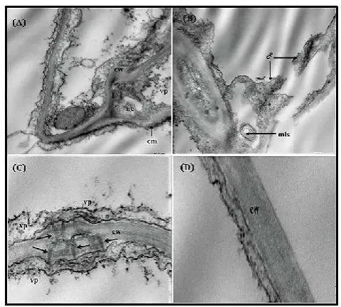
Ultra-thin section of Ch. amaranticolor leaves with OLV-1
and Ch. quinoa artificially inoculated with OLV-2 and those
of healthy ones investigated by electron microscope. Electron microscopy of the mesophyll tissue revealed that cell arrangement was disordered if compared with tissues of healthy plants. Regarding the ultrastructural (cytological) changes observed in OLV-1 infected plants they were obvious in mesophyll layer as it was disorganized and disordered (Fig.3.A) as chloroplasts were miss sharpen , contained abnormal rounded bodies and the cell wall was irregular. In (Fig.3.B) the uneven thickness of the cell wall, disorganization of the cell membrane was observed. A Few number of isometric virus particles were detected in the cytoplasm next to the cell wall. The cell membrane was obliterated and invaginated in several positions (arrows). Narcolization and vacuolation of the cytoplasm is also obvious as a result of virus infection. The nucleus was also affected (Fig.3.C) as it was enlarged, the nuclear membrane was irregular and disturbed.
Degradation of chloroplast membrane is also clear and the chloroplast lost its normal structure if compared with that in cell of healthy Ch. amaranticolor leaf tissues (Fig.3.D). Regarding the cytopathological effect of OLV-2 on infected
Ch. quinoa leaf tissues, numerous changes at the cell and
organelle level were observed .The mesophyll layer was deformed, the different organelles were obliterated .The cell wall was highly affected as it was splatted, showing protrusions into the cytoplasm, lysis of the cell wall was very clear as a result of the cell death and narcolization of the cytoplasm (Fig.4.A.). Full splatting of the cell wall and plasma membrane was remarkable in (Fig.4.B.).
Fig.(3): (A) An electron micrograph of ultrathin section in chenopodium amaranticolor leaf infected with OLV-1 showing that cell arrangement of the mesophyll is disordered .cw=cell wall ,m=mitocondria ,n=nucleus ,er=endoplasmic reticulum ,cst= circular structure and ch=chloroplast (12000X). (B) Magnified part of Fig.3.a, representing the uneven thickening and disorganization of the cell wall on the cell membrane. Few number of isometric virus particles (vp) is observed adjacent to cell membrane. Necrolization and vacuolation of the cytoplasm is obvious. vp=virus particles, nm= nuclear membrane and va= vacuolation (30000X). (C) Magnified part from Fig.3.A, representing the nucleolus of palisade cell as severe dilation and disturbance the nuclear membrane is observed with full disappearance of the nucleus. The chromatin material is condensed preferably. The affected nucleus contained rounded inclusion bodes containing aggregates of virus particles. The mitochondria are also swollen. chm= chromatin material (30000X). (D).Normal chloroplast in mesophyll cell of healthy Ch.amaranticolor leaf. The organized structure of lamina and grana is obvaius.sg=starch granules, lg=lipid granules, g/l=grana and lamina (20000X).
On the other hand tubular like structures attached with the plasmodesmata were observed in the cell wall which permitted the virus particles to cross over from the cell to another and virus particles were arranged on both sides of cell wall (Fig.4.C.). All these changes were not observed in cell wall and cell membrane of healthy cells as shown in (Fig.4.D.). AS olive leavs contained oil droplets and polysaccarides, these materials were an obesticul in visiualization of the ultrathin sections prepared for electron microscopy. So both
Ch.amaranticolor and Ch. quinoa leaf tissues infected with
[image:4.595.312.553.49.245.2]OLV-1and OLV-2 respectavily were used for this purpose. Significant ultrastructural changes occurred in cells of OLV-1
Table 2. Screening of olive viruses infecting olive trees under fields condition in three different locations in Egypt using one step RT-PCR with specific primer for each tested virus
locations cultivars No .of tested samples Olive viruses tested
OLV-1 OLV-2 OLRSV ArMV SLRSV CLRV OLYaV Siwa
Aggazi 30 3 4 0 0 0 0 0
Aggazi shami 40 8 3 1 0 0 0 0
Dulce 20 4 1 1 0 0 0 0
Picual 50 1 10 4 0 1 0 0
Maraqi 20 2 3 2 0 0 0 0
Marsa Matrouh
Manzanillo 35 9 2 1 0 0 0 1
Aggazi 25 3 1 1 0 1 0 0
Khudairy 15 2 4 2 0 0 0 0
Aggazi shami 25 2 3 3 1 0 0 0
Kalamata 20 0 5 1 0 0 0 0
Beheira Picual 15 3 4 0 0 0 0 0
Manzanillo 10 2 1 0 0 0 0 1
kuartena 10 1 1 4 0 0 0 0
Koronaki 15 1 2 2 0 0 0 0
Kalamata 10 1 1 1 0 0 0 0
Total 300 14% 15% 7.7% 0.3% 0.7% 0% 0.7%
[image:4.595.37.287.51.155.2] [image:4.595.62.540.468.643.2]Fig.(4): (A). An electron micrograph of cross section is mesophyll
cell of Chenopodiumquinoa infected with OLV-2. Lysis and
disorganization of cell wall. nacrotization of the cytoplasm and the cell membrane is irregular in shape.virus particles are scattered in the necrotized cytoplasm. cw=cell wall ,nc=necrotized
cytoplasm ,vp=virus particles ,cm=cell membrane (20000X).(B). full splitting of the cell wall. as a result of infection. mayalin like structure is observed in the necrotized cytoplasm. mls=mayline
like structure (20000X). (C).tubular like structures (arrow) attached to the plasmodesmata crossing the cell wall. virus particle are observed on both sides of the cell wall and cell membrane (50000X). (D).cell wall of healthy leaves (50000X)
infected host as compared with that of non-inoculated control plant, when observed under electron microscope. Major alterations are: presence of electron dense rounded virus particles and the cells arrangement of the mesophyll are disordered. They occur in the cytoplasm of mesophyll parenchyma cells and in conducting tissues, either scattered or in ordered paracrystaline arrays and often in association with filaments of proteinaceous nature. Extensive vacuolation of the cell membrane system, from which numerous vesicles appear to derive, with variable sized up to 100 nm in dimeter. these can be observed within the nuclear envelop likely originated from the inner and the outer membrane of the nucleus, freely scattered in the cytoplasm, aggregated near the dyctiosomes and inside the vacuole appearing to protrude from tonoplast .the most part of such abundant vesicular structures contain a fine network of fibrils, usually interpreted as nucleic acid folded strands (Felix et al., 2007).
Cytoplasmic inclusions made up of parallel filaments with a criss- cross pattern or helical structure, often mingled with virus like particles. Occasionally, those filaments were also seen inside the cell nucleus. Small electron dense amorphous inclusions scattered throughout cytoplasm (Castellano et al.,
1987 and Panataleo et al., 2006). OLV-2 is not the only member of the family Bromoviridae to induce the formation of virus containing tubules (Grieco et al., 1999). Comparable structures were previously observed in cells infected with tobacco streak as described by (Martelli and Russo, 1985) and tomato aspermy as described by (Francki et al., 1985) viruses.OLV-2 was found in a cytoplasmic membrane enriched fraction and was detected by immunogold labelling in close proximity to or within plasmodesmata that lacked desmotubules, as recently reported also for AMV (Van der
Wel et al., 1998).
Such tubules may constitute a common but transient feature (which is thus difficult to perceive) of Bromoviridae infection (Zheng et al., 1997). Generally, the described cell alternations are seen in both locally and systemically infected
Chenopodium amaranticolor as well as Chenopodium quinoa
for the most part, the role of the newly induced structure in the infection process or in cell defense. (Castellano et al., 1987 and Felix et al., 2007). Sequencing of PCR-amplified fragment for OLV-1 and OLV-2 were completed to compare the sequence from these isolates with those of other viruses available in GenBank (http://www.ncbi.nlm.nih.gov.). The sequence was performed using the PCR product obtained when primer specific to each virus under study were used.
When the sequences obtained were compared with the coat protein gene of the five strain of OLV-1, 98% similarity with Italy, USA isolates and 96% similarity with Japan isolates were shown. While 94% similarities with Poland and Portugal was obtained in this study (Fig.5) Similarly, the coat protein gene sequences for OLV-2 were done. The coat protein gene sequences of OLV-2 isolate were sharing 98% with USA and 94% with Italy and this isolates is available in the GenBank as shown in (Fig.6).
Fig. (5): Phylogenetic tree generated from the multiple alignment of deduced nucleotide sequence of CP gene for OLV-1 available in
Genbank Italy, USA, Japan, Poland and Portugal and OLV-1 Egyptian isolate. Values at the nodes indicate significance in a
bootstrap analysis
Fig. (6): Phylogenetic tree generated from the multiple alignment of deduced nucleotide sequence of CP gene for OLV-2 available in
Genbank USA, Italy and OLV-2 Egyptian isolate. Values at the nodes indicate significance in a bootstrap analysis
[image:5.595.42.282.51.267.2] [image:5.595.312.556.328.464.2] [image:5.595.309.556.501.620.2]sequences of OLV-2 isolated shared 98% with USA and 94% with Italy identify with other isolates. Same results were obtained from (Grieco et al., 2002) they reported that Blast analysis showed 97% nucleotide identity with isolates from Greece and 98%, 97%, 96%, 94% and 93% with the type isolate for the CP protein respectively. In both proteins, amino acid substitutions were mostly concentrated in the N terminus region. These result demonstrated the successful of application of RT-PCR technique for the detection of OLV-1 and OLV-2 in Egypt. The analysis of retrieved from nucleotide sequence databases of the part of the coat protein gene which can be successfully for the comparison with all known isolates of OLV-1 and OLV-2 available in the Gen Bank.
REFERENCES
Amiot, M.J., Fleuriet, A., Macheix, J.J. 1989. Accumulation of oleuropein derivatives during olive maturation. J.
Phytochemistry V.(28): 67–69.
AlAbdullah, A., El Beaino1, T., Saponari, M., Hussain, H., Digiaro1 M., Paolo G., Martelli, G. 2006. Preliminary survey of olive viruses in Syria. Ninth Arab Congress of
Plant Protection, V (9):19-23.
Anonymous 2015. Agriculture data. In: Agriculture Statistic Data Base. Food and agric., Organization of the United
Nation.org.
Bertolini, E., Fadda, Z., Garcia, F., Celada, B., Olmos, A., Gorris, M. T., Del Rio, C., Caballero, J., Duran-Vila, N., Cambra, M. 1998. Virosis del olivo detectadas en Espana. Nuevos metodoa de diagnostico. Phytoma. Espana. 102 : 191-193.
Bertolini, E., Olmos, A., Carmen Martinez, M.C., Gorris, M. T., Cambra, M.A. 2001. Single-step multiplex RT-PCR for simultaneous and colourimetric detection of six RNA viruses in olive trees. J. of Virol. Methods, 96(1):33-41. Bjelis, M., Loconsole, G., Saponari, M. 2007. Presence of
viruses in Croatian olive groves. Pomologia Croatica, 13(3):165-172.
Castellano, M.A., Franco, A. Di., Martelli, G.P. 1987. Electron microscopy of two Olive viruses in host tissues. J. of
Submicroscopic Cytology, 19, (1): 495-508
De Niro, A., Lombardon, N., Perri, E., Procopio, A., Raffaelli, A., Sindona, G. 1997. Direct identification of phenolic glucosides from olive leaf extracts by atmospheric pressure ionization tandem mass spectrometry. J. Mass
Spectrom, V. (32): 533–541.
Francki, R. I. B., Milne, R. G., Hatta, T. 1985. Cucumovirus group. In An Atlas of Plant Viruses. Boca Raton: CRC Press. V. (2): 53-100.
Faggioli, F., Ferretti, L., Albanese, G., Sciarroni, R., Pasquini, G., Lumia, V., Barba, M. 2005. Distribution of olive tree viruses in Italy as revealed by one-step RT-PCR. J. of
Plant Pathol. V(87): 49-55.
Felix, M. R., Clara, M. I. E., Leitao, F., Fernandez Serrano, J. M. 2001. virus incidence in four Olea europea cultivars evaluated by mechanical inoculation and immunological assays. 4th International Symposium on olive growing,
Bari 2000. Acta Horticulture.
Félix, M.R., Clara, M.I.E. 2002. Virus incidence in four Olea europaea cultivars evaluated by mechanical inoculation and immunological assays. Acta Hortic. 586:721–724. Felix, M. do R. F., Clara, M. I. E. da. 2005. Characterization
and diagnosis of viruses occurring on Olea Europaea. Techniques in diagnosis of plant viruses, V(6):189-226.
Felix, M. do R. F., Cardoso, J. M. S., Oliveira, S., Clara, M. I. E. 2007. Biological and Molecular Characterization of Olive latent virus 1. Plant viruses Global Science Books, 1 (2) : 170-176.
F.A.O 2015. Olive Statistics 2015. Food and agriculture organization of the United Nations.
F.A.O 2016. Olive Statistics 2016. Food and agriculture organization of the United Nations.
Grieco, F., Martelli, G. P., Savino, V. 1995. The nucleotide sequence of RNA3 and RNA4 of olive latent virus 2. J.
Gen.Virol .V(76): 929–937.
Grieco, F., Gallitelli, D. 1999. Multiplex reverse transcriptase polymerase chain reaction applied to virus detection in globe artichoke. J. of Phytopat. 147: 183-185.
Grieco, F., Alkowni, R., Saponari, M., Savino, V., Martelli, G. P. 2000. Molecular detection of olive viruses. Bulletin OEPP/EPPO Bullettin V(30):469-473.
Grieco,F., Parrella, G., Vovlas , C. 2002. An isolate of olive
latent virus 2 infecting castor bean in Greece. J. of Plant
Pathol., 84 (2): 129-131.
Hadidi, A., Candresse, T. 2001. Virus Detection - PCR. In: Maloy O.C., Murray T.D. (eds.). Encyclopaedia of Plant Pathol., pp. 1095-1100. John Wiley & Sons, Inc., New York, USA.
Martelli, G. P., Russo, M. 1985. Virus host relationships: symptomatological and ultra structural aspects in the plant viruses. Polyhedral Virions with Tripartite Genomes. New York: Plenum Press pp:163-205.
Martelli, G.P., Yilmaz, M.A., Savino, V., Baloglu, S., Grieco, F., Güldür, M.E., Greco, N., Lafortezza, R. 1996. Properties of a citrus isolate of Olive latent virus 1 a new necrovirus. European J. of Plant Pat.,102 : 527-536 Martelli, G. P., Grieco, F. 1997. Oleavirus a new genus in the
family Bromoviridae. Archives of Virol., 142(9):1933-6. Martelli, G. P. 1999. Infectious diseases and certification of
olive. Bullefin O€PP/€PPO Bulletin 29 :127-133.
Olmos, A., Cambra, M., Esteban, O., Gorris, M.T., Terrada, E. 1999. New device and method for capture, reverse transcription and nested PCR in a single closed-tube. Nucleic Acids ResearchV (27):1564-1565.
Pantaleo, V., Saponari, M. and Gallitelli, D. 2001. Development Of A Nested PCR Protocol For Detection Of Olive-Infecting Viruses In Crude Extracts. J. of Plant
Pathol.83 (2): 143-146.
Pantaleo, V., Grieco, F., Franco, A. Di; Martelli, G.P. 2006. The role of the C-terminal region of Olive latent virus 1 coat protein in the host systemic infection. Archives of Virol. 151: 1973-1983.
Pereira , P.A., Ferreira, C.F.R; Filipa, M., Patricia, V., Paula, B.A., Rosa, S., Estevinho, L., Albino, B., Alberto J.P. 2007. Phenolic Compounds and Antimicrobial Activity of Olive (Olea europaea L. Cv. Cobrançosa) Leaves. J.
Molecules, 12(5):1153-1162.
Parrella, G., De Stradis, A., Vovlas C. 2007. First report of Olive latent virus 2 in wild castor bean (Ricinus communis) in Italy .ISSN 2044-0588.
Ragozzino, E., Faggioli ,F., Barba, M. 2004. Development of a one tube-one step RT-PCR protocol for the detection of seven viroids in four genera. Apscaviroid, Hostuviroid, Pelamoviroid and Pospiviroid. J. of Virol. Methods
(121):25-29.
Savino, V., Sabanadzovic, S., Scarito, G., Laviola, C., Martelli, G. P. 1996. Due giallumi di possibile origine virale in Sicilia. Informatore Fitopatologico 46 (5): 55-59.
Sabanadzovic, S., Abou-Ghanem, N., La Notte, P., Savino, V., Scarito, G., Martelli, G. P. 1999. Partial molecular characterization and RT-PCR detection of a putative closterovirus associated with olive leaf yellowing. J.of
Plant Pathol. V(81):37-45.
Serce, C. U., Yalcn, S., Gazel, M., Caglayan, K., Faggioli, F. 2007. First report of Olive latent virus 1 from olive trees in Turkey. J. of Plant Pathology., 89(3):S73:S69-S76. Van der Wel, N. N., Goldbach, R. W., van Lent, J. 1998. The
movement protein and coat protein of alfalfa mosaic virus accumulate in structurally modi®ed plasmodesmata. Virology 244:322-329.
Vovlas, C., Parrella, G., Di Franco, A., 2002. Infezioni naturali del virus latente 2 dell’olivo su piante di ricino in Grecia.
Informatore Fitopatologico 7-8: 66-68.
Varanda, C. M. R., Nolasco, G., Clara, M. I., Felix, M. R. 2014. Genetic diversity of the coat protein of olive latent virus 1 isolates. Archives of Virol ., 159(6):1351-1357. Wilson, I.G. 1997. Inhibition and facilitation of nucleic acid
amplification. Appl. Environ. Microbiol., 63: 3741–3751 . Youssef, S., Moawed, S., El-Sayed, M., Shalaby, A.A. 2010.
Detection of olive tree viruses in Egypt by one-step RT-PCR. International Conference on Virus and other Graft Transmissible Diseases of Fruit Crops 427: 51-55.
Zheng, H., Wang, G., Zhang, L. 1997. Alfalfa mosaic virus movement protein induces tubules in plant protoplasts. Molecular Plant Microbe Interactions 10:1010-1014