University of Warwick institutional repository: http://go.warwick.ac.uk/wrap
A Thesis Submitted for the Degree of PhD at the University of Warwick
http://go.warwick.ac.uk/wrap/57641
This thesis is made available online and is protected by original copyright. Please scroll down to view the document itself.
Native mass spectrometry
approaches to study
zinc-binding plasma proteins
Esther Marie Martin
A thesis submitted in partial fulfilment of the
requirements for the degree of
Doctor of Philosophy in Chemistry
Department of Chemistry
University of Warwick
ii
Table of Contents
List of Figures
viiList of Tables
xAcknowledgements
xiDeclaration
xiiiAbstract
xivList of Abbreviations
xviAmino acid Abbreviations
xviii1
Introduction
1.1 Metal ions in biological systems 1
1.2 The importance of Zn2+ 2
1.3 Blood plasma proteins 3
1.4 Human serum albumin (HSA) 4
1.4.1 Structure and function 4
1.4.2 HSA dimer formation 6
1.4.3 HSA as a Zn2+ transporter 7
1.5 Ligand binding to HSA 8
1.5.1 Metal binding sites 8
1.5.2 Fatty acid transport by HSA 15
1.5.3 Fatty acid binding sites 16
1.6 Conformational changes of HSA 17
1.6.1 pH-induced conformational changes 17 1.6.2 Fatty acid-induced conformational changes 18 1.7 Interactive binding of metal ions and fatty acids 20
1.7.1 Evidence that fatty acid binding influences Zn2+ binding
20 1.7.2 Disruption of Site A due to occupation of FA2 22
1.7.3 Biological significance 23
1.8 Histidine rich glycoprotein (HRG) 24
1.8.1 Structure and function 24
1.8.2 The His-rich region of HRG 28
1.8.3 Metal binding to HRG 29
1.8.4 Zn2+ mediates HRG-ligand interactions 32 1.8.5 Could the fatty acid “switch” mechanism be
involved in the regulation of HRG functions?
33
iii
2
Experimental methods
2.1 Materials and chemicals 37
2.2 Methods for defatting HSA 37
2.2.1 Dialysis of HSA 37
2.2.2 Charcoal treatment 38
2.3 Characterisation of HSA 38
2.3.1 SDS-PAGE 38
2.3.2 Determining HSA concentration 39
2.3.3 Assay for free thiol content 39
2.3.4 Reaction with DTNB for ESI-MS analysis 39
2.4 ESI-MS of HSA 40
2.4.1 Desalting with PD-10 column 40
2.4.2 Sample preparation 40
2.4.3 Acquiring data: microTOF 41
2.4.4 Acquiring data: maXis-UHR-TOF 41
2.4.5 Identifying fatty acids in the low mass range 42
2.5 Preparation of HSA complexes 42
2.5.1 Metal ion-bound HSA 42
2.5.2 Fatty acid-bound HSA 43
2.5.3 pH titration with HSA 43
2.6 Determination of metal:protein stoichiometry by ICP-OES 43
2.7 NMR experiments with HSA 44
2.7.1 1-D 1H NMR spectroscopy 44
2.7.2 111Cd-NMR spectroscopy 45
2.8 TWIM-MS of HSA complexes 45
2.8.1 Experimental parameters 45
2.8.2 TWIM-MS calibration: sperm whale myoglobin 46 2.8.3 Estimation of theoretical cross sections using
MOBCAL
46 2.9 Purification of the synthetic peptide HRGP330 46
2.9.1 Reverse phase-high performance liquid chromatography
46
2.9.2 Lyophilisation of peptides 47
2.9.3 Estimating HRGP330 concentration 48
2.10 Metal binding experiments with HRGP330 48
2.10.1 Desalting HRGP330 samples 48
2.10.2 Metal ion titrations by ESI-MS 49
2.10.3 pH titration of Zn5-HRGP330 by ESI-MS 49
2.10.4 CD spectroscopy of HRGP330 50
2.10.5 Comparison of apo- and holo-HRGP330 using TWIM-MS
iv 2.11 Comparison of apo- and holo-HRGP330 by NMR
spectroscopy
51
2.11.1 NMR sample preparation 51
2.11.2 1-D 1H NMR spectroscopy 51
2.11.3 2-D homonuclear [1H, 1H] NMR spectroscopy 51 2.11.4 Chemical shift referencing using residual water 52 2.12 Electrospray-tandem mass spectrometry (ESI-MS/MS) 52 2.13 Interaction of HRGP330 with a heparin dodecasaccharide 53 2.14 Determination of an apparent binding constant for HRGP330 53
2.15 Zn2+ binding to full length HRG 54
2.16 Metal binding to Gly-Gly-His peptide 54
2.16.1 ESI-MS 54
2.16.2 1-D 1H NMR spectroscopy 55
2.17 Metal ion transfer experiments 55
2.17.1 Zn2+ distribution between HSA and HRGP330 55 2.17.2 Separation of HSA and HRGP330 with a MWCO
filter
55
2.17.3 Size exclusion chromatography 56
2.17.3 Cu2+ transfer between Gly-Gly-His and HRGP330 56
2.18 Bioinformatic analysis 56
3
HSA-ligand complexes
3.1 Introduction 58
3.2 Electrospray-ionisation mass spectrometry (ESI-MS) 58
3.2.1 An overview of mass spectrometry 58
3.2.2 Development of ESI-MS for biomolecules 59
3.2.3 Mechanisms of ion formation 60
3.3 Advantages of ESI-MS 61
3.4 Maintaining the native structure of proteins in the gas phase 62
3.4.1 Sample preparation 62
3.4.2 Instrumental conditions 63
3.5 How has ESI-MS previously been applied to albumin? 64
3.6 Results and discussion 66
3.6.1 Native ESI-MS of HSA 66
3.6.2 Incomplete desolvation of the analyte ions 68
3.6.3 NanoESI-MS of HSA 71
3.6.4 Analysis of Zn2+-HSA 72
3.6.5 pH-induced Zn2+ release and unfolding of HSA 74 3.6.6 Analysis of HSA in the presence of fatty acids 76 3.6.7 Binding of PFCAs to HSA in the gas phase: a fatty
acid mimic
v 3.6.9 Analysis of Zn2+ binding in the presence of
myristate
83 3.6.10 Identification of myristic acid in low mass range 85
3.7 Conclusion 87
4
Travelling wave ion mobility mass spectrometry
of HSA
4.1 Introduction 88
4.2 Ion-mobility mass spectrometry 88
4.2.1 Development of IM-MS 88
4.2.2 TWIM-MS 89
4.2.3 A commercially available TWIM-MS instrument: Synapt HDMS
90 4.2.4 Calibration of travelling wave ion mobility data 91
4.3 Results and discussion 92
4.3.1 TWIM-MS of recombinant HSA at physiological pH 92 4.3.2 Comparison of N- and F-forms of HSA. 95 4.3.3 Comparison of the conformations of apo- and
holo- HSA
97 4.3.4 Conformational changes induced by fatty acids 101
4.4 Conclusion 105
5
Metal binding properties of a peptide mimic from
HRG
5.1 Introduction 107
5.2 HRGP330: a peptide mimic of the His/Pro-rich region 107
5.3 Results and discussion 109
5.3.1 Purification of HRGP330 by RP-HPLC 109 5.3.2 Metal binding properties of HRGP330 investigated
by ESI-MS
113 5.3.3 Comparison of Zn2+ binding to intact HRG 116
5.3.4 pH titration of Zn5-HRGP330 117
5.3.5 CD spectroscopy of HRGP330 118
5.3.6 TWIM-MS of HRGP330 120
5.3.7 Comparison of apo- and Zn2+ bound HRGP330 by TWIM-MS
122
5.4 NMR spectroscopy of HRGP330 123
5.4.1 1-D and 2-D NMR spectroscopy of apo-HRGP330 123 5.4.2 1-D and 2-D NMR spectroscopy of Zn-HRGP330 127
5.5 Tandem mass spectrometry techniques 131
5.5.1 An overview of MS/MS 131
vi 5.5.3 Electron Capture Dissociation and Electron
Transfer Dissociation.
132 5.5.4 MS/MS of non-covalent protein complexes:
metalloproteins
134
5.5.5 MS/MS of HRGP330 135
5.5.6 MS/MS of Zn2+-bound HRGP330 138
5.6 Conclusion 147
6
Investigating metal ion distribution between HSA
and HRGP330
6.1 Introduction 149
6.2 Results and discussion 149
6.2.1 Zn2+ transfer occurs between HSA and HRGP330 149 6.2.2 Does Zn2+ transfer occur from HRGP330 to HSA? 151 6.2.3 Effect of fatty acid on Zn2+ distribution 152 6.2.4 Effect of excess HSA on Zn2+ distribution 155 6.2.5 Investigation of metal binding between HSA and
HRGP330 at plasma Zn2+ concentrations
159 6.2.6 Estimation of the apparent binding constant for
Zn2+-HRGP330
162 6.2.7 Cu2+ transfer between Gly-Gly-His and HRGP330 164 6.2.8 Implications of Zn2+ transfer to HRG: effect on
HRGP330 binding to heparin
167
6.3 Conclusion 171
7
Conclusion
7.1 Summary of observations 173
7.1.1 Native ESI-MS of HSA-ligand complexes 173 7.1.2 Metal binding properties of HRGP330 174 7.1.3 Metal ion distribution between HSA and HRGP330 176
7.2 Future directions 179
References
182vii
List of Figures
1
Introduction
1.1 Essential vs. non-essential metal ions 2 1.2 Summary of the biological functions of HSA 5
1.3 X-ray crystal structure of HSA 6
1.4 Structure of the ATCUN motif at the N-terminus of HSA
10 1.5 Zn2+ binding site located at the interface between
domain I and domain II of HSA
13 1.6 X-ray crystal structure of myristate-bound HSA. 16 1.7 Influence of myristate loading on metal binding to
BSA by 111Cd-NMR spectroscopy
21 1.8 Molecular modelling shows binding modes of fatty
acids with different chain lengths to FA2
23 1.9 Multi-domain structure proposed for HRG 27 1.10 Sequence alignment of the HRR of HRG from
various mammalian species
31 1.11 Mechanism of possible HRG regulation by the levels
of plasma fatty acids
34
3
HSA-ligand complexes
3.1 A general overview of a mass spectrometer 59
3.2 Ion formation during ESI-MS 61
3.3 Comparison of the charge state distribution for denatured and native HSA
66 3.4 Deconvoluted spectrum of acid-denatured HSA 68 3.5 Effect of in-source CID on desolvation of protein
complex ions
70 3.6 Observation of higher order oligomers of HSA during
nano-ESI-MS and SDS-PAGE
72 3.7 Native ESI-MS of HSA shows 1 Zn2+ ion bound 73 3.8 Acid-induced dissociation of Zn2+ from HSA 75 3.9 HSA titrated with varying amounts of fatty acid
followed by ESI-MS
77
3.10 Binding of PFOA to HSA by ESI-MS 80
3.11 Simultaneous binding of PFOA and Zn2+ to HSA observed by ESI-MS
82 3.12 Effect of myristate on Zn2+ binding to HSA 83 3.13 Comparison of Zn2+ binding to Gly-Gly-His peptide
by ESI-MS and 1H NMR spectroscopy
85 3.14 Detection of protonated myristic acid in the low m/z
range of HSA
viii
4
Travelling-wave ion mobility mass
spectrometry of HSA
4.1 An outline of the ion mobility cell in the Synapt G2 instrument
91 4.2 Comparison of ATD for the 16+ and 15+ charge
states of apo-HSA at varying cone voltages of 60, 80 and 100 V
94
4.3 ATDs for the 17+ and 18+ charge states of HSA measured at pH 7.4 and pH 4.0
96 4.4 Comparison of ATD for the 17+, 16+, 15+ and 14+
charge states of apo-HSA, Zn2-HSA and Cd2-HSA
98
4.5 Dependence of the estimated collisional cross
section on charge state for apo-HSA, Zn2-HSA and
Cd2-HSA
99
4.6 Comparison of ATD for the 16+ and 15+ charge states of Zn2-HSA at varying cone voltages of 60, 80
and 100 V
100
4.7 Dependence of charge state for the estimated
collisional cross sections of apo-HSA and HSA in the presence of 5 mol. equiv. myristate
102
4.8 Comparison of experimental and theoretical collisional cross sections for HSA complexes
104
5
Metal binding properties of a peptide mimic of
HRG
5.1 Location of HRGP330 in the full-length sequence of HRG
108 5.2 Purification of crude HRGP330 with RP-HPLC 110 5.3 Identification of purified HRGP330 by SDS-PAGE
and ESI-MS
111 5.4 Comparison of apo-HRGP330 spectra from two
different mass spectrometers
112
5.5 Metal binding properties of HRGP330 115
5.6 pH titration of Zn5-HRGP330 118
5.7 Far-UV region CD spectrum of HRGP330 in the presence and absence of Zn2+
119 5.8 ATDs and estimated collisional cross sections of the
charge states observed for apo-HRGP330
121 5.9 Effect of Zn2+ binding on the ATD and collisional
cross section of the 5+ charge state of HRGP330
123 5.10 Stacked plot of 700 MHz 1-D 1H-NMR spectra
showing fingerprint region of HRGP330 obtained at varying temperatures from 5-35 °C
124
5.11 Fingerprint region of [1H,1H] TOCSY and NOESY spectra of HRGP330 obtained at 700 MHz
126 5.12 Stacked plot of 700 MHz 1-D 1H-NMR spectra
showing fingerprint region of HRGP330 in the presence of varying mol. equiv. ZnCl2
ix 5.13 Effect of Zn2+ on TOCSY spectrum of HRGP330 at
700 MHz
130 5.14 Schematic showing the nomenclature of peptide
ions formed from tandem MS/MS experiments
131
5.15 CID spectrum of apo-HRGP330 136
5.16 ETD spectrum of apo-HRGP330 137
5.17 CID spectrum of Zn-HRGP330 139
5.18 Comparison of the CID spectra of apo-HRGP330 (867 m/z), Zn1-HRGP330 (880 m/z) and Zn2
-HRGP330 (893 m/z)
140
5.19 Comparison of experimental and theoretical isotopic patterns for the Zn2+ adducts of y12 and y10
generated from CID
141
5.20 ETD spectrum of Zn-HRGP330 143
5.21 Zn2+ binding to HRGP330 affects the intensity of different ions produced by ETD
144 5.22 Identification of Zn2+-adducts in the ETD spectrum of
Zn1-HRGP330
145
6
Investigating metal ion distribution between
HSA and HRGP330
6.1 ESI-MS of HRGP330 and ICP-OES of HSA as evidence for Zn2+ transfer having occurred
150 6.2 Direct infusion of a 1:1 mixture of dialysed HSA and
Zn-HRGP330
152 6.3 Direct infusion of a 1:1 mixture of Zn-HSA and
HRGP330 in the absence and presence of myristate
153 6.4 HSA:Zn2+ stoichiometry in the presence of 1 mol.
equiv. HRGP330 (± myristate)
154 6.5 Relative intensity of Zn1-HRGP330 following
incubation with Zn-HSA (± myristate)
156 6.6 HSA:Zn2+ stoichiometry (600 µM) in the presence of
20 µM HRGP330 (± myristate)
157 6.7 Elution of HSA and insulin B chain on a
BioSep-SEC-2000 column
159 6.8 Elution of HSA and HRGP330 from a
BioSep-SEC-2000 column
161 6.9 Estimation of the apparent binding constant of Zn2+
and HRGP330 using competition with Zincon
163
6.10 Structure of Gly-Gly-His 165
6.11 Effect of HRGP330 on Cu2+ binding to Gly-Gly-His 166 6.12 Chemical structure of the heparin dodecasaccharide
(dp12) used in this work
168 6.13 ESI-MS shows increased HRGP330:dp12 complex
in the presence of Zn2+
169 6.14 Close-up view of the 5+ charge state of
HRGP330:heparin complex
x
List of Tables
1
Introduction
1.1 Stability constants for metal ion-HSA complexes 9 1.2 Albumin conformations that are dependent on pH 18
1.3 Biological functions of HRG 26
1.4 Stability constants for metal ion-HRG complexes 32
2
Experimental methods
2.1 Gradient used for purification of HRGP330 by RP-HPLC
47
3
HSA-ligand complexes
3.1 Non-covalent albumin complexes observed by ESI-MS
65
4
Travelling-wave ion mobility mass
spectrometry of HSA
4.2C Estimated collisional cross sections of the conformations observed for apo-HSA at each cone voltage
94
4.8B Experimental cross sections for HSA and theoretical values obtained from the PA and EHSS methods
104
6
Investigating metal ion distribution between
HSA and HRGP330
6.1B ICP-OES analysis of the HSA fraction confirms a significant loss of Zn2+ in the presence of
HRGP330
xi
Acknowledgements
Firstly, I would like to extend my gratitude to my supervisors, Dr Claudia Blindauer and Prof Peter Sadler for giving me the opportunity to undertake this research project and for their guidance over the past few years.
A big thank you to all the members of the Blindauer group both past and present for all their help: James, Amira, Maria, Tanvir, Greg, Fran, Jie, Oksana and Jin. I also thank the Dixon group for all our interesting discussions on proteins and in particular to Fay and Mike - all those lattes definitely helped!
I am extremely grateful to all the members of the Chemical Biology Research Facility for their help in the lab and for use of the equipment.
Also thanks must go to the Sadler group for making me feel so welcome on the fourth floor, particularly to Ruth McQuitty for her help with the HPLC.
I would also like to express my gratitude to Andrea Lopez-Clavijo for her mass spectrometry advice and useful discussions about albumin.
Thanks go to Dr Alan Stewart (University of St Andrews) for his valuable ideas on the project and the kind donation of protein samples.
xii I would like to thank all my friends and family for all their love and encouragement (Mom and Dek, Dad and Jo, Em, Jon, Nanny Rose and Grandad John). And to Tom: thanks for your continued reassurance and cups of tea.
xiii
Declaration
I hereby declare that this thesis, submitted in partial fulfilment for the degree of Doctor of Philosophy, represents my own work and has not been previously submitted to this or any other institution for another qualification. Work was carried out between October 2009 and December 2012 under the supervision of Dr Claudia Blindauer and Prof Peter Sadler.
Collaborative work contributes to Chapter 4 and parts of Chapter 5. In summary, ion-mobility mass spectrometry experiments were carried out in collaboration with Dr Frances Kondrat and electron transfer dissociation data with Dr Julia Smith (Bruker, Coventry, UK). In both cases, the data analysis was carried out by the author. Other contributions are explicitly stated throughout.
xiv
Abstract
Human serum albumin (HSA) is a plasma protein that fulfils a wide range of biological functions and is thought to be the major Zn2+ transporter in blood plasma. The high affinity Zn2+ binding site (Site A) has recently been characterised and is located at an interdomain site. In addition to metal binding, HSA is also important in the transport of fatty acids. Previous work has shown that the binding of Zn2+ at Site A and the binding of myristate at fatty acid site 2 are mutually exclusive. It has been predicted that upon fatty acid binding, a conformational change occurs that can disrupt the residues that form Site A. This allosteric interaction could have an impact on the Zn2+ dependent activities of histidine-rich glycoprotein (HRG), a plasma protein involved in blood coagulation. The purpose of this work was to investigate the metal-binding properties of HSA and a peptide derived from HRG using a native MS approach. Furthermore, the possible Zn2+ transfer between the proteins was explored and also whether fatty acids influenced the Zn2+ distribution.
Native ESI-MS was able to detect Zn2+ ions associating with HSA although the interactions with fatty acids appeared to be broken upon entering the gas phase. No apparent loss of Zn2+ from HSA was observed by ESI-MS following incubation with myristate which was confirmed by elemental analysis in solution. Travelling wave ion mobility-MS showed no significant conformational changes between apo-HSA and holo-HSA although Zn2+ appears to have a role in stabilising the domain I/II interface. HSA incubated with myristate showed a larger collisional cross section that is in agreement with the X-ray crystal structures.
xvi
Abbreviations
1-D, 2-D, 3-D One-. two-, three-dimensional
Å Angstrom
ATCUN Amino terminal Cu2+ and Ni2+ binding ATD Arrival time distribution
ATP Adenine triphosphate BSA Bovine serum albumin
CD Circular dichroism spectroscopy
CID Collision induced dissociation fragmentation
DEPC Diethyl pyrocarbonate
DSA Dog serum albumin
DTNB 5,5-dithiobis (2-nitrobenzoic acid) or Ellman’s reagent
DTT Dithiothreitol
ECD Electron capture dissociation fragmentation
EDTA Ethylenediaminetetraacetic acid
ESI-MS Electrospray ionisation-mass spectrometry ETD Electron transfer dissociation fragmentation
FA Fatty acid
GGH Gly-Gly-His peptide
HEPES 4-(2-Hydroxyethyl)piperazine-1-ethanesulfonic acid HMWK High molecular weight kininogen
HRG Histidine-rich glycoprotein
HRGP330 Histidine-rich glycoprotein peptide, residues 330-365 HRGP335 Histidine-rich glycoprotein peptide, residues 335-365 HRR His-rich region
HSA Human serum albumin
ICP-MS Inductively coupled plasma-mass spectrometry
ICP-OES Inductively coupled plasma-optical emission spectroscopy IM-MS Ion mobility-mass spectrometry
ITC Isothermal titration calorimetry LCFA Long chain fatty acid
MCFA Medium chain fatty acid
MS Mass spectrometry
MS/MS Tandem mass spectrometry MWCO Molecular weight cut-off NMR Nuclear magnetic resonance
NOESY Nuclear Overhauser enhancement spectroscopy PFCA Perfluorinated carboxylic acid
xvii pI Isoelectric point
ppb Parts per billion ppm Parts per million PRR Pro-rich region
PTR Proton transfer reaction QTOF Quadrupole-Time-of-Flight
RP-HPLC Reverse phase-high pressure liquid chromatography SCFA Short chain fatty acid
SDS-PAGE Sodium dodecyl sulphate-polyacrylamide gel electrophoresis
SEC Size exclusion chromatography TOCSY Total correlated spectroscopy TOF Time of flight
TWIM-MS Travelling wave ion mobility-mass spectrometry UHR Ultra-high resolution
xviii
Amino acid abbreviations
Single letter code
Three letter code
Full name
A Ala Alanine
C Cys Cysteine
D Asp Aspartic acid
E Glu Glutamic acid
F Phe Phenylalanine
G Gly Glycine
H His Histidine
I Ile Isoleucine
K Lys Lysine
L Leu Leucine
M Met Methionine
N Asn Asparagine
P Pro Proline
Q Gln Glutamine
R Arg Arginine
S Ser Serine
T Thr Threonine
V Val Valine
W Trp Tryptophan
1
Chapter 1
Introduction
1.1 Metal ions in biological systems
Many transition metals play crucial roles in biology where they can enhance or even determine the activities of biomolecules. Approximately 50% of all enzymes require interaction with a metal ion to fulfil their biological function (Waldron et al., 2009). A delicate balance of these elements needs to be maintained as high concentrations can be toxic but too little can be equally damaging and have a detrimental effect on cells.
2
Figure 1.1 Essential vs. non-essential metal ions. The diagram illustrates that the effect of a metal ion on an organism is concentration dependent. Biologically essential metal ions can become toxic at high concentrations and non-essential metal ions can only be tolerated at low concentrations. Adapted from Bertrand (1912).
1.2 The importance of Zn2+
Zn2+ is the second most abundant trace metal ion present in man after iron, with 2-3 g in adult humans (Haase and Rink, 2009). It is considered to be relatively non-toxic to humans (Fosmire, 1990) and studies administering the radioisotope 65Zn to patients showed that 90% of the metal ion is stored in the bones and muscle (Wastney et al., 1986). In biology, Zn2+ exists only in a 2+ oxidation state and does not undergo redox reactions, a consequence of its filled d orbital. Furthermore, it is a borderline Lewis acid which allows it to interact with a variety of ligand donors including nitrogen, sulfur and oxygen. A study of 57 organisms in 2006 indicated that Zn2+-binding proteins are extremely abundant, comprising 4-10% of proteomes (Andreini et al., 2006).
Zn2+ is vital for many cellular processes, and its roles in nature can be broadly placed into three categories: structural, catalytic and regulatory. It is involved in the transmission of genetic information, the development
Concentration Optimal concentration
Good effects (health)
Bad effects
(death) Essential metal ions
3 of the immune system and as a signalling substance (Cousins et al., 2006). On a cellular level it has critical roles in proliferation and apoptosis and therefore is essential for growth (Maret and Sandstead, 2006). Many enzymes require Zn2+ in order to be catalytically active, for example, carbonic anhydrase which allows the interconversion of carbon dioxide and water to bicarbonate and protons to proceed rapidly (Keilin and Mann, 1940). In addition, Zn2+ is required in so-called “Zn2+ finger proteins” where the protein chain arranges itself around the metal ion to form a stable fold. An example of this was first discovered in a transcription factor from Xenopus Laevis, TFIIIA, which contained a series of these small domains which were ordered in the presence of Zn2+ (Miller et al., 1985).
1.3 Blood plasma proteins
4 normally present in healthy plasma may accumulate as a biomarker for a particular disease which further complicates the system (Schaller et al., 2008). This study will focus on two particular plasma proteins: human serum albumin (HSA) and histidine-rich glycoprotein (HRG) with a focus on their metal-binding properties.
1.4 Human serum albumin (HSA) 1.4.1 Structure and function
5
Figure 1.2Summary of the biological functions of HSA.
6
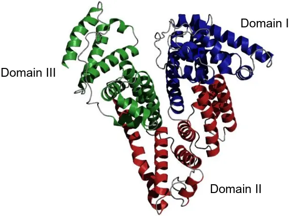
Figure 1.3 X-ray crystal structure of HSA. The three homologous domains are highlighted as follows: blue = domain I, red = domain II and green = domain III. The figure was created using PyMOL v1.3 and based on PDB 1AO6.
1.4.2 HSA-dimer formation
It has been well documented in the literature that HSA can form a dimer that is suggested to form through a covalent disulfide bond between Cys34 of two HSA molecules. Evidence against this, however, is that this residue is buried in a hydrophobic cavity 9.4 Å deep but it has been proposed that the pocket could flatten out to bring the residues closer together in space (Peters, 1995). A synthetic mercury bridged dimer was first produced by Straessle and co-workers in 1954 by linking the Cys34 residue of two HSA molecules, although it showed low stability (Edsall et al., 1954; Straessle, 1954). Other efforts with longer linker molecules to avoid strain on the protein molecules achieved high yields and a dimer that was stable for over a year at room temperature (Komatsu et al., 1999). It is not known in great detail what biological significance a HSA dimer may have, although the possible clinical applications have recently
Domain I
[image:25.595.145.441.77.298.2]7 been reviewed (Taguchi et al., 2012). These include its use as a plasma volume expander to treat conditions such as inflammation and as a drug carrier because it shows prolonged blood retention compared to the monomer.
1.4.3 HSA as a Zn2+ transporter
The concentration of Zn2+ in plasma is estimated to be ca. 15-20 µM (Kiilerich et al., 1980). It is thought that HSA is the major transporter of Zn2+ in blood plasma (Cousins, 1986) and binds approximately 80% of the metal ion present (Foote and Delves, 1984). The remainder is bound by α2-macroglobulin and low molecular weight compounds such as Cys and His which are able to compete for protein-bound Zn2+ (Giroux and Henkin, 1972). Although early studies implicated transferrin in Zn2+ transport this was found to be an insignificant amount (Chesters et al., 1981).
8 appear to be due to enhanced occupancy of the metal ion to HSA (Failla
et al., 1982).
1.5 Ligand binding to HSA 1.5.1 Metal binding sites
HSA has the capacity to bind various metal ions, both essential and toxic, with different specificities at each binding site (Martins and Drakenberg, 1982; Bal et al., 1998). Although there is a lack of detailed structural information, four metal binding sites have been described in the literature:
a) Amino-Terminal Cu2+ and Ni2+ binding motif (ATCUN) b) Site A
c) Site B
d) Free thiol on Cys34
9
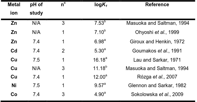
Table 1.1 Stability constants for metal ion- HSA complexes. Metal
ion
pH of study
nc logK1 Reference
Zn N/A 3 7.53b Masuoka and Saltman, 1994
Zn N/A 1 7.10b Ohyoshi et al., 1999
Zn 7.4 1 6.98a Giroux and Henkin, 1972
Cd 7.4 2 5.30a Goumakos et al., 1991
Cu 7.5 1 16.18a Lau and Sarkar, 1971
Cu N/A 3 11.18b Masuoka and Saltman, 1994
Cu 7.4 1 12.00a Rózga et al., 2007
Ni 7.5 1 9.57a Glennon and Sarkar, 1982
Co 7.4 3 4.90a Sokolowska et al., 2009 a. K1 = apparent binding constant of the highest affinity binding site
b. K1= intrinsic binding constant of the highest affinity binding site. These are not
dependent on pH. c. Number of binding sites
Amino-Terminal Cu2+ and Ni2+ binding motif (ATCUN)
10 associated with circulating amino acids (Neumann and Sass-Kortsak, 1967).
In contrast, HSA is the major transporter of Ni2+ with 95% estimated to be bound to the protein in vivo (Lucassen and Sarkar, 1979) although Ni2+ is generally thought to be non-essential for human health. Intriguingly, this ATCUN motif does not occur in some albumins from mammalian species such as dog serum albumin (DSA) and as a result this protein does not show a specific binding site for the first equivalent of Cu2+. This suggests that the mechanism of Cu2+ metabolism could differ across mammalian species (Appleton and Sarkar, 1971).
Figure 1.4 Structure of the ATCUN motif at the N-terminus of HSA. Asp1, Ala2 and His3 are involved in binding and M represents a Cu2+ or Ni2+ ion. Lys4 may also have a role in metal binding as proposed by Sadler et al. (1994)
Co2+ binding to HSA has attracted attention due to the albumin-cobalt binding assay (ACB assay), an FDA-approved test for myocardial ischemia (Bar-Or et al., 2000). In patients with acute coronary syndrome the binding of Co2+ in serum is significantly reduced (Bar-Or et al., 2001)
M
His3 Ala2
Asp1
N
O N
O
O–
O
N H
N protein
11 and this can be detected colorimetrically by measuring complex formation between Co2+ and dithiothreitol (DTT) which has absorbance at 470 nm. Early studies suggested that the N-terminal site was the primary binding site for Co2+ due to the fact that it was shown by potentiometry to interact with peptide mimics of the N-terminus (Lakusta and Sarkar, 1979). Sadler and co-workers (1994) provided further evidence for this by showing that addition of Co2+ to HSA reduced the intensity of the 1H-NMR resonances observed for the N-terminal residues (Asp1, Ala2, His3 and also Lys4). However, it was not clear if the binding mode was the same as the square-planar arrangement formed with Cu2+ and Ni2+. More recent studies have demonstrated that Co2+ actually competes with Zn2+ and Cd2+ for the occupation of Sites A and B and that only the third equivalent will be bound to the N-terminus (Mothes and Faller, 2007; Sokolowska et al., 2009). ITC and 111Cd-NMR experiments have also confirmed that the primary binding site is not the N-terminus as the addition of Co2+ to
111
Cd2+ loaded HSA caused suppression of the peaks for Sites A and B as seen by NMR spectroscopy. This could have implications for the molecular mechanism of the ACB assay as the reduced Co2+ binding of plasma from ischemic patients may be due to elevated fatty acids (Lu et al., 2012a).
Site A
12 metal ion. 111Cd-NMR experiments of HSA yield two signals at ~ 25-30 ppm and another at 110-150 ppm, designated Site B and Site A respectively. Site A has been identified as the high affinity Zn2+ binding site on HSA and an interdomain location was proposed based on analysis of the albumin crystal structures available. The amino acids involved are from two domains: His67 and Asn99 from domain I and His247 and Asp249 from domain II. It is important to note that this ligand set has also been identified in other Zn2+-binding enzymes such as human calcineurin,
E. coli 5-endonucleotidase and kidney bean purple acid phosphatase
13
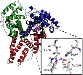
Figure 1.5 Zn2+ binding site located at the interface between domain I and domain II of HSA. Domain I is shown in blue and domain II is shown in red with the Zn2+ ion highlighted in magenta. The close-up image shows the 5-coordinate site composed of His67, Asn99, His247, Asp249 and a coordinating water molecule. The model was created using PyMOL v1.3 and based on the model published by Blindauer et al. (2009).
Cd2+ can also bind to Site A but is readily displaced by Zn2+ as shown by
113
Cd-NMR spectroscopy (Martins and Drakenberg, 1982; Goumakos et al., 1991). Circular dichroism (CD) studies also indicated that it is a weaker binding site for Cu2+ and Ni2+ once the N-terminus has become saturated (Sadler and Viles, 1996) which was also confirmed by the work of Bal et al. (1998). This is consistent with the findings of Masuoka and Saltman (1994) who demonstrated by equilibrium dialysis that the first equivalents of Zn2+ and Cu2+ do not bind to the same site.
Site B
A high affinity Cd2+ site has been identified, Site B, but its location is yet to be fully characterised (Goumakos et al., 1991). The binding of Cu2+ or
His67
Asp249
His247 Asn99
14 Zn2+ to HSA did not reduce its affinity towards Cd2+ which supported the idea of a site that preferentially bound Cd2+ (Goumakos et al., 1991). This was later confirmed by 113Cd-NMR data which showed that the peak for Site B was not supressed by the addition of 0.5-1.5 mol. equiv. of Zn2+ (Sadler and Viles, 1996). It has been proposed that the site consists of all oxygen ligands plus one or no nitrogen ligands, based on the fact that its
111
Cd chemical shift is 30 ppm (Öz et al., 1998; Stewart et al., 2003). A
13
C-NMR study with tripeptides suggested residues Asp35, Glu36 and His37 as good candidates for this binding site (Lakusta et al., 1980).
Free thiol located on Cys34
The final metal binding site that can be discussed is the only free thiol on the protein located at Cys34. The remaining 34 Cys residues are involved in the formation of disulfide bonds which are a characteristic trait of the albumin family. The sulfur has a high affinity for ‘soft’ acids for example Au+, Ag+ and Hg2+. Previous workers have reported that metallodrugs can bind at this site, for example Pt2+ anticancer drugs (Pizzo et al., 1988; Esposito and Najjar, 2002) and also Au+ antiarthritic drugs (Christodoulou
et al., 1994). Although Sadler and co-workers later showed that it was
15 1.5.2 Fatty acid transport by HSA
16 1.5.3 Fatty acid binding sites
Experimental evidence has indicated that fatty acid binding stabilises HSA against denaturation (Kragh-Hansen, 1981). Under normal physiological conditions, 0.1-2 moles of fatty acids are complexed to HSA, but seven sites have been identified by X-ray crystallography (Curry et al., 1998). Five of these sites are proposed to be of high to moderate affinity where the molecules are anchored into the protein by polar interactions (Bhattacharya et al, 2000). There is no evidence of such interactions at the other two sites and therefore these are thought to be weaker binding sites. Figure 1.6 shows the distribution of the high affinity binding sites occupied by myristate molecules. FA1 is located in subdomain IB and FA2 is at the interface between IA and IIA. FA3 and FA4 are both located in subdomain IIIA and FA5 is in subdomain IIIB. Sites FA6 and FA7 are not shown in Figure 1.6, but FA6 lies at the interface between IIA and IIB and FA7 in subdomain IIA (Curry, 2004).
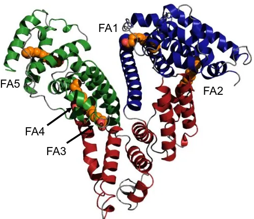
Figure 1.6 X-ray crystal structure of myristate-bound HSA. The protein structure is shown in the same colour scheme as previous figures. Myristate molecules are shown to occupy FA sites 1-5 (orange). The figure was created using PyMOL v1.3 and based on PDB 1BJ5 (Curry et al., 1998).
FA1
FA2
[image:35.595.184.434.494.710.2]17 For many years, efforts have been directed towards quantifying the affinity of various fatty acids for HSA, even though difficulties occur as they are not readily soluble in aqueous media. Simard and co-workers carried out an NMR titration of HSA with 13C-labelled palmitate, which revealed that FA sites 2, 4 and 5 are the high affinity binding sites whereas 1, 3, 6 and 7 are the low affinity binding sites (Simnard et al., 2006). This is most likely because sites 2, 4 and 5 provide the most enclosed environment and the aliphatic chain can sit in the pocket in almost a linear conformation (Van der Vusse, 2009).
1.6 Conformational changes of HSA
1.6.1 pH-induced conformational changes
18
Table 1.2 Albumin conformations that are dependent on pH
Abbreviation Name pH of transition Characteristics
E Extended 2.7 Fully expanded form with disulfide bonds intact F Fast 4.3 Fast migrating form
observed on gels N Neutral/Normal 7.0 Normal heart-shaped
conformation observed at physiological pH. B Basic 8.0 Subtle change in molecule
as it loses rigidity; physiologically relevant A Aged 10.0 Slower migrating form
observed on gels
Dockal and co-workers demonstrated by CD spectroscopy and fluorescence measurements that during the N to B transition, domains I and II experienced an isomerisation in tertiary structure whereas domain III was unaffected (Dockal et al., 2000). The biological relevance of these isoforms has yet to be fully determined, although they are thought to be important for ligand binding and release.
1.6.2 Fatty acid-induced conformational changes
19 chemical cross-linking and mass spectrometry has also been used to probe the effect of unsaturated fatty acid binding to HSA and the results correlated well with the changes observed in the crystal structures (Huang et al., 2005).
It is unclear whether these conformational changes have physiological significance, but one theory is that it could allow HSA receptors to differentiate between HSA molecules loaded with fatty acid and those that are fatty acid-free. This could have important implications for the delivery of fatty acids to cells (Curry et al., 1999). Overall, it is debatable whether HSA protein receptors on cells exist but there has been some evidence put forward (Schnitzer and Oh, 1994; de Château et al., 1996; Tiruppathi
et al., 1996; Luiken et al., 1997). The conformational change could also
20 1.7 Interactive binding of metal ions and fatty acids
1.7.1 Evidence that fatty acid binding influences Zn2+ binding
Evidence to date that fatty acids influence the occupation of Site A has been obtained from combinations of 111Cd-NMR spectroscopy, 1H-NMR spectroscopy, isothermal titration calorimetry (ITC) and molecular modelling. Zn2+ is spectroscopically inactive, therefore it can usefully be replaced with isostructural Cd2+ in order to gain information about metal binding.
21
Figure 1.7 Influence of myristate loading on metal binding to BSA by 111Cd-NMR spectroscopy. For dialysed BSA two peaks are observed: one for Site A and one for Site B. In the presence of myristate these peaks are significantly affected. (Lu et al., 2012b).
Further evidence for a relationship between fatty acids and Zn2+ was gained from ITC. Titrations with Zn2+ in the presence of 5 mol. equiv. octanoate gave similar data as titrations carried out in the absence of fatty acid. This was consistent with 1H-NMR data showing simultaneous binding of Zn2+ and octanoate as although the H1 resonances for His67 and His247 were affected it was concluded that octanoate binding to FA2 does not abolish Zn2+ binding to BSA (Lu et al., 2012b). In contrast, analysis of BSA-Zn2+ in the presence of myristate by ITC revealed that Zn2+ binding is dramatically decreased. This suggested that the chain length of the fatty acid is an important factor. One of the most important outcomes of ITC was the observation that Zn2+ binding was affected by the addition 1-2 molar equivalents of myristate which are physiologically relevant levels in plasma (Lu et al., 2012b).
111Cd
A
B
BSA + 5 Myr
Dialysed BSA
111Cd
A
B
BSA + 5 Myr
[image:40.595.214.402.74.247.2]22 1.7.2 Disruption of Site A due to occupation of FA2
X-ray crystallography of fatty acid-bound albumin showed that the occupation of site FA2 was mainly responsible for the conformational change observed. Crucially, this is the only fatty acid site that is located between two domains (Curry et al., 1998) and is also next to Site A, the major Zn2+ binding site. At this site, the carboxylate end of the molecule interacts with Arg257 and Ser287 in subdomain IIA and the aliphatic tail interacts with Tyr150 in subdomain IB. In order to accommodate the fatty acid anion, domains I and II have to move by 10 Å to form a cavity that is large enough. This causes significant disruption of the Zn2+ binding site as the His247/Asp249 pair from domain II and the His67/Asn99 pair from domain I are moved apart by 4-6 Å (Stewart et al., 2003).
Molecular modelling has given an insight into how fatty acids with different chain lengths could have an impact on the Zn2+ binding site. A model with octanoate bound shows that both the short-chain fatty acid and Zn2+ could be accommodated in their binding sites without the need for any conformational changes (Figure 1.8 B). As a result Site A is still observed to be intact as the C8 chain is too short to act as a pin between the two half sites. In contrast, when myristate is bound, a conformational change has to occur to accommodate the longer fatty acid chain and Zn2+ binding is lost (Figure 1.8 A, Lu et al., 2012b).
23
Figure 1.8 Molecular modelling shows binding modes of fatty acids with different chain lengths to FA2. When myristate is bound in FA2, Site A is disrupted (A) due to a significant conformational change whereas when octanoate occupies the site, the Zn2+ -binding site is still observed to be intact (B). Adapted from (Lu et al., 2012b).
1.7.3 Biological significance
The experimental observations indicate that fatty acids could be a way of regulating Zn2+ speciation in blood plasma. HSA in healthy plasma carries 0.1-2 fatty acid molecules per protein, but some disease states including obesity, diabetes, cancer or cardiovascular disease are characterised by elevated fatty acid levels (Richieri et al., 1993). This is also a symptom of analbuminemia, which is a deficiency of HSA. Therefore this allosteric fatty acid “switch” mechanism may be a reason for shifts in Zn2+
distribution in plasma. The consequences of Zn2+ displacement from HSA are currently unknown, but predictions can be made. Zn2+ is potentially toxic to cells, so therefore an elevated fatty acid concentration could cause an increase in the unbound Zn2+ concentration in plasma which could be detrimental. A so-far unexplored link is the possibility that an increase in “free” Zn2+ concentration could have an impact on the
A B
24 activities of other proteins that are Zn2+-dependent such as histidine-rich glycoprotein (Stewart et al., 2009). This is the basis for this project and the hypothesis will be discussed later in this Chapter.
1.8 Histidine rich glycoprotein (HRG) 1.8.1 Structure and function
HRG is a 75 kDa plasma protein that was first isolated from human serum in 1972 (Heimburger et al., 1972) and belongs to the cystatin superfamily (Koide and Odani, 1987). It is synthesised in the liver and is present in blood at relatively abundant concentrations of 1.5-2 µM. The local concentration, however, could increase when it is released from activated platelets or close to thrombocytes activated during coagulation (Leung et al., 1983). During pregnancy HRG levels fall to half their normal concentration and then gradually return to normal after birth (Morgan et al., 1978a). It has been suggested that this may be related to delivering essential metal ions to the foetus during development (Morgan, 1981). At birth, the HRG concentration is 20% of that in adults and then increases with age (Corrigan et al., 1990). Thrombophilia (or hypercoagulability), a condition that increases the risk of blood clots, has been shown to be a consequence of a HRG deficiency (Shigekiyo et al., 1998).
25 heme (Morgan, 1978b; Katagiri et al., 1987), Zn2+ (Morgan, 1978b), plasminogen (Lijnen et al., 1980), fibrinogen (Leung, 1986),
thrombospondin (Leung et al., 1984), vasculostatin (Klenotic et al., 2010) immunoglobulin (Gorgani et al., 1997) and heparin (Lijnen et al., 1983). Table 1.3 summarises the roles of HRG including comments on the supporting experimental evidence. With regards to the immune system, HRG is involved in the clearance of circulating immune complexes (ICs) whereby antibodies form complexes with target antigen and facilitate the clearance of invading microorganisms (Poon et al., 2010). If ICs are not cleared, deposition at tissues can occur, a consequence of which are diseases such as arthritis and vasculitis (Poon et al., 2011).
26
Table 1.3Biological functions of HRG.
Function Comments Reference(s)
Angiogenesis inhibitor
Tumour angiogenesis was reduced in mice in the presence of HRG and tumour growth was reduced by >60 %.
A peptide derived from the HRR showed antiangiogenic activity
Olsson et al., 2004
Dixelius et al., 2006
Anticoagulant Mice that were HRG-deficient showed increased blood coagulation and clotting
Tsuchida-Straeten
et al., 2005
Antifibrinolytic effect
HRG deficient mice showed greater spontaneous fibrinolytic activity in clotted blood
Tsuchida-Straeten
et al., 2005
Antibacterial
A peptide derived from the HRR, (GHHPH)4, had antibacterial effect against E. faecalis in the presence of Zn2+
HRG-deficient mice were more susceptible to S. pyogenes bacteria.
Rydengård et al., 2006
Shannon et al., 2010
Antifungal HRG has significant antifungal action against Candida
Rydengård et al., 2008
Apoptotic and necrotic cell clearance
A complex consisting of both HRG and immunoglobulin G (IgG) was characterised and found necessary to aid necrotic cell uptake by monocytes
HRG binds to cytoplasmic ligands exposed in necrotic cells – this interaction is mediated by the N-terminal domain.
Poon et al., 2010
Jones et al., 2005a
Endotoxin-neutralising effects
A 25-mer peptide from the HRR was shown to be an endotoxin
lipopolysaccharide (LPS) antagonist Bosshart and Heinzelmann., 2003 Formation of immune complexes
HRG regulates the binding of
monomeric IgG and IC to monocytes.
HRG binds to IgG and C1q as shown by ELISA
Gorgani et al., 1999
Gorgani et al., 1997
Regulates T-cell adhesion
HRG binds strongly to human T-cells with the interaction enhanced by Zn2+
27 No structure exists for HRG, however, suggestions have been made as to its domain structure from experimental observations. The different domains are illustrated in Figure 1.9 which is based on the models proposed in previous studies (Borza et al., 1996; Poon et al., 2011). The proposed structure contains 2 N-terminal regions which show homology to cystatin-like domains: N1 which consists of residues 1-112 and N2 from residues 113-225. The HRR occurs from residues 330-389 in the centre and is flanked by Pro-rich regions (PRRs). Finally, there is a C-terminal domain from residues 440-507. Four intradomain and two interdomain disulfide bonds are distributed across the protein as well as six predicted N-glycosylation sites at various Asn residues. Plasmin readily cleaves the multi-domain protein into various fragments ranging from 9-67 kDa and after 30 minutes none of the intact protein remained in patients undergoing thrombolytic therapy (Smith et al., 1985).
Figure 1.9Multi-domain structure proposed for HRG. There are two cystatin-like N-terminal domains (pink) and a His-rich region (HRR) in the centre of the molecule
(green). The HRR is flanked by Pro-rich regions (PRRs; blue) and finally a C-terminal
domain with a disulfide bond linking it back to the N-terminus. Carbohydrate recognition sites and disulfide bonds are highlighted. This is based on models by Borza et al. (1996) and Poon et al. (2011).
N1
N2
PRR1
HRR
PRR2
C
1 112 113 225
238 303
330 389
398 431
440 507
= carbohydrate attachment (Asn45, Asn69, Asn107, Asn184, Asn326, and Asn327)
28 A 30 kDa fragment containing the HRR is released upon cleavage while the N- and C-terminal domains remain linked by disulfide bond Cys6-Cys486 (Borza et al., 1996). Far UV CD spectroscopy revealed that the N-terminal regions are mainly comprised of β-sheet and some α-helices which are similar to the structure of cystatin. The C-terminal region and both the HRR and PRR regions show a lack of regular secondary structure which can be attributed to their high Pro content. In fact, the C-terminal appears to be predominantly random coil while a polyproline II helix has been suggested for the HRR and PRR which will be discussed in more detail below (Borza et al., 1996).
1.8.2 The His-rich region of HRG
29 proteins than prokaryotic proteins and a reason for these could be that these proteins evolve quicker than those with non-repetitive units (Marcotte et al., 1998). In order for HRG to exert its anti-angiogenic properties the HRR is proteolytically released from the protein (Olsson et al., 2004) and smaller protein fragments have been found in preparations of HRG and identified by SDS-PAGE, which supports this idea (Kluszynski et al., 1997).
The HRR and PRR lack conventional secondary structure such as α-helices or β-sheet, probably due to the high Pro content. Borza and co-workers carried out CD-spectroscopy on the HRR region obtained from proteolytic cleavage, which indicated the formation of a polyproline II helix (Borza et al., 1996). This type of secondary structure is characterised by a large negative peak at 203 nm and a small positive peak at 226 nm. This structure formation is due to a lack of basic and hydrophobic amino acids which precludes more compact protein folding.
1.8.3 Metal binding to HRG
30 bound to HRG in vivo. Metal ion titrations and equilibrium dialysis have indicated that as many as 10 Zn2+ ions can bind to rabbit HRG (Morgan, 1981) and human HRG (Horne et al., 2001) with the His residues being involved in metal coordination (Morgan, 1981). Evidence for His involvement has been demonstrated by chemically modifying the His residues with diethyl pyrocarbonate (DEPC) which caused HRG to lose the ability to bind both Zn2+ (Morgan, 1981; Burch et al., 1987) and heparin (Borza and Morgan, 1998). At physiological pH the imidazole ring is partially deprotonated therefore it is readily available for metal binding. It is likely that Zn2+ binding modifies the protein structure of HRG in order to mediate interactions with other biomolecules (Jones et al., 2005b). The His residues provide a good “anchorage point” for metal ions, particularly in unstructured proteins where the residues are far more accessible (Rowinska-Zyrek et al., 2013). It is thought that the extended conformation of the HRR allows easier metal ion access to the imidazole rings of the His residues. It has also been demonstrated that the binding of Zn2+ to the HRR is cooperative, so that the binding of the first Zn2+ ion could make it more favourable for subsequent Zn2+ ions to bind (Morgan, 1981).
31 capacity to bind more metal ions. Moreover, the repeating unit GHHPH is predominant in bovine, human, mouse and rat HRG whereas GPPPH is more common in rabbit HRG.
Figure 1.10 Sequence alignment of the HRR of HRG from various mammalian species. Amino acids are coloured according to their chemical properties: hydrophobic (red), acidic (blue), basic (magenta), hydroxyl/sulfhydryl/amines (green). Symbols represent conservation of amino acids: fully conserved (*), conservation between groups with strongly similar properties (:) and conservation between groups of less similar properties (.).
In contrast, there is very little homology between the linker regions connecting the HRR and PRR thus highlighting the importance of these unusual domains with regards to the functionality of HRG (Hulett and Parish, 2002).
The binding affinity of HRG for metal ions has been addressed by a number of research groups as summarised in Table 1.4, These indicate that HRG binds Zn2+ with logK~ 5-6which suggests it is a weaker metal binder than HSA. Although the binding of Zn2+ and Cd2+ showed a similar binding affinity, Morgan (1981) demonstrated that Zn2+ is the strongest binder of the two as it was able to inhibit heme binding to a much greater extent than Cd2+. The general consensus is that affinity data are difficult to obtain for HRG and these values may be underestimated due to the HUMAN PLLPMSCSSCQHATFGTNGAQRHSHNNNSS---DLHPHKHHSHEQHPHGHHPHAHHPH 354 BOVINE LPFPPPGLRCPHPPFGTKGNHRP---PHDHSSDE--- 263 RABBIT PLSPPFRPRCRHRPFGTNETHRFPHHRISVNI-IHRPPPHGHHPHGPPPHGHHPHGPPPH 343 RAT PQLPPGYP----PHSGANRTHRPSYNHSCNEHPCHGHRPHGHHPHSHHPPGHHSHGHHPH 346 MOUSE PQMLPGHS----GPSGTNRSHRPPHNHSCNEHPCHGQHPHGHHPHGQHPHGHHPHGQHPH 346 *:: :* ** * ..
32 fact that binding of Zn2+ and Cd2+ to the protein show cooperativity. Additionally, most work has been carried out using rabbit HRG, as the protein is more abundant in rabbit serum, but this has a longer HRR region compared to the human form.
Table 1.4 Stability constants for metal ion-HRG complexes
Metal ion pH of study Species logK Reference
Cu 7.4 Rabbit 6 Morgan, 1981 Cd 7.4 Rabbit 6a Morgan, 1981 Zn 7.4 Rabbit 6a Morgan, 1981 Zn 7.1 Rabbit 5 Guthans and Morgan,
1982
Zn 7.4 Rabbit 6-6.6 Burch and Morgan, 1985 Zn 7.4 Human 5.4 Horne et al., 2001
a
Values must be taken with caution as due to cooperative binding of these metal ions the apparent Kd was qualitatively determined from competition experiments. However,
these are still in good agreement with other estimations.
1.8.4 Zn2+ mediates HRG-ligand interactions
33 tropomyosin with a suggested mechanism involving HRG becoming positively charged upon Zn2+ binding which facilitates its interaction with negatively charged tropomyosin (Doñate et al., 2004). Furthermore, the more recently discovered antibacterial activity of HRG was facilitated by low pH and Zn2+ (Rydengård et al., 2007). It has also been revealed that Zn2+ greatly increased the binding of HRG to platelets. As high concentrations of Zn2+ have been detected in activated platelets (Gorodetsky et al., 1993) this is a physiologically relevant function that could occur at blood clotting sites (Horne et al., 2001).
1.8.5 Could the fatty acid “switch” mechanism be involved in the regulation of HRG functions?
Elevated fatty acid levels may well result in an increase in plasma Zn2+ concentration through the fatty acid “switch” mechanism described above. An increase in the plasma Zn2+ concentration could allow more of the metal ion to associate with HRG and consequently trigger its interactions with its functional partners (Stewart et al., 2009). The proposed interplay between these two proteins is summarised in Figure 1.11. Even though HSA binds to Zn2+ with a greateraffinity than HRG, the binding constants are not completely dissimilar so even small alterations in fatty acid levels could influence whether Zn2+ is bound to HSA and HRG. This suggested mechanism is biologically relevant as free fatty acids have been shown to be elevated during diseases such as cancer, diabetes and obesity (Brown
34
Figure 1.11 Mechanism of possible HRG regulation by the levels of plasma fatty acids. An increase in fatty acid levels could affect the Zn2+ distribution in blood plasma and cause more Zn2+ to associate with HRG (Stewart et al., 2009).
Additionally, an increase in fatty acids is also observed with myocardial ischemia, a heart disorder where a decrease in blood flow to the heart reduces its oxygen supply and hence fatty acid consumption (Hendrickson et al., 1997). When this is taken together with the fact that the local pH also drops during ischemia (Xia et al., 1996), which could cause HRG to become more protonated, then a scenario where fatty acids may affect the activities of HRG is even more viable.
This model is based on the assumption that HSA is the major Zn2+ carrier in plasma due to its abundance and HRG would only be Zn2+ bound if conditions in plasma became abnormal. However, there is conflicting evidence for this which needs to be taken into account. Indeed, Guthans and Morgan (1982) showed by equilibrium dialysis that when HSA was in 10-fold excess over HRG, 88 % of the Zn2+ present was found to be bound to rabbit HRG. Altering the HRG:HSA ratio to 1:100 still resulted in
Zn 1-2 equiv fatty acid HRG
+
HSA HSA 2-5 equiv fatty acid HRG Zn Functional complex+
Zn 1-2 equiv fatty acid HRG+
HSA HSA 2-5 equiv fatty acid HRG Zn Functional complex+
35 equal concentrations of Zn2+ (29 µM) associating with both HSA and HRG. This led to the suggestion that HRG could also been an important competitor for Zn2+ in plasma although that fatty acid content of HSA used in this study is unclear.
1.9 Research motivation and aims
The aim of this work is to study the interactive binding of metal ions and fatty acids to albumin and to explore its biological significance. In particular native mass spectrometry will be used as a new approach to investigate metal ion binding to HSA directly. Overall, structural details on the biomolecular interactions of HRG are lacking although there have been numerous studies linking it to important physiological functions. The possible link between the fatty acid “switch” mechanism and the HRG-Zn2+ interaction could have implications for Zn2+ transport in blood plasma although there is no experimental evidence for this as yet. The reported binding constants for HSA and HRG give no clear answer as to whether Zn2+-dependent HRG-complex formation would only occur as a result of elevated fatty acids and therefore this issue needs to be addressed.
Objectives:
36
Can Zn2+ binding to HSA be observed in the gas phase? Do fatty
acids have any effect on this?
2. To utilise travelling wave- ion mobility mass spectrometry as a novel technique to study HSA complexes (Chapter 4).
Can any conformational changes be detected in the presence of metal
ions or fatty acids?
3. To identify a suitable peptide mimic of the HRR of HRG and study its metal binding properties. There is currently very little structural information on HRG and its biomolecular interactions therefore this work will attempt to address this (Chapter 5).
What are the metal binding properties of the HRGP330 peptide and
can these be related to its biological functions? Do any structural
changes occur upon metal binding?
4. To investigate the distribution of metal ions between the peptide mimic of HRG and HSA and explore if fatty acids have an effect on this (Chapter 6).
Is the peptide mimic of HRG able to compete with HSA for metal ions?
37
1.
Chapter 2
Experimental Methods
2.1 Materials and chemicals
Wherever possible, reagents of the highest quality were used. If not stated, chemicals were obtained from either Fisher Scientific (UK) or Sigma Aldrich (UK).
2.2 Methods for defatting HSA 2.2.1 Dialysis of HSA
38 2.2.2 Charcoal treatment
The method used was based on that described (Chen, 1967) with slight modifications. HSA in distilled water was acidified to pH 3.0 using microlitre aliquots 0.1 M HCl. An equal weight of activated charcoal was added and the mixture stirred on ice for 1 hour. Charcoal was removed from the solution by centrifugation (Sorvall RC6 floor standing centrifuge) at 30,000 x g for 20 minutes at 2 °C. The solution was filtered through a 0.22 µm filter and neutralised by addition of 0.2 M NaOH. Desalting into the appropriate buffer was achieved using a PD-10 column.
2.3 Characterisation of HSA 2.3.1 SDS-PAGE
Two kits were used: the NuPAGE® system from Invitrogen and Biorad. Pre-cast gels NuPAGE® gels (10 well, 4-12% gradient Bis-Tris gels) were assembled in the electrophoresis apparatus (OmniPAGE mini, Geneflow). Ten microlitres of SeeBlue2® pre-stained molecular weight marker was loaded into the first well. Samples were mixed 1:1 with NuPAGE 4 x LDS sample buffer and loaded onto additional sample wells. Gels were run at 200 mV for 35 minutes and then stained overnight with Coomassie Blue solution (0.25 g/L Coomassie Blue R-250, 10% acetic acid and 50% methanol). Gels were destained in distilled water overnight with agitation.
39 buffer and loaded onto the gel. Gels were allowed to run for 35 minutes at 200 mV. Staining was achieved as described previously.
2.3.2 Determining HSA concentration
Protein concentration was estimated by measuring the absorption at 279 nm and using a value of A279 (1 mg/ml, 1 cm) = 0.556 for HSA (Sadler et
al., 1994).
2.3.3 Assay for free thiol content
A modified colorimetric assay was used to determine the free thiol concentration (Ellman, 1958). HSA samples (100-200 µL) were diluted with 2.6 ml of 0.1 M Tris-Cl (pH 7.05 containing 1 mM EDTA) and 200 µL of 2.5 mM DTNB (dissolved in 50 mM ammonium acetate, pH 5.0, 1 mM EDTA) was added. Milli-Q water was used to make the total sample volume up to 3 ml. The solution was left to react for 10-15 minutes at room temperature before measuring the absorbance at 412 nm (Biomate 3 spectrophotometer, Fischer Scientific). An 800 µM cysteine stock, containing 1 mM EDTA, was used to prepare standards of known thiol concentration. Thiol concentration was calculated through calibration using these standard solutions. This was then divided by the protein concentration to give the free thiol content of the HSA sample.
2.3.4 Reaction with DTNB for ESI-MS analysis
(5,5'-Dithio-40 bis(2-nitrobenzoic acid) were added to a 30 µM solution of HSA and incubated at room temperature for an hour. Excess DTNB was removed using a PD-10 column. Confirmation that the free thiol of Cys-34 had been covalently modified was obtained using ESI-MS (theoretical mass of HSA-NTB = 66,635 Da).
2.4 ESI-MS of HSA
2.4.1 Desalting with PD-10 column
Lyophilised samples were reconstituted in 10 mM ammonium acetate (pH 7.4). The PD-10 column (Sephadex® G-25, Amersham Biosciences) was equilibrated with the appropriate buffer (25 ml) and loaded with 2.5 ml of protein sample. Elution was achieved with 3.5 ml of the buffer solution. The column was washed with a further 25 ml of buffer, and stored at 4 °C. If required, protein eluate fractions were concentrated using Amicon® Ultra-4 centrifugal filter units (Millipore) with a 30 kDa MWCO.
2.4.2 Sample preparation
41 2.4.3 Acquiring data: microTOF
Mass spectra were recorded on a microTOF instrument (Bruker Daltonics, Germany) operating in the positive mode. Samples were introduced into the syringe pump with injection into the instrument at a flow rate of 240
µL/hour. The following parameters were used: source temperature = 195 °C; capillary exit = 150 V; skimmer 1 = 50 V; skimmer 2 = 23.5 V; hexapole RF = 450 V; hexapole 1 = 24 V; hexapole 2 = 20.9 V; transfer time = 88 µs. Data were acquired for 1.2-1.5 minutes over an m/z range of 1500-5000. Raw data were averaged and deconvoluted using Bruker Daltonics Data Analysis v3.3.
2.4.4 Acquiring data: maXis-UHR-TOF