RESEARCH ARTICLE
Changes in free amino acid concentrations and associated gene
expression profiles in the abdominal muscle of kuruma shrimp
(Marsupenaeus japonicus) acclimated at different salinities
Hiroki Koyama1, Nanami Mizusawa2, Masataka Hoashi2, Engkong Tan3, Ko Yasumoto2, Mitsuru Jimbo2, Daisuke Ikeda2, Takehiko Yokoyama2, Shuichi Asakawa3, Sanit Piyapattanakorn4and Shugo Watabe2,*ABSTRACT
Shrimps inhabiting coastal waters can survive in a wide range of salinity. However, the molecular mechanisms involved in their acclimation to different environmental salinities have remained largely unknown. In the present study, we acclimated kuruma shrimp (Marsupenaeus japonicus) at 1.7%, 3.4% and 4.0% salinities. After acclimating for 6, 12, 24 and 72 h, we determined free amino acid concentrations in their abdominal muscle, and performed RNA sequencing analysis on this muscle. The concentrations of free amino acids were clearly altered depending on salinity after 24 h of acclimation. Glutamine and alanine concentrations were markedly increased following the increase of salinity. In association with such changes, many genes related to amino acid metabolism changed their expression levels. In particular, the increase of the expression level of the gene encoding glutamate-ammonia ligase, which functions in glutamine metabolism, appeared to be associated with the increased glutamine concentration at high salinity. Furthermore, the increased alanine concentration at high salinity was likely associated with the decrease in the expression levels of the the gene encoding alanine-glyoxylate transaminase. Thus, there is a possibility that changes in the concentration of free amino acids for osmoregulation in kuruma shrimp are regulated by changes in the expression levels of genes related to amino acid metabolism.
KEY WORDS: Alanine-glyoxylate transaminase, Glutamate-ammonia ligase, RNA-seq analysis, Osmolytes, Osmoregulation
INTRODUCTION
Shrimps belong to the subphylum Crustacea, which forms a large and diverse group in invertebrates, and some of them have exploited their niche using adaptation to different temperatures as an isolation factor (David, 2014; Jorde et al., 2015; Martin and Davis, 2001). Several shrimps inhabiting coastal areas can survive in a wide range of salinity, by changing intracellular free amino acid concentrations to maintain osmotic pressures (Camien et al., 1951; Freire et al., 2008; Henry et al., 1980; McNamara et al., 2004). It has been reported that the concentrations of total free amino acids were
increased in the muscles of crayfish Procambarus clarkii and kuruma shrimp,Marsupenaeus japonicus, following the increase of environmental salinity, and changes were largely due to those of glycine andL-alanine (Abe et al., 2005; Okuma and Abe, 1994). Therefore, the two amino acids are considered to be important osmolytes for these invertebrates (Abe et al., 1999, 2005; Fujimori and Abe, 2002; Okuma and Abe, 1994). Another experiment indicated that the concentrations of total free amino acids were decreased in the muscle of Pacific white shrimp, Litopenaeus vannamei, acclimated at low salinity, whereas those of glycine and L-serine were increased in the hemolymph and were associated with a decrease in the osmotic pressure in the muscle, suggesting that tissue amino acids were released into the hemolymph to lower the osmolarity of the tissue (Shinji et al., 2012).
Despite such results, the acclimation to different salinities may also be regulated by the particular genes. Suppression subtractive hybridization and real-time PCR revealed the relationship between environmental salinity and gene expression levels. For instance, black tiger shrimp,Penaeus monodon, (Shekhar et al., 2013, 2014), Pacific white shrimp (Gao et al., 2012; Sun et al., 2011) and ridgetail white prawn,Exopalaemon carinicauda(Li et al., 2015), increased the expression levels of the gene encoding the Na+/K+-ATPaseα -subunit in various tissues such as gills, gut, hepatopancreas and antennal glands exposed to both high and low salinities. Na+/K+ -ATPase is known to be one of the ion transporters that exchanges ions between the cytoplasm and the hemolymph to maintain inorganic ion concentrations in shrimp (Boudour-Boucheker et al., 2014; Faleiros et al., 2010; Havird et al., 2014; Holliday, 1985). Therefore, Na+/K+-ATPase plays an important role in osmoregulatory systems at both high and low salinities. Black tiger shrimp exposed to high salinity also increased the expression levels of genes encoding intracellular fatty acid binding proteins in gut tissues (Shekhar et al., 2013), whereas Pacific white shrimp decreased those encoding hemocyanin, chitinase, ecdysteroid-regulated protein, trypsin and chymotrypsin 1 in the hepatopancreas (Gao et al., 2012; Sun et al., 2011).
RNA sequencing (RNA-seq) analysis has been demonstrated to be a powerful method to examine the effects of salinity or temperature on gene expression levels in several invertebrates (Huang et al., 2017; Lv et al., 2013; Meng et al., 2013; Santos et al., 2014; Sellars et al., 2015; Zhao et al., 2012). It has been reported that the swimming crab Portunus trituberculatus, acclimated for 10 days at different salinities, changed the expression levels of osmoregulation-related genes such as those encoding ion transporters and amino acid metabolism-related proteins in their gills (Lv et al., 2013).
As mentioned above, many genes including ion and amino acid transporters seem to participate in acclimation of crustaceans to the
Received 25 August 2017; Accepted 11 April 2018
1Department of Bioresource Science, Graduate School of Biosphere Science,
Hiroshima University, Higashi-hiroshima 739-8528, Japan.2Department of Marine
Biochemistry, School of Marine Bioscience, Kitasato University, Kanagawa 252-0373, Japan.3Department of Aquatic Bioscience, Graduate School of
Agricultural and Life Sciences, The University of Tokyo, Tokyo 113-8657, Japan.
4Center of Excellence for Marine Biotechnology, Department of Marine Science,
Faculty of Science, Chulalongkorn University, Bangkok 10330, Thailand.
*Author for correspondence (swatabe@kitasato-u.ac.jp)
S.W., 0000-0002-0037-7778
Journal
of
Experimental
salinity change. However, the molecular mechanisms involved have remained unclear, as the regulatory mechanisms underlying changes of free amino acid concentrations are not well understood. Thus, it is necessary to identify the relationship of the concentrations of free amino acids with the expression levels of the genes related to amino acid metabolism using crustaceans exposed to different salinities. It is also important to examine the expression levels of genes in crustaceans acclimated at different salinities.
In the present study, we targeted kuruma shrimp as experimental animals, as they are widely cultured and a commercially available, important species. Because kuruma shrimp inhabit inland bays or brackish-water regions and can survive in a wide range of salinity, it is considered that they are suitable animals with which to examine the osmoregulation system. We acclimated the shrimp at different salinities and determined free amino acid concentrations in their abdominal muscle to identify the amino acid related to osmoregulation. In addition, we performed RNA-seq analysis on the same shrimp using a next-generation sequencer to identify the genes related to the salinity response.
MATERIALS AND METHODS Animals
Approximately 40 specimens of kuruma shrimp were obtained from Matsumoto Suisan Co., Ltd, an aquaculture company in Miyazaki Prefecture, Japan. They were cultured in outdoor ponds under conditions of approximately 3.0% salinity. The ranges of water temperature and pH were from 10°C to 31°C and from 7.5 to 9.0, respectively, through the year. The dissolved oxygen concentration was greater than 4.0 mg l−1. The shrimp were transported to Kitasato University in Kanagawa Prefecture, Japan, under cold and wet conditions with sawdust. First, we acclimated the shrimp at 3.4% salinity, because wild specimens inhabit approximately this salinity, although the salinities in the culture ponds were approximately 3.0%. The shrimp were acclimated in recirculating water tanks with filtration systems at 25°C for 3 days. Then, they were divided into three groups using 60 liter tanks at 1.7%, 3.4% and 4.0% salinities. Shrimp were fed commercially available pellets for shrimpad libitumunder a 14 h:10 h light:dark cycle. After acclimating at 25°C for 6, 12, 24 and 72 h, three specimens were collected each from the three tanks. The body lengths and masses of kuruma shrimp are shown in Table 1. Kuruma shrimp at different molting states were mixed, whereas
male and female could not be identified owing to their premature stage in this experiment.
Determination of free amino acid concentrations
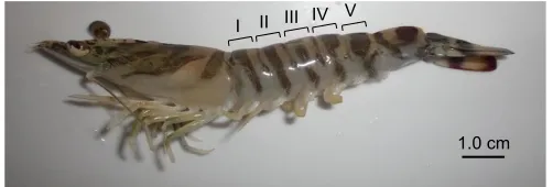
The second abdominal segments (Fig. 1) of three kuruma shrimp each from different salinity tanks were dissected after different periods of acclimation. The abdominal muscle was then collected and homogenized individually with eight volumes of 10% perchloric acid (w/w), and centrifuged at 12,000g for 20 min at 4°C. The resulting supernatant was neutralized with an appropriate amount of 12 mol l−1 and 2 mol l−1 KOH, and centrifuged at 12,000gfor 20 min at 4°C to collect the supernatant containing free amino acids. Free amino acids were derivatized with O-phthalaldehyde and 3-mercaptopropionic acid, and their concentrations were determined using a high performance liquid chromatography LC-2000 series (Jasco, Tokyo, Japan) with a reverse-phase column TSK gel ODS-80Ts (length, 250 mm; inside diameter, 46 mm; Tosoh, Tokyo, Japan). Mobile phase A consisted of 50 mmol l−1sodium acetate buffer ( pH 5.63) and mobile phase B consisted of 20% 50 mmol l−1sodium acetate buffer ( pH 5.63) plus 80% absolute methanol (v/v). Amino acids were eluted at room temperature with a linear gradient from A:B=100:0 to A:B=10:90 in 75 min at a flow rate of 1.0 ml min−1. Excitation and emission wavelengths to detect derivatized amino acids were 340 and 450 nm, respectively. The present method cannot distinguish betweenL- andD-amino acids.
Water content
The fourth and fifth abdominal segments (Fig. 1) of three specimens each from different salinity tanks were dissected after various periods of acclimation. Then, the abdominal muscle was collected and minced individually. The water content was measured with an MA35 moisture meter (Sartorius, Göttingen, Germany) according to the manufacturer’s instructions.
Construction of cDNA libraries
Total RNA was extracted from the abdominal muscles in the third abdominal segments (Fig. 1) of each of three specimens acclimated at different salinities for 24 h, using ISOGEN II solution (Nippon Gene, Tokyo, Japan) according to the manufacturer’s instructions, where 30 µg of total RNA from each of three specimens acclimated at 1.7%, 3.4% or 4.0% salinity were mixed. Total RNA was treated with DNase I (Takara, Otsu, Japan) to digest contaminated genomic DNA (gDNA) according to the manufacturer’s instructions, although we did not check any possible DNA remaining. Then, mRNA was purified using the Poly(A)+Isolation Kit from Total RNA (Nippon Gene). Subsequently, complementary DNA (cDNA) libraries were constructed from purified messenger RNA (mRNA) using the Ion Total RNA-Seq Kit v2 (Life Technologies, Carlsbad, CA, USA) according to the manufacturer’s instructions. The average size of each cDNA library was determined with a 2100 Bioanalyzer (Agilent Technologies, Santa Clara, CA, USA). An List of symbols and abbreviations
FPKM fragments per kilobase of transcript per million fragments gDNA genomic DNA
MHC myosin heavy chain
MYH myosin heavy chain (gene family) RNA-seq RNA sequencing
[image:2.612.50.561.656.730.2]SERCA sarco/endoplasmic reticulum Ca2+-ATPase
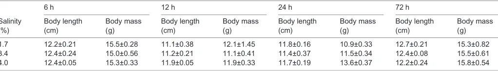
Table 1. Body length and mass of kuruma shrimp acclimated at different salinities
Salinity (%)
6 h 12 h 24 h 72 h
Body length (cm)
Body mass (g)
Body length (cm)
Body mass (g)
Body length (cm)
Body mass (g)
Body length (cm)
Body mass (g)
1.7 12.2±0.21 15.5±0.28 11.1±0.38 12.1±1.45 11.8±0.16 10.9±0.33 12.7±0.21 15.3±0.82 3.4 12.4±0.24 15.0±0.56 11.2±0.21 11.1±0.41 11.4±0.37 11.5±0.34 12.4±0.08 15.5±0.61 4.0 12.4±0.05 15.3±0.33 11.9±0.05 11.9±0.33 11.7±0.19 13.6±0.37 12.2±0.24 15.8±0.54
Values are given as means±s.d. (n=3).
Journal
of
Experimental
acclimation period of 24 h was selected because significant differences in free amino acid concentrations were observed at this period between kuruma shrimp acclimated to different salinities, as described in Results.
Sequencing
cDNA libraries prepared as above were treated with an Ion PGM Sequencing 200 Kit (Life Technologies) supplied with an Ion 318 chip (Life Technologies) according to the manufacturer’s instructions. Sequencing was performed using an Ion PGM next-generation sequencer (Life Technologies). Sequencing data were subjected to the Maser analysis platform provided by the National Institute of Genetics in Japan.
Statistical analysis
Data were analyzed with one-way or two-way ANOVA, and differences shown in ANOVA were analyzed with the Tukey’s method. To determine whether the assumptions of a normal distribution and homogeneity of variance were met, to qualify for parametric testing using ANOVA, we used the Kolmogorov– Smirnov test and Bartlett’s test, respectively. Because data for alanine and glutamine concentration did not conform to a normal distribution, they were processed using natural logarithm and Box– Cox power transformations prior to ANOVA, respectively (Box and Cox, 1964; Clark et al., 2016; Cyr et al., 1998; Little et al., 2013). The statistical analysis was also carried out using Student’s t-tests.
RESULTS
Free amino acid concentration
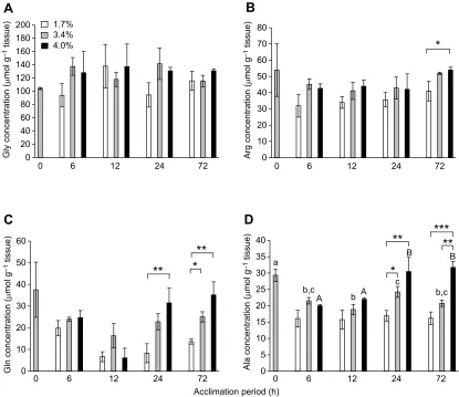
The concentration of total free amino acids extracted from the abdominal muscle in the starting samples of kuruma shrimp (0 h) after acclimating at 3.4% salinity for 72 h was 260.7±30.6 µmol g−1 tissue (Fig. 2). The most abundant free amino acid was glycine, followed by arginine, glutamine and alanine in most cases. ANOVA analysis revealed that the concentration of total free amino acids at 3.4% salinity did not change significantly during a further 72 h of acclimation (P>0.05). The average concentration in the shrimp acclimated for 24 h at 1.7% salinity was significantly lower than that at 4.0% salinity as well as that at 3.4% salinity (Student’s t-test, P<0.05). After 72 h, the concentration of total free amino acids at 4.0% salinity was significantly higher than that at 1.7% salinity (P<0.01) and 3.4% salinity (P<0.05). The concentration at 3.4% salinity was also significantly higher than that at 1.7% salinity (P<0.05). The concentration of total free amino acids was also higher at 3.4% than at 1.7% salinity after acclimating for 6 h (P<0.05).
The major free amino acids were glycine, arginine, glutamine and alanine, irrespective of different salinities and acclimation periods (Table S1). Changes in the major free amino acids during
acclimation were compared individually. No statistical differences were observed for glycine at any salinity (Fig. 3A), although the concentration of glycine was the highest among all free amino acids. The concentration of arginine, with the second largest concentration, showed a significant difference between the shrimp only at 1.7% and 4.0% salinity after 72 h (Fig. 3B).
Marked changes were observed in the concentration of glutamine, as shown in Fig. 3C. The statistical analysis did not show any significant differences for the shrimp at 3.4% salinity for any acclimation period, although the concentration at 0 h was apparently higher than that at 6–72 h. Student’st-test revealed that the concentration at 4.0% salinity was significantly higher than that at 1.7% salinity after acclimating for 24 and 72 h (P<0.01). The concentration at 3.4% salinity was also significantly higher than that at 1.7% salinity after 72 h. ANOVA analysis demonstrated that the concentration at 1.7% salinity after 6 h was significantly higher than that after 12 h, whereas the concentration at 4.0% salinity after 12 h was significantly lower than that after 6, 24 and 72 h at the same salinity.
The concentration of alanine showed changes similar to those of glutamine, as shown in Fig. 3D. Highly significant differences (P<0.01) were observed in the concentration between the shrimp at 1.7% and 4.0% salinity after 24 and 72 h as well as between shrimp at 3.4% and 4.0% salinity after 72 h. The difference between the shrimp at 1.7% and 3.4% salinity was also significant (P<0.05). ANOVA analysis demonstrated that alanine concentrations after 24 and 72 h were significantly higher than those after 6 and 12 h for the shrimp at 4.0% salinity. Taken together, these data show that the concentrations of glutamine and alanine changed clearly after acclimating kuruma shrimp at 1.7% and 4.0% salinity.
Water content
Fig. 4 shows changes in the water content of kuruma shrimp acclimated at different salinities. The water content was 74.9% initially. ANOVA analysis demonstrated that the water content was not significantly changed when the shrimp were acclimated for 72 h at 3.4% and 4.0% salinity. In contrast, the water content for the shrimp acclimated for 24 h at 1.7% salinity was significantly higher than that after 6 and 72 h. The differences in water content after acclimating for the same period at different salinities were significant between 1.7% and 4.0% salinity after
I
[image:3.612.49.300.58.143.2]1.0 cm II III IV V
Fig. 1. Kuruma shrimp.I–V correspond to the number of individual abdominal segments.
**
*
*
*
*
*
0 50 100 150 200 250 300 350
0 6 12 24 72
T
otal amino acid concentration
(
µ
mol g
−
1 tissue)
Acclimation period (h)
[image:3.612.316.560.59.218.2]1.7% 3.4% 4.0%
Fig. 2. Concentrations of free amino acids in the second abdominal segments of kuruma shrimp acclimated at different salinities for various periods up to 72 h.Open, grey and black bars indicate the concentrations in the shrimp acclimated at 1.7%, 3.4% and 4.0% salinity, respectively. Significance by Student’st-test at *P<0.05, **P<0.01.
Journal
of
Experimental
6 h (P<0.01), 12 h (P<0.05), 24 h (P<0.01) and 72 h (P<0.01). Significant differences were also observed between the shrimp at 1.7% and 3.4% salinity after 12, 24 and 72 h (P<0.05).
However, within an acclimation period, no significant differences in water content were observed between the shrimp at 3.4% and 4.0% salinity.
0 20 40 60 80 100 120 140 160 180 200
0 6 12 24 72
Gly concentration (
µ
mol g
–1
tissue)
0 10 20 30 40 50 60 70 80
0 6 12 24 72
Arg concentration (
µ
mol g
–1
tissue)
*
1.7% 3.4% 4.0%
A
B
0 10 20 30 40 50 60
0 6 12 24 72
Gln concentration (
µ
mol g
–1
tissue)
Acclimation period (h)
**
**
*
C
0 5 10 15 20 25 30 35 40
0 6 12 24 72
Ala concentration (
µ
mol g
–1
tissue)
**
***
**
*
a
b,c
b b,c
c
A A
B B
[image:4.612.99.514.58.417.2]D
Fig. 3. Concentrations of amino acids in the second abdominal segments of kuruma shrimp acclimated at different salinities for various periods up to 72 h.(A) Glycine, (B) arginine, (C) glutamine and (D) alanine. Open, grey and black bars indicate the concentrations of glycine in the shrimp acclimated at 1.7%, 3.4% and 4.0% salinities, respectively. Significance by Student’st-test at *P<0.05, **P<0.01, ***P<0.001. Different letters indicate significant differences by ANOVA. Lowercase and uppercase letters indicate the differences among alanine concentrations at 3.4% and 4.0% salinity, respectively.
Acclimation period (h) 70
72 74 76 78 80 82
0 6 12 24 78
1.7% 3.4% 4.0%
**
*
W
a
ter content (%)
b
a,b
a
a
**
*
**
*
0
*
Fig. 4. Water content in the fourth and fifth abdominal segments of kuruma shrimp acclimated at different salinities for various periods up to 72 h. Open, grey and black bars indicate the water content in shrimp acclimated at 1.7%, 3.4% and 4.0% salinity, respectively. Significance by Student’st-test at *P<0.05, **P<0.01. Different letters indicate significant
differences by ANOVA.
Journal
of
Experimental
[image:4.612.48.373.543.731.2]The concentrations of total free amino acids were inversely proportional to the water content, with a significant relative coefficient value ofr=−0.54076 (P<0.01).
RNA-seq analysis
To minimize any possible individual variations, we mixed mRNA prepared each from three specimens with the same amount (30 µg) for all sampling points, as described in the Materials and methods. Table S2 shows the average sizes of the constructed cDNA libraries for the shrimp acclimated for 24 h at 1.7, 3.4 and 4.0% salinity, together with corresponding Ion PGM sequencing data. RNA-seq data for the shrimp acclimated at 1.7% and 4.0% salinity were subjected to MA plot analysis (Wang et al., 2010), together with those for the shrimp acclimated at 3.4% salinity as a reference. At 1.7% salinity, the numbers of genes with expression levels that were increased more than twofold and decreased less than 50% than those at 3.4% salinity were 8696 and 5367, respectively. In contrast, the corresponding numbers at 4.0% salinity compared with those at 3.4% salinity were 3407 and 3683, respectively.
Tables S3–S6 show the genes with expression levels that were increased by more than 10-fold or decreased less than 10% during acclimation for 24 h at 1.7% or 4.0% salinity compared with those at 3.4% salinity as a reference, together with gene names, fragments per kilobase of transcript per million fragments (FPKM) values and fold change. A total of 2065 genes showed increased expression levels at 1.7% salinity, among which 124 genes were identified (Table S3). In contrast, 2618 genes had decreased expression levels
at 1.7% salinity, among which 113 genes were identified (Table S4). Meanwhile, the numbers of genes with expression levels that were increased and decreased at 4.0% salinity were 444 and 302, respectively, among which 16 and 31 genes were identified, respectively (Tables S5 and S6).
Gene expression profiles in glutamine- and alanine-related metabolic pathways
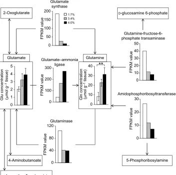
Glutamine-related metabolic pathways are shown in Fig. 5, where changes in the expression levels of the genes encoding glutamate synthase, glutamate–ammonia ligase, glutaminase, glutamine– fructose-6-phosphate transaminase and amidophosphoribosyltransferase determined by RNA-seq analysis for the shrimp acclimated for 24 h at different salinities are depicted, together with changes in the concentrations of glutamine and glutamate after the same acclimation period (Fig. 3C). The expression levels of these genes, except that encoding glutamate–ammonia ligase, were decreased in association with the increase of salinity. In contrast, the expression levels of the gene encoding glutamate–ammonia ligase were increased following the increase of salinity.
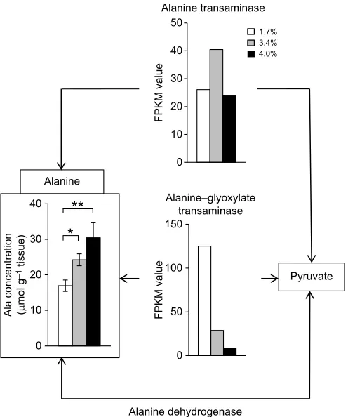
Alanine-related metabolic pathways are shown in Fig. 6, where changes in the expression levels of the genes encoding alanine transaminase and alanine–glyoxylate transaminase determined by RNA-seq analysis for the shrimp after 24 h at different salinities are depicted, together with changes in the concentration of alanine after the 24 h acclimation period at different salinities (Fig. 3D). The expression levels of the gene encoding alanine
Glutamate
D-glucosamine 6-phosphate
5-Phosphoribosylamine 0
50 100 150 200
FPKM value
0 100 200 300
FPKM value
0 40 80 120
FPKM value
0 10 20 30 40 50
FPKM value
0 10 20 30
FPKM value
0 10 20 30 40 Glutamine
0 1 2 3 4 5
Glu concentration (µmol g
–1
tissue)
Gln concentration (µmol g
–1
tissue)
Glutamate synthase
Glutamate–ammonia ligase
Glutaminase
Glutamine-fructose-6-phosphate transaminase
Amidophosphoribosyltransferase 2-Oxoglutarate
4-Aminobutanoate
L-1-pyrroline-5-carboxylate
**
[image:5.612.49.402.375.725.2]1.7% 3.4% 4.0%
Fig. 5. Genes and metabolites related to glutamine metabolism.Open, grey and black bars indicate the concentrations of glutamine and its related metabolites or the FPKM values of the genes related to glutamine metabolism in kuruma shrimp acclimated for 24 h at 1.7%, 3.4% and 4.0% salinity, respectively.
Journal
of
Experimental
dehydrogenase were decreased following the increase of salinity, whereas those of the gene encoding alanine–glyoxylate transaminase were higher at 3.4% salinity than at 1.7% and 4.0% salinity. Unfortunately, the gene encoding alanine dehydrogenase could not be detected in the present RNA-seq analysis.
Expression profiles of genes other than those related to amino acid metabolism
RNA-seq analysis revealed that many genes other than those related to amino acid metabolism exhibited altered expression levels when the shrimp were acclimated for 24 h at different salinities (Tables S3–S6). The expression levels of the genes encoding myosin heavy chain (MYH) type 1 (MYH1) and MYH2 were increased at 1.7% salinity (Table S3). The expression levels of MYH3 were decreased at 1.7% salinity (Table S4) and increased at 4.0% salinity (Table S5).
The expression levels of the genes encoding sarco/endoplasmic reticulum Ca2+-ATPase (SERCA) and ATP synthase subunit 9 mitochondrial precursor were decreased at 1.7% salinity. In contrast, the expression levels of the gene encoding Na+/K+ -ATPaseα-subunit were increased at 1.7% salinity (Table S3).
DISCUSSION
In order to understand the mechanisms involved in the acclimation of kuruma shrimp to different environmental salinities, we determined the concentrations of free amino acids in the abdominal muscle of shrimp acclimated at different salinities for various time periods (up to 72 h). As shown in Fig. 2, the concentrations of total free amino acids were changed following the alteration of acclimating salinity. It has
been reported that the concentrations of total free amino acids in kuruma shrimp were increased following the increase of salinity over 4 days (Abe et al., 2005). We obtained similar results in the present study. In addition, the present study demonstrated that it took 24 h to change the concentrations of free amino acids following the alteration of salinity.
Fig. 3 shows changes in the concentrations of major amino acids–glycine, arginine, glutamine and alanine. The concentration of glycine did not change significantly (Fig. 3A), although previous investigations observed an increase in the glycine concentration in the muscles of kuruma shrimp and crayfish (Okuma and Abe, 1994; Abe et al., 2005). It has also been reported that the concentration of alanine increased in adductor muscle of hard clamMeretrix lusoria at high salinity, whereas that of glycine did not increase (Okuma et al., 1998). It was not clear why the concentration of glycine did not change depending on salinity in the present study. The concentration of arginine also did not change significantly following the increase of salinity (Fig. 3B), whereas those of glutamine and alanine were increased at high salinity and decreased at low salinity after 24 h of acclimation (Fig. 3C,D). Therefore, in the present study, these two amino acids were found to possibly act as osmolytes, taking at least 24 h to adjust the cellular osmotic pressure to the environmental salinity. We previously reported changes in the accumulation of metabolites in the brackish water clamCorbicula japonicaexposed to different salinities, where the concentration ofL-alanine was also increased in association with the increase of environmental salinity, although that ofL-glutamine did not change significantly (Koyama et al., 2015; Okamoto et al., 2012). Therefore, it is considered that alanine is an osmolyte common to invertebrates with open vascular systems.
The increase in water content was almost proportional to the decrease in the concentration of total free amino acids (r=−0.54076). Although the concentrations of free amino acids were increased following the decrease in water content, the difference in water content between the shrimp acclimated for 72 h at 1.7% and 4.0% salinity (5%) was much less than the difference in the concentration of total free amino acids between the same shrimp (30%). Thus, the increase in the concentration of total free amino acids following the increase of salinity is not likely the simple effect of the decrease in water content, but seems attributable to the acclimation of kuruma shrimp to high salinity by increasing the concentration of free amino acids, as reported previously (Okuma and Abe, 1994; Abe et al., 2005).
In the present study, we could partly explain the accumulation of glutamine and alanine at high salinity by the functions of the genes participating in metabolism of the respective amino acids (Figs 5 and 6).
Fig. 5 shows the metabolic pathways of glutamine and its related compounds. The concentrations of glutamine and glutamate and the FPKM value of five genes are also shown. Kuruma shrimp acclimated at high salinity increased the expression level of the gene encoding glutamate–ammonia ligase (Fig. 5). Therefore, it is considered that glutamine was synthesized from glutamate by this enzyme in response to high salinity, increasing the concentration of glutamine. In contrast, the expression levels of four other enzymes – glutamate synthase, glutaminase, glutamine–fructose-6-phosphate transaminase and amidophosphoribosyltransferase–were increased at low salinity (Fig. 5). These results suggest that glutamine was catabolized to glutamate,D-glucosamine 6-phosphate or 5-phosphoribosylamine by the four abovementioned enzymes, thus decreasing the concentration of glutamine at this low salinity.
Alanine
Pyruvate
0 10 20 30 40
Ala concentration (µmol g
–1
tissue)
0 50 100 150
FPKM value
0 10 20 30 40 50
FPKM value
Alanine transaminase
Alanine–glyoxylate transaminase
Alanine dehydrogenase
**
*
[image:6.612.51.299.57.354.2]1.7% 3.4% 4.0%
Fig. 6. Genes and metabolites related to alanine metabolism.Open, grey and black bars indicate the concentration of alanine or the FPKM values of the genes related to alanine metabolism in kuruma shrimp acclimated for 24 h at 1.7%, 3.4% and 4.0% salinities, respectively.
Journal
of
Experimental
Fig. 6 shows the metabolic pathways of alanine and its related compounds. As in the case of glutamine, the concentration of alanine was increased following the increase of environmental salinity after 24 h (Fig. 3D). The FPKM value of the alanine– glyoxylate transaminase gene was increased at low salinity, whereas that of the alanine transaminase gene was not changed markedly at different salinities. Thus it seems that alanine was catabolized to pyruvate by the alanine–glyoxylate transaminase gene at low salinity, decreasing the concentration of alanine at this low salinity. However, we could not detect the transcripts encoded by the alanine dehydrogenase gene. Thus, the mechanisms involved in changes of the alanine concentration at different salinities remain unclear. In this regard, it has been reported that pyruvate was not detected in the gill and foot muscle of brackish water clam, although the concentration of alanine was increased at high salinity (Koyama et al., 2015). Pyruvate, once accumulated at low salinity, might have been quickly catabolized into another substance.
The expression levels of the genes encoding MYH1 and MYH2 were increased at 1.7% salinity (Table S3). MYH is the major muscle protein and two genes encoding MYH, MHC1 and MHC2, which correspond to the genes encoding MYH1 and MYH2, respectively, have been reported to be expressed in the abdominal muscle of kuruma, black tiger and Pacific white shrimp (Koyama et al., 2012a,b). These are expressed only in flexor muscle (MHC1) and in both flexor and extensor muscles (MHC2) in anaerobic metabolism. In the present study, the expression levels of MYH1 at 3.4% salinity were approximately 1.5-fold more than that of MYH2, and the expression levels of MYH2 at 1.7% salinity were approximately 3-fold more than that of MYH1. The expression levels of MYH3 were decreased at 1.7% salinity (Table S4) and increased at 4.0% salinity (Table S5). The gene encoding MYH3 has been reported asMHC3to be expressed in the pleopod muscle having aerobic metabolism of kuruma shrimp and black tiger shrimp (Koyama et al., 2012a, 2013). However, the relationship between MYH gene expression level and osmoregulation remains unclear.
SERCA plays an important role in regulating the calcium concentration in the cytoplasm (Clapham, 1995). It has been reported that the expression levels of SERCA in the muscle of Pacific white shrimp acclimated at high salinity (40 psu) were 9.7-fold higher than those acclimated at low salinity (20 psu) (Wang et al., 2013). Although the expression levels of the gene encoding SERCA were decreased at 1.7% salinity, we did not observe an increase at 4.0% salinity in this study (Table S4). It has been reported that European lobsterHomarus gammarusacclimated at low salinity (2.21%) from seawater (3.50%) increased their Ca2+-ATPase and Na+/Ca2+ exchange activities in epipodites (2.6- and 1.2-fold, respectively) and branchiostegites (1.6- and 1.2-fold, respectively), and decreased these activities in gills (0.30- and 0.52-fold, respectively) (Flik and Haond, 2000). Thus, the activity of SERCA might be changed depending on salinity and may modulate the calcium concentration in kuruma shrimp. The expression levels of the ATP synthase subunit 9 mitochondrial precursor gene were also decreased at 1.7% salinity (Table S4). Such decreased expression levels of this gene have been reported in the gills of Pacific white shrimp acclimated at low salinity (Gonçalves-Soares et al., 2012).
The expression levels of the Na+/K+-ATPase α-subunit gene were increased at 1.7% salinity (Table S3). Such enhanced expression of the Na+/K+-ATPase α-subunit gene has been reported in the gills of black tiger shrimp acclimated at low salinity (Shekhar et al., 2013). In addition, when cinnamon shrimp,Macrobrachium amazonicum, which live in freshwater,
were acclimated at 2.5% salinity, the expression level of the Na+/K+-ATPaseα-subunit gene and Na+/K+-ATPase activity in their gills, along with their hemolymph osmolality and chloride ion concentration, were increased, and their hemolymph osmolality and chloride ion concentration were also increased within 24 h (Faleiros et al., 2010). It has also been reported that freshwater prawnMacrobrachium rosenbergiiacclimated at high salinity (two-thirds seawater and full seawater) increased the concentrations of total free amino acids, sodium ion and chloride ion in their hemolymph (Huong et al., 2001). In the case of blue crab, Callinectes sapidus, acclimated at low salinity, the expression level of the Na+/K+-ATPaseα-subunit gene and Na+/K+-ATPase activity in their gills were also increased (Lucu and Towle, 2003). It is thus suggested that ion transporters such as Na+/K+-ATPase play an important role in adjusting the cellular osmotic pressure to the environmental salinity together with changes in free amino acid concentrations.
Although we did not carry out real-time PCR to confirm the data obtained from global gene expression analysis by RNA-seq in the present study, we previously demonstrated that the RNA-seq data were satisfactorily verified by real-time PCR experiments with our work on global gene expression analyses of muscle tissues from medaka acclimated to low and high environmental temperatures (Ikeda et al., 2017) and of gill tissues from normal and thermally selected strains of rainbow trout (Tan et al., 2012).
In conclusion, we examined the concentrations of free amino acids and gene expression profiles of kuruma shrimp acclimated at different salinities. A number of genes changed their expression levels in response to changes in environmental salinity. In addition, the concentrations of free amino acids were considered to be regulated by various genes related to amino acid metabolism. For instance, the concentration of glutamine was increased at high salinity in association with the increase of the expression level of glutamate–ammonia ligase. The concentration of alanine was increased at high salinity in association with the decrease of the expression level of alanine–glyoxylate transaminase. In future studies, further investigation is required regarding the participation
of D-amino acids and their related enzymes in the adaptation of
shrimp to environmental salinity change.
Acknowledgements
Matsumoto Suisan Co., Ltd, Miyazaki Prefecture, Japan, is greatly acknowledged for their kind supply of live specimens of kuruma shrimp. We are also grateful to Dr Toshinao Ineno from Kindai University for providing beneficial information about the culture condition of kuruma shrimp.
Competing interests
The authors declare no competing or financial interests.
Author contributions
Conceptualization: S.W.; Methodology: K.Y., M.J., D.I., S.W.; Software: S.A.; Validation: D.I., S.P.; Formal analysis: H.K., E.T., T.Y.; Investigation: H.K., N.M., M.H., E.T., T.Y.; Data curation: H.K., N.M., S.W.; Writing original draft: H.K.; Writing -review & editing: K.Y., M.J., T.Y., S.P., S.W.; Supervision: S.A., S.W.; Project administration: S.W.; Funding acquisition: S.W.
Funding
This work was partly supported by a Grant-in-Aid from the Japan Society of Promotion of Science (JSPS) for Scientific Research (S) (S.W., no. 19108003), by the JSPS-NRCT Asian CORE University Program granted to the Tokyo University of Marine Science and Technology and by The Towa Foundation for Food Research.
Data availability
RNA-seq data are available in the DNA Data Bank of Japan database under the
accession number DRA 006082.
Journal
of
Experimental
Supplementary information
Supplementary information available online at
http://jeb.biologists.org/lookup/doi/10.1242/jeb.168997.supplemental
References
Abe, H., Okuma, E., Amano, H., Noda, H. and Watanabe, K.(1999). Role of free D-and L-amino acids in the muscle of crayfish during seawater acclimation.Comp.
Biochem. Physiol.109A, 191-197.
Abe, H., Yoshikawa, N., Sarower, M. G. and Okada, S.(2005). Distribution, metabolism, and physiological function of free D-amino acids in invertebrates.
Vitamin Soc. Japan79, 79-86.
Boudour-Boucheker, N., Boulo, V., Charmantier-Daures, M., Grousset, E., Anger, K., Charmantier, G. and Lorin-Nebel, C.(2014). Differential distribution of V-type H+-ATPase and Na+/K+-ATPase in the branchial chamber of the palaemonid shrimpMacrobrachium amazonicum.Cell Tissue Res.357, 195-206.
Box, G. E. P. and Cox, D. R.(1964). An analysis of transformations.J. Roy. Stat.
Soc. Ser. B.26, 211-252.
Camien, M. N., Sarlet, H., Duchâteau, G. and Florkin, M.(1951). Non-protein amino acids in muscle and blood of marine and fresh water Crustacea.J. Biol.
Chem.193, 881-885.
Clapham, D. E.(1995). Calcium signaling.Cell80, 259-268.
Clark, J. E., Osborne, J. W., Gallagher, P. and Watson, S.(2016). A simple method for optimising transformation of non-parametric data: an illustration by reference to cortisol assays.Hum. Psychopharmacol.31, 259-267.
Cyr, D. G., Idler, D. R., Audet, C., McLeese, J. M. and Eales, J. G.(1998). Effects of long-term temperature acclimation on thyroid hormone deiodinase function, plasma thyroid hormone levels, growth, and reproductive status of male Atlantic
cod,Gadus morhua.Gen. Comp. Endocrinol.109, 24-36.
David, C. J.(2014). Geographical distribution of pelagic decapod shrimp in the Atlantic Ocean.Zootaxa3895, 301-345.
Faleiros, R. O., Goldman, M. H. S., Furriel, R. P. M. and McNamara, J. C.(2010). Differential adjustment in gill Na+
/K+
- and V-ATPase activities and transporter mRNA expression during osmoregulatory acclimation in the cinnamon shrimp
Macrobrachium amazonicum (Decapoda, Palaemonidae). J. Exp. Biol. 213,
3894-3905.
Flik, G. and Haond, C.(2000). Na2+ and Ca2+
pumps in the gills, epipodites and branchiostegites of the European lobsterHomarus gammarus: effects of dilute sea water.J. Exp. Biol.203, 213-220.
Freire, C. A., Onken, H. and McNamara, J. C.(2008). A structure-function analysis of ion transport in crustacean gills and excretory organs.Comp. Biochem. Physiol.
151A, 272-304.
Fujimori, T. and Abe, H.(2002). Physiological roles of free D- and L-alanine in the crayfishProcambarus clarkiiwith special reference to osmotic and anoxic stress responses.Comp. Biochem. Physiol.131A, 893-900.
Gao, W., Tan, B., Mai, K., Chi, S., Liu, H., Dong, X. and Yang, Q.(2012). Profiling of differentially expressed genes in hepatopancreas of white shrimp (Lipopenaeus
vannamei) exposed to long-term low salinity stress. Aquaculture 364-365,
186-191.
Gonçalves-Soares, D., Seiffert, W. Q., Schlindwein, A. D., Toledo-Silva, G., Zanette, J., Marques, M. R. F. and Bainy, A. C. D.(2012). Identification of differentially transcribed genes in shrimp Litopenaeus vannamei exposed to osmotic stress and challenged with WSSV virus. Comp. Biochem. Physiol.
7D, 73-81.
Havird, J. C., Santos, S. R. and Henry, R. P.(2014). Osmoregulation in the Hawaiian anchialine shrimpHalocaridina rubra(Crustacea: Atyidae): expression of ion transporters, mitochondria-rich cell proliferation and hemolymph osmolality during salinity transfers.J. Exp. Biol.217, 2309-2320.
Henry, R. P., Mangum, C. P. and Webb, K. L.(1980). Salt and water balance in the oligohaline clam, Rangia cuneata. II. Accumulation of intracellular free amino acids during high salinity adaptation.J. Exp. Zool.211, 11-24.
Holliday, C. W.(1985). Salinity induced changes in gill Na,K-ATPase activity in the mud fiddler crab,Uca pugnax.J. Exp. Zool.233, 199-208.
Huong, D. T. T., Yang, W. J., Okuno, A. and Wilder, M. N.(2001). Changes in free amino acids in the hemolymph of giant freshwater prawn Macrobrachium
rosenbergiiexposed to varying salinities: relationship to osmoregulatory ability.
Comp. Biochem. Physiol.128A, 317-326.
Huang, W., Ren, C., Li, H., Huo, D., Wang, Y., Jiang, X., Tian, Y., Luo, P., Chen, T. and Hu, C.(2017). Transcriptomic analysis on muscle tissues ofLitopenaeus
vannameiprovide the first profile insight into the response to low temperature
stress.PLoS ONE12, e0178604.
Ikeda, D., Koyama, H., Mizusawa, N., Kan-no, N., Tan, E., Asakawa, S. and Watabe, S.(2017). Global gene expression analysis of the muscle tissues of medaka acclimated to low and high environmental temperatures.Comp. Biochem.
Physiol.24D, 19-28.
Jorde, P. E., Søvik, G., Westgaard, J.-I., Albretsen, J., André, C., Hvingel, C., Johansen, T., Sandvik, A. D., Kingsley, M. and Jørstad, K. E. (2015). Genetically distinct populations of northern shrimp,Pandalus borealis, in the Northern Atlantic: adaptation to different temperatures as an isolation factor.Mol.
Ecol.24, 1742-1757.
Koyama, H., Akolkar, D. B., Shiokai, T., Nakaya, M., Piyapattanakorn, S. and Watabe, S.(2012a). The occurrence of two types of fast skeletal myosin heavy chains from abdominal muscle of kuruma shrimpMarsupenaeus japonicusand their different tissue distribution.J. Exp. Biol.215, 14-21.
Koyama, H., Akolkar, D. B., Piyapattanakorn, S. and Watabe, S.(2012b). Cloning, expression, and localization of two types of fast skeletal myosin heavy chain genes from black tiger and Pacific white shrimps.J. Exp. Zool.317A, 608-621.
Koyama, H., Piyapattanakorn, S. and Watabe, S.(2013). Cloning of skeletal myosin heavy chain gene family from adult pleopod muscle and whole larvae of shrimps.J. Exp. Zool.319A, 268-276.
Koyama, H., Okamoto, S., Watanabe, N., Hoshino, N., Jimbo, M., Yasumoto, K. and Watabe, S.(2015). Dynamic changes in the accumulation of metabolites in brackshi water clamCorbicula japonicaassociated with alternation of salinity.
Comp. Biochem. Physiol.181B, 59-70.
Li, J., Peng, M., Liu, P., Chen, P. and Li, J.(2015). The roles of Na+/K+-ATPaseα -subunit gene from the ridgetail white prawnExopalaemon carinicaudain response to salinity stresses.Fish Shellfish Immunol.42, 264-271.
Little, A. G., Kunisue, T., Kannan, K. and Feebacher, F.(2013). Thyroid hormone actions are temperature-specific and regulate thermal acclimation in zebrafish
(Danio rerio).BMC Biol.11, 26.
Lucu, Č. and Towle, D. W.(2003). Na++K+-ATPase in gills of aquatic crustacea.
Comp. Biochem. Physiol.135A, 195-214.
Lv, J., Liu, P., Wang, Y., Gao, B., Chen, P. and Li, J.(2013). Transcriptome analysis ofPortunus trituberculatusin response to salinity stress provides insights into the molecular basis of osmoreguration.PLoS ONE8, e82155.
Martin, J. W. and Davis, G. E.(2001). An updated classification of the recent Crustacea.Nat. History Museum Los Angeles Country Sci. Ser.39, 1-124.
McNamara, J. C., Rosa, J. C., Greene, L. J. and Augusto, A.(2004). Free amino acid pools as effectors of osmotic adjustment in different tissues of the freshwater shrimpMacrobrachium olfersii(Crustacea, Decapoda) during long-term salinity acclimation.Mar. Freshw. Behav. Physiol.37, 193-208.
Meng, J., Zhu, Q., Zhang, L., Li, C., Li, L., She, Z., Huang, B. and Zhang, G.
(2013). Genome and transcriptome analyses provide insight into the euryhaline adaptation mechanism ofCrassostrea gigas.PLoS ONE8, e58563.
Okamoto, S., Yamaguchi, H., Koyama, H., Nakaya, M., Yoneda, C. and Watabe, S.(2012). Comparison of extractive components and taste of soup from brackish water clamCorbicula japonicain different habitat waters of the Hinuma River system.Nippon Suisan Gakkaishi78, 444-453.
Okuma, E. and Abe, H.(1994). Total D-amino and other amino acids increase in the muscle of crayfish during seawater acclimation.Comp. Biochem. Physiol.109A, 191-197.
Okuma, E., Watanabe, K. and Abe, H.(1998). Distribution of free D-amino acids in bivalve mollusks and the effects of physiological conditions on the levels of D- and L-alanine in the tissues of the hard clam,Meretrix lusoria.Fish. Sci.64, 606-611.
Santos, C. A., Blanck, D. V. and de Freitas, P. D.(2014). RNA-seq as a powerful tool for penaeid shrimp genetic progress.Front. Genet.5, 298.
Sellars, M. J., Trewin, C., McWilliam, S. M., Glaves, R. S. E. and Hertzler, P. L.
(2015). Transcriptome profiles ofPenaeus(Marsupenaeus)japonicusanimal and vegetal half-embryos: identification of sex determination, germ line, mesoderm, and other developmental genes.Mar. Biotechnol.17, 252-265.
Shekhar, M. S., Kiruthika, J. and Ponniah, A. G. (2013). Identification and expression analysis of differentially expressed genes from shrimp
(Penaeus monodon) in response to low salinity stress.Fish Shellfish Immunol.
35, 1957-1968.
Shekhar, M. S., Kiruthika, J., Rajesh, S. and Ponniah, A. G.(2014). High salinity induced expression profiling of differentially expressed genes in shrimp (Penaeus
monodon).Mol. Biol. Rep.41, 6275-6289.
Shinji, J., Okutsu, T., Jayasankar, V., Jasmani, S. and Wilder, M. N.(2012). Metabolism of amino acids during hyposmotic adaptation in the white leg shrimp,
Lipopenaeus vannamei.Amino Acids43, 1945-1954.
Sun, H., Zhang, L., Ren, C., Chen, C., Fan, S., Xia, J. J., Lin, H. and Hu, C.(2011). The expression of Na, K-ATPase inLitopenaeus vannameiunder salinity stress.
Mar. Biol. Res.7, 623-628.
Tan, E., Wongwarangkana, C., Kinoshita, S., Suzuki, Y., Oshima, K., Hattori, M., Ineno, T., Tamaki, K., Kera, A., Muto, K. et al.(2012). Global gene expression analysis of gill tissues from normal and thermally selected strains of rainbow trout.
Fish. Sci.78, 1041-1049.
Wang, L., Feng, Z., Wang, X., Wang, X. and Zhang, X.(2010). DEGseq: an R package for identifying differentially expressed genes from RNA-seq data.
Bioinfomatics26, 136-138.
Wang, Y., Luo, P., Zhang, L., Hu, C., Ren, C. and Xia, J.(2013). Cloning of sarco/ endoplasmic reticulum Ca2+-ATPase (SERCA) gene from white shrimp,
Litopenaeus vannameiand its expression level analysis under salinity stress.
Mol. Biol. Rep.40, 6213-6221.
Zhao, X., Yu, H., Kong, L. and Li, Q.(2012). Transcriptomic responses to salinity stress in the Pacific oysterCrassostrea gigas.PLoS ONE7, e46244.