Original Article
Up-regulation of miR-221 mediates the
pioglitazone-induced inhibition of vascular
endothelial cell proliferation
Ben Huang, Wenshuo Wang, Ye Yang, Shuyang Lu, Jiayu Zheng, Lai Wei, Chunsheng Wang Department of Cardiovascular Surgery, Zhongshan Hospital, Fudan University, Shanghai, China
Received October 27, 2015; Accepted December 24, 2015; Epub February 1, 2016; Published February 15, 2016 Abstract: Given that angiogenesis plays a vital role in development of cancer, a becoming anti-tumor strategy is anti-angiogenic. Although the anti-angiogenic effect of pioglitazone has been known already, the detailed molecular mechanism, which accounts for the endothelial cells (ECs) to proliferation inhibited by pioglitazone, is limited, partic-ularly concerning their posttranscriptional regulation involving miRNA. In present study, we provide the mechanistic link between pioglitazone and its anti-angiogenic activity. Primarily, using immumofluorescence and CCK-8 assay, we find pioglitazone inhibits the ECs proliferation. During this inhibiting process, 12 microRNAs (miRNAs) show significant differences at relative gene abundance in which miR-221 presents a highest up-regulated expression. In following experiment, we demonstrate that expression of miR-221 mediated by pioglitazone positively correlate with anti-angiogenic action of pioglitazone. At last, we discover that pioglitazone augments the reduction of p27 mRNA level in ECs by up-regulating miR-221. Collectively, we have exploited a valuable mechanism by which up-regulation of miR-221 contributes to the pioglitazone-induced inhibition vascular EC proliferation, which raise a possibility that inhibition of miR-221 may be a potential strategy for therapeutic intervention in excessive vascular EC proliferation.
Keywords: Pioglitazone, miR-221, vascular endothelial cell, angiogenesis
Introduction
The process of angiogenesis is that pre-exist-ing vasculature produces the new vessels, in which endothelial cells (ECs)-dependent prolif-eration, migration, and differentiation are main-ly responsible for this process [1, 2]. Multiple normal physiological processes including em- bryonic development, wound healing and men-strual cycle rely on angiogenesis [3]. Besides, angiogenesis plays a vital role in numerous pathological conditions, especially in develop-ment of cancer [4]. In the vast majority of cancers, blood vessel growth is motivated and these vessels are unnatural in almost all aspects of their structure and function, thereby nourishing cancers [5]. Given that, a becom- ing concept is that anti-angiogenic strategies through inhibiting vessel growth and normaliz-ing vessels are potential therapeutic interven-tion in cancers, which were consistently evi-denced in recent studies [6-8]. Thus, current main task is still to further screen the anti-angiogenic agents although some functional agents have been determined [9].
dual anti-tumour and anti-angiogenic effect of miRNA, their value to cancer therapy remains to be established.
Besides miRNA, pioglitazone, a prescription drug of class thiazolidinedione, has also been reported to associate with anti-angiogenesis [19]. Traditionally, pioglitazone was utilized for the therapy of type-II diabetes mellitus in clini-cal because of their ability to reverse insulin resistance [20]. In spite of this application, pio-glitazone have also been presented a compre-hensive package of biological activities such as anti-inflammatory [21], anti-proliferative [22] and anti-tumor effects [23-25]. More impor-tantly, the anti-cancer effect of pioglitazone is positively related to its anti-angiogene- sis [19] and may be regulated through peroxi-some proliferator-activated receptor-γ (PPAR-γ)-dependent or -in(PPAR-γ)-dependent way [19, 23-26]. Although the potential tumour and anti-angiogenic activity of pioglitazone has been speculated, molecular mechanism by which pioglitazone acts is poorly understood, particu-larly concerning their posttranscriptional regu-lation involving miRNA. Here we attempted to investigate whether the miRNAs are involved in the anti-angiogenic effect of pioglitazone in human microvascular endothelial cell line HMEC-1.
Materials and methods
Cell culture
The human microvascular endothelial cell line HMEC-1 was cultured in Dulbecco’s modified eagle medium (DMEM, Hyclone) supplemented with 10% (V/V) FBS (Hyclone), 10 ng/ml epider-mal growth factor and 1% penicillin-streptomy-cin at 37°C, 5% CO2 incubator. Cells grown in 60 mm cell culture dish were allowed approxi-mately to reach 85% confluence. The culture medium was changed every two day and then were rinsed and removed from the dishes by incubating them with a trypsin-EDTA solution (Hyclone), and harvested in a 15 mL centrifuge tube for subsequent study.
MiRNA mimic
Chemically modified double-stranded RNAs engineered to mimic the endogenous mature miR-221 and negative control miRNA were purchased from Ambion. miRNA mimics were transfected using RNAi Max (Invitrogen)
accord-ing to the manufacturer’s indication at 0.3 or 3 nM as directed. MiR-221 inhibitor (2’OMe-miR-221) were chemically synthesized and purified by high-performance liquid chromatog-raphy (Gene Pharma, Shanghai, China). HMEC-1 transfected with scrambled 2’-OMe oligonu-cleotides (scramble) were used as a negative inhibitor control. These oligonucleotides were transfected using Lipofectamine 2000 (Invit- rogen) according to the manufacturer’s direc-tion at 300 pmol as indicated.
Cell proliferation assay
HMEC-1 cells were directly cultured at a density of 1×10⁵ cells/well in 6 well plates. For experi-ment with the effect of pioglitazone on prolifer-ation, the cells cultured overnight were incu-bated with DMSO (negative control) and piogli-tazone (prepared in DMSO) respectively for indicated time at 37°C. For experiment with administrations of miR-221 mimic, negative mimic, miR-221 inhibitor, negative inhibitor or synergy of each of above-mentioned molecules with pioglitazone, after overnight cultivation, the cells were stimulated with these reagents for indicated concentration and time. Cell prolif-eration was primarily investigated by immuno-fluorescence. For immunofluorescence obser-vation, we incubated HMEC-1 cells with anti-fade mounting medium containing carboxyfluo-rescein diacetate (CFDA, Invitrogen) for cyto-plasmic staining and DAPI (Invitrogen) for nucle-ar staining, followed by analysis with a laser scanning confocal microscope (Leica 224). Cell proliferation was analyzed additionally by using CCK-8 assay kit (Dojindo) according to manufacturer introductions. In particular, HMEC-1 cells were incubated in the medium containing DMSO or pioglitazone in 96-well plates. After that, 5 µl CCK-8 reagent was added to each well and incubated at 37°C for 1 h. The cell numbers were evaluated by measurement of absorbance at 450 nm. Proliferation assays for cells pretreated with miR-221 mimic, negative mimic, miR-221 inhib-itor, negative inhibitor or each of above-men-tioned molecules plus pioglitazone were also assessed by utilizing CCK-8 assay. All the experiments were carried out in triplicate.
MiRNAs array screening
expression profiling of miRNA was investigated by miRNA microarray analysis using the hu- man miRNA array probes including 15 mature human miRNAs. These miRNAs are 7a, let-7b, let-7d, miR-1, miR-221, miR-222, miR-122, miR-375, miR-24, miR-133, miR-126, miR-16, miR-21, miR-155 and miR-320.
Quantitative RT-PCR assay
For quantitative RT-PCR assay, RNA was isolat-ed from HMEC-1 cells using the TRIzol reagent according to the manufacturer’s instructions. 4 µg of total RNA were provided to generate the first-strand cDNAs by using commercially avail-able kits (Applied Biosystems). All subsequent PCR reactions were carried out using the 7 Universal PCR Master Mix (Applied Biosystems). Thermal cycling and fluorescence detection of mRNA were analyzed by 7500 real-time PCR System (Applied Biosystems). To normalize mRNA concentrations, mRNA levels of β-actin gene were identified in parallel for each sam-ple, and relative mRNA level of Notch1 was adjusted by standardization based on the β- actin transcriptional levels. Samples for each experimental condition were run in triplicate.
Statistical analysis
All data were expressed as mean ± standard deviation (SD) and subjected to analysis of vari-ance (ANOVA) to assess the treatment effects by using SPSS 13.0 software. The Student t
test was used to determine the statistically sig-nificant differences in numbers with two signifi-cant levels (0.05 and 0.01).
Results
Pioglitazone inhibits the efficient proliferation of HMEC-1 cell
To understand the anti-angiogenic effect of pioglitazone in ECs, HMEC-1 cell was used to treat. Cell proliferation was measured by utiliz-ing the immunofluorescence. Prior to incubat-ing the pioglitazone, the identical cell popula-tion was plated on coverslips (Figure 1A). After that, pioglitazone was used to stimulate the cells and then immune stained with CFDA and DAPI. Interestingly, the results showed that cell numbers between untreated and DMSO-treated group had no difference, however, HMEC-1 cell treated with pioglitazone present-ed a significant rpresent-eduction in the cell population in contrast to untreated and DMSO treated group (Figure 1A). Then, we explored the cell proliferation using CCK-8 assay, as shown in Figure 1B. Similarly, the cell proliferation rate was differentially decreased in the HMEC-1 cell treated with pioglitazone compared with untreated and DMSO-treated cells (P<0.01), indicating that pioglitazone efficiently sup-pressed the proliferation of HMEC-1 cell.
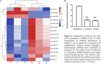
Pioglitazone induces the miR-221 expression
[image:3.612.92.522.73.192.2]treated cells (Figure 2A). Out of them, two con-taining miR-221 and miR-222 were up-regulat-ed in pioglitazone-treatup-regulat-ed cells while ten includ-ing miR-1, miR-122, miR-375, miR-24, miR-133, 126, 16, 21, 155 and miR-320 were down-regulated. Among these 12 miRNAs, we found that miR-221 presented the most significant difference in pioglitazone-treated cells in contrast to DMSO-pioglitazone-treated cells. Then, we further confirmed the miR-221 ex- pression in pioglitazone-treated cells from transcriptional level. As Figure 2B showed, the expression of miR-221 was significantly improved in HMEC-1 cells treated with piogli-tazone compared to the untreated and DMSO-treated cells, respectively (P<0.01). This finding suggested that pioglitazone have an ability to promote the miR-221 expression in HMEC-1 cell and miR-221 may be a central downstream target of pioglitazone.
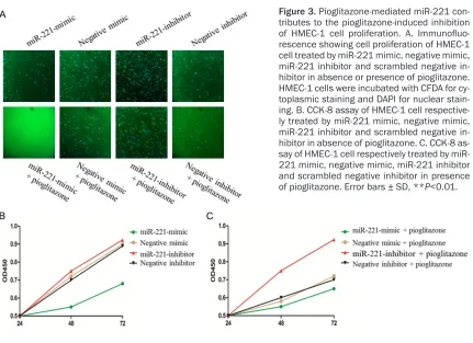
miRNA-221 contributes to pioglitazone-in-duced inhibition of EC proliferation
Next, to examine the mechanism that links miR-221 to anti-angiogenic activity of pioglitazone, miR-221 mimic oligonucleotides, nontargeting negative control mimic, miR-221 inhibitor and scrambled negative inhibitor were transfected
[image:4.612.95.524.74.347.2]tor. These observations indicate that a piogli-tazone-mediated miRNA, miR-221, play a sig-nificantly positive role in pioglitazone-induced inhibition of HMEC-1 cell proliferation.
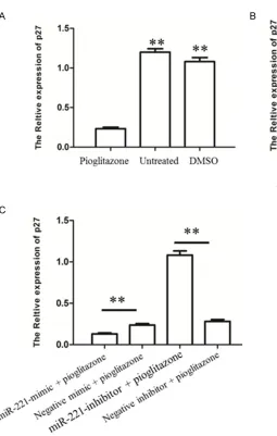
Pioglitazone-mediated miR-221 reduces the p27 expression in HMEC-1 cell
In a follow-up experiment, we evaluated wheth-er the pioglitazone have a capacity to regulate the expression of p27 in HMEC-1 cells through up-regulation of miR-221. To this end, we pri-marily detected the transcriptional expression of p27 in HMEC-1 cell in response to piogli-tazone treatment, as shown in Figure 4A. The untreated and DMSO-treated HMEC-1 cell rela-tively highly expressed p27, while the expres-sion of p27 markedly reduced in HMEC-1 cell treated by pioglitazone (P<0.01). This finding suggested that pioglitazone have an ability to inhibit the p27 expression in ECs. Given that miR-221 mimic and miR-221 inhibitor could also respectively suppress and enhance the p27 mRNA level (P<0.01, Figure 4B), present-ing the targetpresent-ing activity of miR-221 on p27 in ECs, it is therefore intriguing to suggest that the effect of pioglitazone on expression of p27 is likely to be accomplished by up-regulation of miR-221. As expected, pioglitazone-stimulated
cells treated by miR-221 inhibitor had a specific restoration in expression of p27 as compared to pioglitazone-stimulated cells treated by scrambled inhibitor (P<0.01, Figure 4C). Mean- while, the transcriptional level of p27 was fur-ther reduced in cells response to miR-221 mimic plus pioglitazone treatment in contrast to negative mimic plus pioglitazone treatment (P<0.01, Figure 4C). Altogether, these data indi-cate that pioglitazone boosts the reduction of p27 expression in vitro by up-regulating miR- 221.
Discussion
[image:5.612.94.525.65.378.2]vide the mechanistic link between pioglitazone and its inhibiting function of cell proliferation. The core discovery in our study is the potent ability of exogenous pioglitazone to regulate the expression of miRNA, such as miR-221, in HMEC-1 cell. This is a novel finding, since previ-ous studies did not disclose how any miRNA might be reliable for pioglitazone-induced inhi-bition of cell proliferation. Currently, we pre- sent that miR-221 contributes to pioglitazone-induced inhibition of cell proliferation, demon-strating a plausible miR-221-regulated mecha-nism. The cell proliferation regulated by miRNA and pioglitazone have been extensively evi-denced in the literature [12-19]. However, the amazed capacity of pioglitazone to facilitate the up-regulation of miR-221 and in turn reduc-es cell proliferation in ECs was previously un- recognized. Additionally, previous studies indi-cated that silencing of tumor suppressor gene
p27 is an important role of miR-221 [27, 28]. Given that, a becoming hypothesis is that pio-glitazone treatment may lead to the down-regu-lation of p27. This is consistent with current observation that pioglitazone-treated cells rep-resent the declined transcriptional level of p27 through up-regulation of miR-221 (Figure 4), which historically leaded to cancer cell prolifer-ation, exhibiting a prognostic significance in human cancer [27, 28]. This apparent contra-diction has raised a conundrum regarding the role of p27 in pioglitazone-mediated inhibition of ECs proliferation and therefore need to be further investigated in future.
[image:6.612.96.351.64.465.2]Besides miR-221, we also found that other 11 miRNAs containing miR-222, miR-1, miR-122, miR-375, miR-24, miR-133, miR-126, miR-16, miR-21, miR-155 and miR-320 were differen-tially altered in pioglitazone-treated ECs. Similar to miR221, miR-222 also has anti-angiogenic
effect for ECs in vitro [13, 14]. miR-1/126 involve in the cross-talk between the muscle and vasculature to regulation of developmental angiogenesis [29]. miR-122 functions as a tu- mor suppressor in liver cancer partly because of its anti-angiogenic potential in ECs [30]. By directly targeting vascular endothelial grow- th factor (VEGF), miR-16 is a key negative regu-lator of angiogenic signaling [31] while activa-tion of miR-21 induces tumor angiogenesis through enhancing VEGF expression [32]. Re- cent reports reveal that overexpression of miR-155 contributes to placental angiogenesis [33], and miR-320 augments the degradation of insulin-like growth factor (IGF)-1 in mediating angiogenesis in diabetic rats [34]. However, the potential role of remaining miRNA, miR-24, miR-375 and miR-133, in angiogenesis awaits future studies although it has been shown to regulate apoptosis and inhibit cell proliferation [35-37]. Therefore, a suitable possibility is that EC angiogenesis inhibited by pioglitazone is likely realized in part by altering the expression of these miRNAs, and certainly this inference requires to be further checked in subsequent study.
Taken all together, we represents substantial improve toward elucidating the miRNA-regulat-ed mechanism corresponding to pioglitazone-mediated anti-angiogenesis. These findings raise a possibility that inhibition of miR-221 may be a potential strategy for therapeutic intervention in excessive vascular EC prolifer- ation.
Disclosure of conflict of interest
None.
Address correspondence to: Drs. Lai Wei and Chunsheng Wang, Department of Cardiovascular Surgery, Zhongshan Hospital, Fudan University, 180 Fenglin Road, Shanghai 200032, China. Tel: +86-13818176656; Fax: +86-21-64041990; E-mail: laiwei2015@126.com (LW); Tel: +86-1380197- 8935; Fax: +86-21-64041990; E-mail: chunsheng-wang12@163.com (CSW)
References
[1] Carmeliet P. Angiogenesis in life, disease and medicine. Nature 2005; 438: 932-936. [2] Folkman J. What is the evidence that tumors
are angiogenesis dependent? J Natl Cancer Instit 1990; 82: 4-7.
[3] Carmeliet P and Jain RK. Angiogenesis in can-cer and other diseases. Nature 2000; 407: 249-257.
[4] Folkman J. Angiogenesis in cancer, vascular, rheumatoid and other disease. Nat Med 1995; 1: 27-30.
[5] Carmeliet P and Jain RK. Principles and mech-anisms of vessel normalization for cancer and other angiogenic diseases. Nat Rev Drug Dis- cov 2011; 10: 417-427.
[6] Mazzone M, Dettori D, Leite de Oliveira R, Loges S, Schmidt T, Jonckx B, Tian YM, Lana- han AA, Pollard P and Ruiz de Almodovar C. Heterozygous Deficiency of PHD2 Restores Tumor Oxygenation and Inhibits Metastasis via Endothelial Normalization. Cell 2009; 136: 839-851.
[7] Hamzah J, Jugold M, Kiessling F, Rigby P, Man- zur M, Marti HH, Rabie T, Kaden S, Gröne HJ and Hämmerling GJ. Vascular normalization in Rgs5-deficient tumours promotes immune de-struction. Nature 2008; 453: 410-414.
[8] Stockmann C, Doedens A, Weidemann A, Zhang N, Takeda N, Greenberg JI, Cheresh DA and Johnson RS. Deletion of vascular endothe-lial growth factor in myeloid cells accelerates tumorigenesis. Nature 2008; 456: 814-818. [9] Crawford Y and Ferrara N. VEGF inhibition:
in-sights from preclinical and clinical studies. Cell Tissue Res 2009; 335: 261-269.
[10] Urbich C, Kuehbacher A and Dimmeler S. Role of microRNAs in vascular diseases, inflamma-tion, and angiogenesis. Cardiovasc Res 2008; 79: 581-588.
[11] Ambros V. The functions of animal microRNAs. Nature 2004; 431: 350-355.
[12] Kuehbacher A, Urbich C, Zeiher AM and Dim- meler S. Role of Dicer and Drosha for endo- thelial microRNA expression and angiogene-sis. Circulat Res 2007; 101: 59-68.
[13] Suárez Y, Fernández-Hernando C, Pober JS and Sessa WC. Dicer dependent microRNAs regulate gene expression and functions in hu-man endothelial cells. Circulat Res 2007; 100: 1164-1173.
[14] Poliseno L, Tuccoli A, Mariani L, Evangelista M, Citti L, Woods K, Mercatanti A, Hammond S and Rainaldi G. MicroRNAs modulate the an-giogenic properties of HUVECs. Blood 2006; 108: 3068-3071.
[15] Venturini L, Battmer K, Castoldi M, Schultheis B, Hochhaus A, Muckenthaler MU, Ganser A, Eder M and Scherr M. Expression of the miR-17-92 polycistron in chronic myeloid leukemia (CML) CD34+ cells. Blood 2007; 109: 4399-4405.
[17] Kong W, He L, Richards E, Challa S, Xu C, Permuth-Wey J, Lancaster J, Coppola D, Sellers T and Djeu J. Upregulation of miRNA-155 pro-motes tumour angiogenesis by targeting VHL and is associated with poor prognosis and tri-ple-negative breast cancer. Oncogene 2013; 33: 679-689.
[18] Yamakuchi M, Lotterman CD, Bao C, Hruban RH, Karim B, Mendell JT, Huso D and Lowen- stein CJ. P53-induced microRNA-107 inhibits HIF-1 and tumor angiogenesis. Proc Natl Acad Sci U S A 2010; 107: 6334-6339.
[19] Keshamouni VG, Arenberg DA, Reddy RC, Newstead MJ, Anthwal S and Standiford TJ. PPAR-γ activation inhibits angiogenesis by blocking ELR+ CXC chemokine production in non-small cell lung cancer. Neoplasia 2005; 7: 294-301.
[20] Durbin R. Thiazolidinedione therapy in the pre-vention/delay of type 2 diabetes in patients with impaired glucose tolerance and insulin resistance. Diabetes Obes Metab 2004; 6: 280-285.
[21] Consoli A and Devangelio E. Thiazolidinediones and inflammation. Lupus 2005; 14: 794-797. [22] Schmidt S, Moric E, Schmidt M, Sastre M,
Feinstein DL and Heneka MT. Anti-inflammatory and antiproliferative actions of PPAR-γ ago-nists on T lymphocytes derived from MS pa-tients. J Leukoc Biol 2004; 75: 478-485. [23] Li MY, Deng H, Zhao JM, Dai D and Tan XY.
PPARgamma pathway activation results in apoptosis and COX-2 inhibition in HepG2 cells. World J Gastroenterol 2003; 9: 1220-1226. [24] Yoshizumi T, Ohta T, Ninomiya I, Terada I,
Fushida S, Fujimura T, Nishimura G, Shimizu K, Yi S and Miwa K. Thiazolidinedione, a peroxi-some proliferator-activated receptor-γ ligand, inhibits growth and metastasis of HT-29 hu-man colon cancer cells through differentiation-promoting effects. Int J Oncol 2004; 25: 631-639.
[25] Keshamouni VG, Reddy RC, Arenberg DA, Joel B, Thannickal VJ, Kalemkerian GP and Standiford TJ. Peroxisome proliferator-activat-ed receptor-γ activation inhibits tumor progres-sion in non-small-cell lung cancer. Oncogene 2004; 23: 100-108.
[26] Hazra S, Batra RK, Tai HH, Sharma S, Cui X and Dubinett SM. Pioglitazone and rosiglitazone decrease prostaglandin E2 in non–small-cell lung cancer cells by up-regulating 15-hydroxy-prostaglandin dehydrogenase. Mol Pharmacol 2007; 71: 1715-1720.
[27] Fornari F, Gramantieri L, Ferracin M, Veronese A, Sabbioni S, Calin G, Grazi G, Giovannini C, Croce C and Bolondi L. MiR-221 controls CDKN1C/p57 and CDKN1B/p27 expression in human hepatocellular carcinoma. Oncogene 2008; 27: 5651-5661.
[28] Davis BN, Hilyard AC, Nguyen PH, Lagna G and Hata A. Induction of microRNA-221 by platelet-derived growth factor signaling is critical for modulation of vascular smooth muscle pheno-type. J Biol Chem 2009; 284: 3728-3738. [29] Stahlhut C, Suárez Y, Lu J, Mishima Y and
Giraldez AJ. miR-1 and miR-206 regulate an-giogenesis by modulating VegfA expression in zebrafish. Development 2012; 139: 4356-4365.
[30] Bai S, Nasser MW, Wang B, Hsu SH, Datta J, Kutay H, Yadav A, Nuovo G, Kumar P and Ghoshal K. MicroRNA-122 inhibits tumorigenic properties of hepatocellular carcinoma cells and sensitizes these cells to sorafenib. J Biol Chem 2009; 284: 32015-32027.
[31] Sun CY, She XM, Qin Y, Chu ZB, Chen L, Ai LS, Zhang L and Hu Y. miR-15a and miR-16 affect the angiogenesis of multiple myeloma by tar-geting VEGF. Carcinogenesis 2012; bgs333. [32] Liu LZ, Li C, Chen Q, Jing Y, Carpenter R, Jiang
Y, Kung HF, Lai L and Jiang BH. MiR-21 induced angiogenesis through AKT and ERK activation and HIF-1α expression. PLoS One 2011; 6: e19139.
[33] Zhang Y, Diao Z, Su L, Sun H, Li R, Cui H and Hu Y. MicroRNA-155 contributes to preeclampsia by down-regulating CYR61. Am J Obstet Gyne- col 2010; 202: 466.e461-466.e467.
[34] Wang X, Qian R, Zhang W, Chen S, Jin H and Hu R. MicroRNA-320 Expression In Myocar- dial Microvascular Endothelial Cells And Its Relationship With Insulin-Like Growth Factor-1 In Type 2 Diabetic Rats. Clin Exp Pharmacol Physiol 2009; 36: 181-188.
[35] Lal A, Navarro F, Maher CA, Maliszewski LE, Yan N, O’Day E, Chowdhury D, Dykxhoorn DM, Tsai P and Hofmann O. miR-24 Inhibits cell pro-liferation by targeting E2F2, MYC, and other cell-cycle genes via binding to “seedless” 3’ UTR microRNA recognition elements. Mol Cell 2009; 35: 610-625.
[36] Chang Y, Yan W, He X, Zhang L, Li C, Huang H, Nace G, Geller DA, Lin J and Tsung A. miR-375 inhibits autophagy and reduces viability of he-patocellular carcinoma cells under hypoxic conditions. Gastroenterology 2012; 143: 177-187, e178.