Original Article
Analysis of mRNA profile in different strategy of bladder
cancer carcinoma by deep sequencing
Xiaoyuan Xu1, Xinping Wang1, Bin Fu2, Lirong Meng3, Bin Lang3
1Key Laboratory of System Bio-medicine of Jiangxi Province, Jiujiang University, Jiujiang 332000, China; 2
Depart-ment of Urology, The First Affiliated Hospital of Nanchang University, Nanchang 330006, China; 3School of Health
Sciences, Macao Polytechnic Institute, Macao, China
Received October 18, 2015; Accepted November 28, 2015; Epub February 1, 2016; Published February 15, 2016
Abstract: Harboring gene mutations is an essential step that causes the onset and progression of tumors, and diverse phenotypes of tumors. Analyzing the bladder cells with different malignance is a significant method seek-ing possible genes that lead to tumor development, malignant transformation and migratseek-ing differentiation. This study aimed to find the key genes controlling the progression of bladder cancer. We performed a large-scale RNA sequencing on SV-HUV-1, RT4 (low grade bladder cancer), T24 (malignant bladder cancer), and 5637 (malignant bladder cancer) cells. We screened the differentially expressed genes (DEGs) in comparison pairwise between SV-HUV-1, RT4 and T24; as well as compared DEGs between SV-SV-HUV-1, RT4 and 5637. Further, we performed trend analysis approach on DEGs with k-mean cluster. Gene ontology and pathway analysis were performed on DEGs to observe the biological functions. The molecular signature of SV-HUV-1-RT4-5637 was found to be distinct to that of SV-HUV-1-RT4-T24. In SV-HUV-1-RT4-5637, the majority downregulated DEGs were enriched in Notch pathway were greatly decreased. In the SV-HUV-1-RT4-T24, profile3 showed insignificant expression in SV-HUV-1 and RT4, but remarkably decreased in T24. Profile3 gene mainly enriched in the biological processes of fat metabolism, the gradual increased DEGs in SV-HUV-1-RT4-T24 enriched in the mTOR and HIF-a pathway. In summary, this study revealed that Notch pathway played pivotal roles in the formation process of bladder cancer and may be a potential target for the treatment of bladder cancer; reveal molecular mechanisms of different biological characteristics of various bladder cancers.
Keywords: Bladder cancer, RNA-seq, notch pathway, differentially expressed genes, fatty acid metabolic process
Introduction
Bladder cancer is one of the most common malignancies, which can threaten to human health and has raised the fourth position among malignancies in western countries [1, 2]. When the tumor developed clinically detect-able, tumor genome would acquire numerous mutations [3], the genome mutation would lead to shift of the whole cell gene expression struc-ture, and these abnormal expressions of genes often promoted the incidence and develop-ment, malignant transformation, invasion and differentiation of the tumor, as well as deter-mined the different biological traits such as dif-ferent malignancy, sensitivity to drugs, and clinical prognosis. Analysis of the changes of gene expression profiles in different stages of
bladder cancer cells progress plays an impor-tant role in tumor biology. The existing research-es mainly focus on the culture of tumor linresearch-es, to study the function of individual genes [4]. This study would analyze expression profiles of blad-der cancer cells with diverse malignance through RNA sequencing, attempted to chang-es of gene exprchang-ession as a whole in bladder cancer progression and find the key pathway of tumor formation.
adenocarci-mRNA profile in urinary bladder cancer
noma, and head and neck cancer [7-9]. Due to the diversity of Notch signaling pathway func-tions, the opinions of its function in bladder cancer was diverse, suggesting it might be a cancer-promoting gene while might also have anticancer activity [10]. Under different circum-stance, it plays a distinct role, positive or nega-tive impact on the proliferation and differentia-tion of cells and apoptosis [10]. Therefore, exploring its role indifferent states of bladder cancer is necessary.
Abnormal metabolic pathway is an important symbol of a tumor [11]. Oncogenes and tumor suppressor genes can normally regulate meta-bolic pathways, and different gene mutations of a tumor have different metabolic states [12, 13]. We analyzed the abnormal gene in SV-HUV-1-RT4-T24 progression and found several abnormal expressions involved in multiple met-abolic pathways, especially a large number of mTOR and HIF-a pathway expression in advan- ced bladder cancer.
Our research addressed to various stages of differentially expressed genes (DEGs) in prog-ress and development of bladder cancer cells, combining trend analysis with functional assays, to explain the role of genes differen-tially expressed in the development of tumori-genesis. Through large-scale sequencing, we first analyzed the trend of differential expres-sion gene SV-HUV-1, RT4 and 5637. GO func-tion and pathway analysis showed that there was little difference in expression of SV-HUV-1, RT4, but a steep increase of DEGs in 5637 which mainly concentrated in the Notch signal-ing pathway. The escalatsignal-ing SV-HUV-1-RT4-T24 gene mainly concentrated in mTOR and HIF-1 pathway. Finally, we demonstrated the variation of gene expression in related pathways by RT-PCR.
Materials and methods
Cell lines
Bladder cell lines SV-HUV-1, RT4, 5637, and T24 were all purchased from ATCC and cultured in RPMI-1640 medium containing 10% fetal serum bovine (FBS) at 37°C in an incubator with 5% CO2. When the mixture confluent reach-es to 80%, 107 tumor cells were collected for the application of RNA sequencing.
RNA extraction and sequencing
Different bladder cancer cells were counted in 3 × 10⁶ and cellular RNA was extracted by TRziol (Invitrogen Life Technologies, Carlsbad, CA, USA). The amount and integrity of RNA were detected using ND-1000 spectrophotometer (Nanodrop) and Agilent Bioanalyzer 2100. Illumina RNA-Seq chips were the platform for hybridization and sequencing.
Differentially expressed genes screening
The DEGs in different groups were selected using bioinformatics methods. The genes grad-ually reducing with the increase of malignancy were assorted as group 0; those RT4 reduced, 5637 or T24 had little difference to RT4 as group 1; those RT4 was lower than SV-HUV-1, 5637 or T24 had little difference to SV-HUV-1 as group 2; those RT4 had little difference to SV-HUV-1, 5637 or T24 decreased significantly as group 3; those RT4 had little difference to SV-HUV-1, 5637 or T24 significantly increase das group 4; those RT4 was higher than SV-HUV-1, 5637 or T24 had little difference to SV-HUV-1 as group 5; those RT4 increased, 5637 or T24 had little difference to RT4 as group 6; those RT4 expression was higher than SV-HUV-1, 5637 or T24 was higher than RT4 as group 7.
Gene ontology (GO) database and DAVID web tool v6.7 were employed to analyze the biologi-cal functions of selected DEGs [14, 15] in each group. False discovery rate (FDR) < 0.2 was considered as significantly different.
RT-PCR analysis
RT-PCR analysis was used to analyze the expressions of DEGs to confirm the changes of mRNA in different tumor cells. RNA in tumor cells was extracted, reverse transcribed, then quantified with specific primers PCR. RT-PCR was performed under Real-Time PCR System with cycling conditions of 94°C for 10 min, fol-lowed by 94°C for 15 s and 58°C for 30 s, 72°C for 20 s, 45 cycles in total. The relative expres-sion levels of mRNA were calculated by 2-ΔΔCT. Specific primer sequences are as follow. Results
DEGs in bladder epithelial cells
T24 to analyze the changes of their expression profiles. SV-HUV-1 was considered as normal bladder epithelial cell line, RT4 was low-grade urothelial or benign cell, 5637 and T24 were two different high-grade bladder cancer cell lines. Sequenced genes were comprehensive, including expression of non-coding RNA (Table S1).
Cluster analysis of DEGs
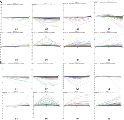
We compared SV-HUV-1-RT4-5637 DEGs pair-wise (Tables S2-S4). DEGs were divided into 8 groups; grouping method is seen in ‘approach’. We obtained 336 DEGs in group 0, 1165 DEGs in group 1, 640 DEGs in Group 2, 288 DEGs in
group 3, 702 DEGs in group 4, 768 DEGs in group 5, 366 DEGs in group 6, 350 DEGs in group 7 (Figure 1A and Table S5).
DEGs in SV-HUV-1-RT4-T24 were clustered (Tables S6-S8), and DEGs were divided into 8 groups. We obtained 188 DEGs in group 0, 533 in group 1, 143 in Group 2,124 in group 3, 345 in group 4, 369 in group 5, 111 in group 6, 131 in group 7 (Figure 1B and Table S9).
Functional analysis of DEGs
In order to further explore the function of each differential gene, we would carry out the GO data analysis on DEGs in each group, as shown in the results, DEGs in group 0 in
[image:3.612.93.518.72.484.2]mRNA profile in urinary bladder cancer
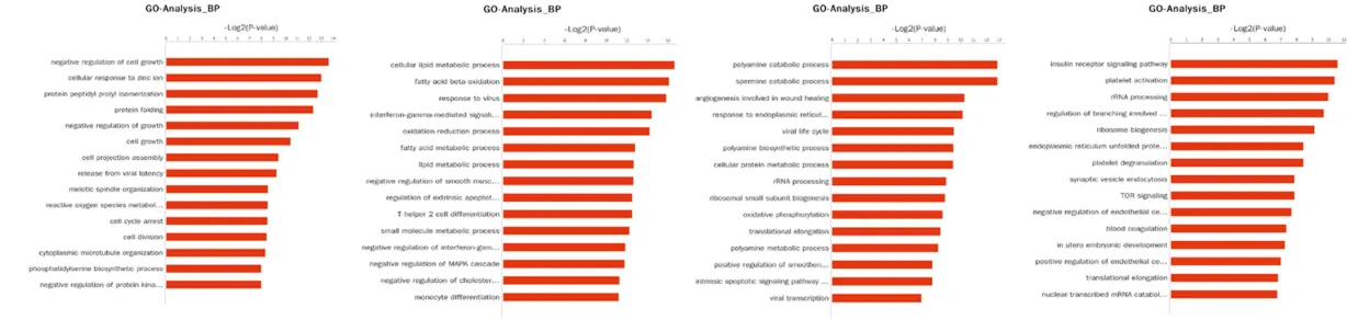
RT4-5637 were mainly concentrated in gene expression, DNA damage repair pathways, RNA metabolic pathways; group 1 mainly concen-trated in the cell cycle and cell division; group 2 mainly concentrated in protein translocation and chromatin regulation; group 3 concentrat-ed in the gene expression; Notch receptors in group 4 processed the highest score; group 5 concentrated in the apoptotic pathway; group 6 concentrated in JNKK activation and cell adhe-sion; group 7 was IL-1 receptor signal and ribo-some biogenesis (Figure 2A).
Meanwhile, we conducted GO gene differential expression data analysis on each group of SV-HUV-1-RT4-T24, as shown in the results, DEGs in group 0 inSV-HUV-1-RT4-T24 mainly concentrated in mitochondrial ATP synthesis and respiratory oxidation of anion chain; group 1 mainly concentrated in mitosis; group 2 main-ly concentrated in mitosis and cell differentia-tion; group 3 concentrated in fatty acid metab-olism; group 4 concentrated in cell proliferation regulation; group 5 concentrated in lipid metab-olism and interferon inducing cell signaling pathways; group 6 concentrated in polyamine catabolism; group 7 concentrated in platelet activation and insulin receptor signaling path-way (Figure 2B).
Pathway analysis of DEGs
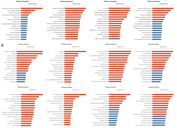
In order to further understand the pathways of DEGs in each group, we performed gene path-way analysis for SV-HUV-1-RT4-5637 and SV-HUV-1-RT4-T24 (Figure 3A and 3B). Through analysis, we found that each group of genes were consistent with GO analysis pathway in profile 4 in SV-HUV-1-RT4-5637, and genes causing 5637 increase were mostly concen-trated in the Notch signaling pathway. We found that those genes gradually increased in profile 7 of the SV-HUV-1-RT4-T24 were mainly concen-trated in HIF-1 and mTOR, which all prompted that T24 and metabolism genes had some relevance.
RT-PCR analysis
RT-PCR was performed to verify the DEGs in RNA-seq. In order to further validate the results of our RNA-seq, we cultured the SV-HUV-1, RT4, 5637 and T24 cells, and extracted their RNA, and expression levels of these genes were vali-dated in messager RNA level. Results showed
that Jagged 1 (JAG1), Histone Deacetylase 5 (HDAC5), JAG2, Recombination Signal Binding Protein for Immunoglobulin Kappa J Region (RBPJ) was significantly increased in Notch sig-naling pathway in SV-HUV-1-RT4-5637. In SV-HUV-1, RT4, 5637, we detected genes EIF4 and EBP1 in HIF-a pathway and VEGFB in mTOR pathway. With the increasing degree of malig-nancy of bladder cancer, the expression of these genes gradually increased.
Discussion
It has been widely known that genomic tions leads to the expression structure muta-tions in bladder cancer, and heterogeneity is an important feature of bladder cancer [15]. mRNA and DNA chip technology [16] have been used to detect abnormal expression of genes in blad-der cancer cells, related prognosis marks [17-19], and parsed the biological nature of bladder cancer [20, 21]. However, due to the self-limita-tion of chip technology such as narrow cover-age [22], more and more studies tended to use second-generation sequencing technology for the changes of expression profiles in bladder cancer [23]. The next generation RNA sequenc-ing technology not on lycan detects mRNA but also discover non-coding RNA. In this study, we first used the next generation sequence to ana-lyze the gene expression profiles in bladder cancer cell lines of different degree of malig-nancy, attempted to discover the key cancer progression pathway and molecules through trend analysis and GO, pathway analysis and other functions.
mRNA profile in urinary bladder cancer
mRNA profile in urinary bladder cancer
significantly increased expression in the SV-HUV-1-RT4-T24, suggesting that these developments might involve in the develop-ment of bladder cancer.
In future experiments, we will detect the chang-es of differential exprchang-ession genchang-es on genome and protein level, combining with genome sequencing and protein detection experiments. Then combined with tumor function experi-ments, such as apoptosis, proliferation, drug reactions, we will explain the functions of detected differentially expressed genes, pro-vide appropriate therapeutic targets for per-sonalized treatment of bladder cancer [32-34]. In conclusion, the data presented in this study revealed that Notch pathway plays pivotal roles in the formation process of bladder cancer and may be a potential target for the treatment of bladder cancer, reveal molecular mechanisms of different biological characteristics of various bladder cancers. Our study may provide theo-retical basis for the future exploration of Notch pathway in bladder cancer.
Acknowledgements
This research was supported by the Science and Technology Development Fund of Macao (Grant No. 064/2012/A).
Disclosure of conflict of interest
None.
Address correspondence to: Dr. Bin Lang, School of Health Sciences, Macao Polytechnic Institute, R. de Luís Gonzaga Gomes, Macao, China. Tel: +86-00853-85993440; E-mail: [email protected]
References
[1] Siegel R, DeSantis C and Jemal A. Colorectal cancer statistics, 2014. CA Cancer J Clin 2014; 64: 104.
[2] Kirkali Z, Chan T, Manoharan M, Algaba F, Busch C, Cheng L, Kiemeney L, Kriegmair M, Montironi R and Murphy WM. Bladder cancer: epidemiology, staging and grading, and diag-nosis. Urology 2005; 66: 4-34.
[3] Lawrence MS, Stojanov P, Polak P, Kryukov GV, Cibulskis K, Sivachenko A, Carter SL, Stewart C, Mermel CH and Roberts SA. Mutational het-erogeneity in cancer and the search for new
cancer-associated genes. Nature 2013; 499: 214-218.
[4] Nicolas M, Wolfer A, Raj K, Kummer JA, Mill P, Van NM, Hui CC, Clevers H, Dotto GP and Radtke F. Notch1 functions as a tumor sup-pressor in mouse skin. Nat Genet 2003; 33: 416-421.
[5] Lefort K, Dotto GP. Notch signaling in the inte-grated control of keratinocyte growth/differen-tiation and tumor suppression. Semin Cancer Biol 2004; 14: 374-386.
[6] Klinakis A, Lobry C, Abdel-Wahab O, Oh P, Haeno H, Buonamici S, Van DWI, Cathelin S, Trimarchi T and Araldi E. A novel tumour-sup-pressor function for the Notch pathway in my-eloid leukaemia. Nature 2011; 473: 230-233. [7] Shouse GP, Nobumori Y and Liu X. A B56γ mu-tation in lung cancer disrupts the p53-depen-dent tumor suppressor function of protein phosphatase 2A. Oncogene 2010; 29: 3933-3941.
[8] Wang NJ, Sanborn Z, Arnett KL, Bayston LJ, Liao W, Proby CM, Leigh IM, Collisson EA, Gordon PB, Jakkula L, Pennypacker S, Zou Y, Sharma M, North JP, Vemula SS, Mauro TM, Neuhaus IM, Leboit PE, Hur JS, Park K, Huh N, Kwok PY, Arron ST, Massion PP, Bale AE, Haussler D, Cleaver JE, Gray JW, Spellman PT, South AP, Aster JC, Blacklow SC, Cho RJ. Loss-of-function mutations in Notch receptors in cu-taneous and lung squamous cell carcinoma. Proc Natl Acad Sci U S A 2011; 108: 17761-17766.
[9] Stransky N, Egloff AM, Tward AD, Kostic AD, Cibulskis K, Sivachenko A, Kryukov GV, Lawrence MS, Sougnez C and Mckenna A. The Mutational Landscape of Head and Neck Squamous Cell Carcinoma. Science 2011; 333: 1157-1160.
[10] Lobry C. Oncogenic and tumor suppressor functions of Notch in cancer: it’s NOTCH what you think. J Exp Med 2011; 208: 1931-1935. [11] Ahn CS and Metallo CM. Mitochondria as
bio-synthetic factories for cancer proliferation. Cancer Metabol 2015; 3: 1-1.
[12] Deberardinis RJ, Sayed N, Ditsworth D and Thompson CB. Brick by brick: metabolism and tumor cell growth. Curr Opin Genet Dev 2008; 18: 54-61.
[13] Kroemer G and Pouyssegur J. Tumor cell me-tabolism: cancer’s Achilles’ heel. Cancer Cell 2008; 13: 472-482.
[14] Harris MA. The Gene Ontology (GO) database and informatics resource. Nucleic Acids Res 2004; 32: D258-D261.
gene lists using DAVID bioinformatics resourc-es. Nat Protoc 2009; 4: 44-57.
[16] Xu A, Wang C and Sun S. Screening candidate genes associated with bladder cancer using DNA microarray. Mol Med Rep 2014; 10: 3087-3091.
[17] Eissa S, Matboli M, Hegazy MGA, Kotb YM and Essawy NO. Evaluation of urinary microRNA panel in bladder cancer diagnosis: relation to bilharziasis. Transl Res 2015; 165: 731-739. [18] Semilia M, Hennenlotter Jr, Pavone C, Bischoff
T, Kühs U, Gakis G, Bedke J, Stenzl A, Schwentner C and Todenhöfer T. Expression patterns and prognostic role of transketolase-like 1 in muscle-invasive bladder cancer. World J Urol 2015; 33: 1403-1409.
[19] Kim PH, Cha EK, Sfakianos JP, Iyer G, Zabor EC, Scott SN, Ostrovnaya I, Ramirez R, Sun A and Shah R. Genomic predictors of survival in patients with high-grade urothelial carcinoma of the bladder. Eur Urol 2014; 67: 198-201. [20] Choudhary D, Hegde P, Voznesensky O,
Choudhary S, Kopsiaftis S, Claffey KP and Pilbeam CC. Increased expression of L-selectin (CD62L) in high-grade urothelial carcinoma: A potential marker for metastatic disease. Urol Oncol 2015; 33: 387, e17-27.
[21] Teo MT, Dyrskjøt L, Nsengimana J, Buchwald C, Snowden H, Morgan J, Jensen JB, Knowles MA, Taylor G and Barrett JH. Next-generation se-quencing identifies germline MRE11A variants as markers of radiotherapy outcomes in mus-cle-invasive bladder cancer. Ann Oncol Offic J Eur Soc Med Oncol 2014; 25: 877-883. [22] Ozsolak F and Milos PM. RNA sequencing:
ad-vances, challenges and opportunities. Nat Rev Genet 2011; 12: 87-98.
[23] Liu Y, Noon AP, Aguiar CE, Shen J, Kuk C, Ilczynski C, Ni R, Sukhu B, Chan K and Barbosa-Morais NL. Next-generation RNA sequencing of archival formalin-fixed paraffin-embedded uro-thelial bladder cancer. Eur Urol 2014; 66: 982-986.
[24] Maraver A, Fernandez-Marcos PJ, Cash TP, Mendez-Pertuz M, Dueñas M, Maietta P, Martinelli P, Muñoz-Martin M, Martínez-Fernández M, Cañamero M, Roncador G, Martinez-Torrecuadrada JL, Grivas D, de la Pompa JL, Valencia A, Paramio JM, Real FX, Serrano M. NOTCH pathway inactivation pro-motes bladder cancer progression. J Clin Invest 2015; 125: 824-830.
[25] Ai X, Jia Z, Liu S, Wang J and Zhang X. Notch-1 regulates proliferation and differentiation of human bladder cancer cell lines by inhibiting expression of Kruppel-like factor 4. Oncol Rep 2014; 32: 1459-1464.
[26] Reedijk M, Pinnaduwage D, Dickson BC, Mulligan AM, Zhang H, Bull SB, O’Malley FP, Egan SE and Andrulis IL. JAG1 expression is as-sociated with a basal phenotype and recur-rence in lymph node-negative breast cancer. Breast Cancer Res Treat 2008; 111: 439-448. [27] Peixoto P, Castronovo V, Matheus N, Polese C,
Peulen O, Gonzalez A, Boxus M, Verdin E, Thiry M, Dequiedt F and Mottet D. HDAC5 is required for maintenance of pericentric heterochroma-tin and controls cell cycle progression of hu-man cancer cells. Cell Death Differ 2012; 19: 1239-52.
[28] Sasnauskienė A, Jonušienė V, Krikštaponienė A, Butkytė S, Dabkevičienė D, Kanopienė D, Kazbarienė B and Didžiapetrienė J. NOTCH1, NOTCH3, NOTCH4, and JAG2 protein levels in human endometrial cancer. Medicina 2014; 50: 14-18.
[29] Lv Q, Shen R and Wang J. RBPJ inhibition im-pairs the growth of lung cancer. Tumour Biol 2015; 36: 3751-3756.
[30] Afonso J. Phospho-mTOR in non-tumour and tumour bladder urothelium: Pattern of expres-sion and impact on urothelial bladder cancer patients. Oncol Lett 2014; 8: 1447-1454. [31] Peng Y. Angiogenin interacts with ribonuclease
inhibitor regulating PI3K/AKT/mTOR signaling pathway in bladder cancer cells. Cell Signal 2014; 26: 2782-2792.
[32] Proctor I, Stoeber K and Williams GH. Biomarkers in bladder cancer. Histopathology 2010; 57: 1.
[33] Proctor MJ, Talwar D, Balmar SM, O’Reilly DS, Foulis AK, Horgan PG, Morrison DS and Mcmillan DC. The relationship between the presence and site of cancer, an inflammation-based prognostic score and biochemical pa-rameters. Initial results of the glasgow inflam-mation outcome study. Bri J Cancer 2010; 103: 870-876.