Original Article
Effect of RAB25 gene on proliferation of human breast
cancer cell line MCF-7
in vivo
and
in vitro
Xiaobin Zhang, Yunfei Lu, Guixin Shen, Jiehua Li
Department of Gastrointestinal and Gland Surgery, The First Affiliated Hospital of Guangxi Medical University, Nanning, China
Received January 6, 2016; Accepted March 20, 2016; Epub April 1, 2016; Published April 15, 2016
Abstract: Rab25 is a newly discovered member of the Rab11 subfamily of the Rab protein family. The purpose of this study was to investigate the proliferation of derivatives of the human breast cancer cell line MCF-7 with differ-ent levels of expression of the Rab25 gene and their tumorigenicity in nude mice. The effect of differdiffer-ent expression levels of the Rab25 gene on various biological activities of the human breast cancer cells was also investigated. Cells in the logarithmic phase of growth were harvested and subcutaneously implanted into nude mice to evaluate tumor induction. The mRNA expression of Rab25 in tumors in nude mice was evaluated using quantitative RT-PCR. The results showed that stable expression of the Rab25 gene after transfection significantly increased the prolifera-tion activity of MCF-7 breast cancer cells. The tumorigenicity in nude mice was also increased. In addiprolifera-tion, interfer-ence with the expression of the Rab25 gene significantly inhibited the proliferation of MCF-7 breast cancer cells and attenuated their tumorigenicity in nude mice. Finally, Rab25 was discovered to be an oncogene. A high level of expression could directly increase the proliferation and invasion abilities of breast cancer cells and their tumori-genicity in nude mice. The RNA interference technology is expected to provide new approaches for tumor targeted gene therapy of breast cancer.
Keywords: Breast cancer, Rab25 gene, human breast cancer cell line MCF-7, biological activity, nude mice
Introduction
The Rab25 protein is a newly discovered mem-ber of the Rab11 subfamily of the Rab protein family. The Rab25 gene is located at 1q21.2 and encodes a low molecular weight protein consisting of 213 amino acid residues [1]. Many domestic and international studies have shown that Rab25 expression is upregulated in a vari-ety of epithelial malignant tumors. In recent years, expression of Rab25 has been exten-sively reported in breast cancer, gastric cancer, colon cancer, esophageal cancer, and ovarian cancer [2]. However, there are different opin-ions about the biological functopin-ions of Rab25 in breast cancer. Current studies are mostly at the cellular level; there are few studies of the effect of Rab25 on the occurrence and development of solid tumors. By upregulating and silencing Rab25 gene expression in MCF-7 breast cancer cells, we established a series of MCF-7 deriva-tives with different Rab25 expression levels to observe the effect of Rab25 expression on breast cancer xenografts in nude mice and to
investigate the effect of the Rab25 gene on the proliferation of the cells in vivo and in vitro. Materials and methods
Materials
from the Shanghai Experimental Animal Center, Chinese Academy of Sciences. The PCR reagent kit was purchased from Promega. The Trizol RNA extraction kit, the LipofectamineTM 2000
transfection reagent kit, and the primers for Rab25 mRNA and β-actin were purchased from Invitrogen. The primers for Rab25 DNA were synthesized by Shanghai Sangon Biotech.
Cell culture technique and methods
A series of cell lines previously constructed by our group, including normal parental MCF-7 cells, Rab25 transfected cells, cells transfect-ed with Rab25 siRNA, and cells transfecttransfect-ed with the empty vector were recovered from storage [3]. The cells were cultured and pas-saged, and some cells were prepared as cell suspensions to detect cell proliferation and adjust cell concentrations. Cell suspensions were centrifuged at a low speed, mixed thor-oughly with DMEM, and transferred to a 100 ml culture flask. Cells were cultured in a 5% CO2 incubator at 37°C. The culture medium was replaced the next day, and the incubation was continued. After the cell confluence reached 90%, the cells were subcultured. After several passages, some cells were cultured for amplifi-cation and some cells were cryopreserved to ensure a source of cells to facilitate subse-quent studies.
Detection of Rab25 in the cell lines studied
The Trizol method was used for the extraction of total RNA from MCF-7 and the three deriva-tives. The target fragment (152 bp) of the Rab25 gene was amplified using the RT-PCR reagent kit as shown below. Primers were designed according to the Rab25 coding sequences (GM ID57111) in GenBank: up- stream, 5’-CCATCACCTCGGCGTACTATC-3’ and downstream, 5’-TTTGTTACCCACGAGCATGAC-3’. RT-PCR products from the four cell lines were detected using 1% agarose gels.
Amplification of the full length Rab25 cDNA. Rab25 total RNA from the parental MCF-7 cells was used as a template to amplify the full-length Rab25 cDNA (649 bp). PCR primers were designed according to the Rab25 mRNA sequence (NCBI Accession No. NM_020387) provided by GenBank. The primers were as fol-lows: upstream, 5’-AAGATGGGGAATGGAACTGA- 3’; downstream, 5’-AAGGTCAGAGGCTGATGCA- AC-3’. RT-PCR was performed, and the prod-ucts were detected using 1% agarose gels.
Detection of cell proliferation activity using the MTT assay. The cells were divided into four groups: parental cells, cells transfected with the recombinant Rab25 plasmid, cells trans-fected with Rab25 siRNA, and cells transtrans-fected with the empty vector. Cells in the logarithmic phase of growth were prepared as single-cell suspensions using DMEM containing 10% fetal bovine serum (FBS) and inoculated onto 96-well culture plates. The blank control group was in serum-free L-15 culture medium. Each well con-tained 0.2 ml, and the culture time was one to six days, after which 20 µl 0.5% MTT was added. After 4 h, the supernatants in the wells were aspirated, and 150 µl DMSO was added to each well. The plates were vortexed for 10 min, and the absorbance value (A value) of each well was determined at 490 nm using a plate reader. Cell growth curves were plotted using time as the horizontal axis and A490 as the vertical axis. Experiments were repeated three times, and three replicate wells were used. The mean value was used as the final result.
Inoculation
A total of 32 four- to six-week-old female BALB/c nude mice with a body weight of 18-22 g were randomly divided into four groups with eight animals in each group. Each group was inocu-lated with parental MCF-7 cells, Rab25 trans-fected cells, Rab25 siRNA transtrans-fected cells, or empty vector transfected cells. The skin of the axilla of each mouse was disinfected using povidone-iodine complex. Tumor cell suspen-sions were aspirated using a syringe with a no. 6 needle and injected subcutaneously into the axilla of each nude mouse. Each inoculation site was injected with 0.2 ml containing approx-imately 107 live cells. After the needle was
with-drawn, the site was clipped using tweezers to prevent leakage of tumor cells.
Observation and measurement
tumor volume was calculated, and the growth curves of the xenograft tumors were plotted. The results were statistically analyzed.
Calculation and fluorescence analysis of quan-titative RT-PCR standard curves of Rab25 mRNA in xenograft tumors in a series of nude mice.
The cDNA from MCF-7 cells that showed Rab25 gene expression was removed from a -80°C ultra-low freezer and used as a standard. β-Actin was used as the quantitative template for the internal control. The following primers were used:
Rab25: upstream, 5’-GGGTTGAGGGCATTGAGC- 3’, downstream, 5’-GAGGTATTTGTGATAGGGCA- TG-3’, probe, 5’-AGATTGGTCTTCCCCACACCTGA- TT-3’; β-actin: upstream, 5’-GTCATCACCATTGGC- AATGAG-3’, downstream, 5’-CGTCACACTTCATG- ATGGAGTT-3’, probe, 5’-TCCTGGGCATGGAGTCC- TG-3’.
The amplification length was 881 bp. The 5’ and 3’ ends of the probes were labeled with fluorescein FAM and TAMRA, respectively. The standard cDNA and the internal control tem-plate (β-actin) were diluted at the ratios of 1:10-108. The reactions contained 25 μl, including
10 μl buffer, 1 μl of primers, fluorescence probe, and dNTPs, 2.5 U Taq polymerase, and 2 μl cDNA from the reverse transcribed products (equivalent to 100 ng). Real-time quantitative RT-PCR was performed. The reaction condi-tions were 94°C denaturation for 4 min fol-lowed by 40 cycles of 94°C for 30 s, 55°C for 30 s, and 70°C for 30 s. With the temperature controlled at 79°C, the A260/A280 absorbance values of RNA were measured. The amplifica-tion curves were plotted using the number of cycles as the horizontal axis and the values of the fluorescence signals as the vertical axis. The threshold values (CT, the minimum number of cycles required for the fluorescence signal in the reaction tube to reach the set threshold value) were confirmed according to the amplifi-cation curve. Two standard curves were plotted using logarithmic values of the gradient con-centrations of Rab25 mRNA and β-actin as the horizontal axis and CT values as the vertical axis. The levels of Rab25 and β-actin in the samples were calculated from the standard curves according to the CT values. Three dupli-cate tubes were used for each sample for detection, and the mean value was calculated.
The double standard curve method was per-formed for relative quantitation. The levels of the Rab25 gene and the internal quantitative template β-actin were measured to estimate the level of Rab25 gene relative to β-actin. Finally, comparisons of the relative levels of the samples in all the experimental groups were performed, and the Rab25 mRNA expression in the tumors was determined. In addition, the CT values for the samples and the β-actin internal control were compared to ascertain whether the linear amplification efficiency (E) of the two genes was basically consistent (E = 10-1/K-1),
where K is the slope of the standard curve, and to control for possible experimental error.
Statistical methods
The data are presented as the mean ± the stan-dard deviation (_x ± s). The SPSS13.0 software was used for the analysis of variance (ANOVA) and for the t test. Values of P < 0.05 indicated statistical significance.
Results
Observation under a microscope of the four groups of cells
The MCF-7 cell line and three derivatives pre-pared in previous studies were recovered from storage and cultured. Cells from the parental MCF-7 cell line, cells transfected with Rab25, and cells transfected with the empty vector were viewed with an inverted microscope and did not show any significant differences. These cells all showed monolayer growth, regular mor-phology, and polygonal or long spindle shapes. The cell nuclei were round or oval, the cyto-plasm was transparent and bright, and the den-drites were long. However, cells transfected with Rab25 siRNA showed irregular shapes, increased cell body size, wider intercellular spaces, decreased brightness of the cyto-plasm, increased number of dendrites (which were short and thick), and decreased cell attachment, indicating a decrease in cell growth (Figure 1).
Analysis of Rab25 expression in MCF-7 and its derivatives
with the recombinant plasmid pDNR-Dual-Rab25 or the empty vector (Figure 2).
Amplification of full-length of Rab25 cDNA
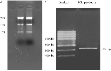
The total RNA of MCF-7 cells was extracted and used as the template. As shown in (Figure 3A), the brighter band was the 28S RNA, and the relatively weaker band below it was the 18S RNA. This result indicated that the extracted RNA was intact and without DNA contamina-tion. After RT-PCR amplification, a single spe-cific band (649 bp) was observed (Figure 3B), which was consistent with the length of the Rab25 gene sequence provided by GenBank.
Results of cell proliferation assays
Absorbance values (A values) of the cultured cells were measured. The A values for MCF-7 cells with different levels of Rab25 are shown in (Table 1). The growth curve of each cell line was plotted based on the A values (Figure 4). Figure 4 shows that the proliferation of cells transfect-ed with Rab25 increastransfect-ed rapidly starting from day 3 (P < 0.05) and that the proliferation of cells transfected with Rab25 siRNA was signifi-cantly slower (P < 0.05). The proliferation of cells transfected with the empty vector was not significantly different from that of the parental cells or cells transfected with Rab25 siRNA (P > 0.05).
Growth of the different cell lines in nude mice
[image:4.612.90.527.72.263.2]The tumor incidence in mice for all the cell lines after inoculation was 100%. The incubation Figure 1. A: Cells from the parental MCF-7 cell line, cells transfected with Rab25, and cells transfected with the empty vector were viewed with an inverted microscope and did not show any significant differences. These cells all showed monolayer growth, regular morphology, and polygonal or long spindle shapes. The cell nuclei were round or oval, the cytoplasm was transparent and bright, and the dendrites were long. B: Cells transfected with Rab25 siRNA showed irregular shapes, increased cell body size, wider intercellular spaces, decreased brightness of the cytoplasm, increased number of dendrites (which were short and thick), and decreased cell attachment, indicating a decrease in cell growth.
[image:4.612.92.257.360.616.2]period for tumor growth in nude mice inoculat-ed with the parental MCF-7 cells, cells trans-fected with Rab25, and cells transtrans-fected with the empty vector was approximately seven to
[image:5.612.91.521.70.343.2] [image:5.612.90.525.414.476.2]increased with time and that there were local ulcers. In contrast, the mice inoculated with cells transfected with Rab25 siRNA only devel-oped small, palpable subcutaneous tumors, Figure 3. A: Electrophoresis results of total RNA extracted from MCF-7 cells. B: Electrophoresis results of the PCR products of the Rab25 cDNA which show that the extracted total RNA was intact and without DNA contamination. B indicates that the length of the detected gene was consistent with the Rab25 gene sequence provided by GenBank.
Table 1. Detection by absorbance of the effect of Rab25 on MCF-7 cells (_x ± s)
Groups 1 d 2 d 3 d 4 d 5 d 6 d 7 d
Normal parental MCF-7 cells 0.15 ± 0.02 0.18 ± 0.03 0.22 ± 0.02 0.26 ± 0.01 0.32 ± 0.01 0.28 ± 0.02 0.26 ± 0.03 Rab25 transfected cells 0.15 ± 0.02 0.18 ± 0.03 0.34 ± 0.01 0.46 ± 0.02 0.54 ± 0.03 0.62 ± 0.02 0.58 ± 0.01 Cells transfected with Rab25 siRNA 0.16 ± 0.02 0.17 ± 0.02 0.19 ± 0.04 0.23 ± 0.03 0.24 ± 0.02 0.20 ± 0.01 0.21 ± 0.03 Cells transfected with the empty vector 0.16 ± 0.03 0.18 ± 0.02 0.22 ± 0.03 0.26 ± 0.02 0.33 ± 0.02 0.29 ± 0.01 0.31 ± 0.01
Figure 4. Growth curves of all the cell lines based on A values. The prolifera-tion of cells transfected with Rab25 was significantly more rapid than that of cells transfected with the empty vector, the parental cell line group, or the interference group.
[image:5.612.89.384.453.621.2]and the tumor growth was slow (Figure 5). The animals were sacrificed after six weeks. The mean tumor volumes of mice inoculated with the parental cell line, cells transfected with Rab25, or cells transfected with the empty vec-tor were all larger than 100 mm3, whereas the
mean tumor volume of mice inoculated with
surements of Rab25 expression levels in xeno-graft tumors induced by derivatives of MCF-7
[image:6.612.90.523.71.397.2]Reactions with four groups of samples and the standard were performed at the same time. Each sample was measured three times. The mean CT values obtained for Rab25 mRNA and Figure 5. Gross morphology of xenograft tumors in nude mice induced by a series of breast cancer cells at week 6. A: Parental cell line group; B: Transfection group; C: Interference group; D: Empty vector group. It was visible to the unaided eye that the transfection group had the largest tumors.
Table 2. Weight and volume of xenograft tumors in nude mice in all groups (_x ± s, n = 8)
Groups Volume (mm³) Tumor weight (g)
Normal parental MCF-7 cells 1073.12 ± 419.59 1.196 ± 0.307 Rab25 transfected cells 2232.25 ± 621.56 1.855 ± 0.346 Cells transfected with Rab25 siRNA 246.50 ± 73.83 0.656 ± 0.053 Cells transfected with the empty vector 1052.60 ± 336.75 1.146 ± 0.185
Note: Results were analyzed using analysis of variance (ANOVA) for completely ran-domized design. Pairwise comparison of volume and weight of xenograft tumors was performed using the LSD method. N: number of samples. Significance level: α = 0.05. F = 31.362 and 30.827. Comparison between the transfection group and the parental cell line group, P < 0.01; comparison between the interference group and the parental cell line group, P < 0.01; comparison between the empty vector transfection group and the parental cell line group, P > 0.05.
cells transfected with the Rab25 siRNA was < 250 mm3. The records of the
tumor weights and volu- mes of the xenograft tu- mors from the nude mice in each group are shown in (Table 2). Growth curves of the xenograft tumors in nude mice with different Rab25 expression levels are plotted according to data from the weekly mea-surements (Figure 6).
[image:6.612.92.391.485.552.2]mea-β-actin were separately introduced into the regression equation of the standard curve to calculate the individual amount of the initial template. β-actin was used as the control refer-ence gene to normalize the results from all the samples (RNA calibration). The quantitative result for Rab25 mRNA divided by the quantita-tive result for β-actin was the calibrated value. To facilitate the comparison of expression lev-els of samples between groups, the Rab25 mRNA expression level in tumors from mice inoculated with the parental cell line was set to “1” to calculate the relative levels in other
[image:7.612.93.394.76.262.2]tiple steps. Currently, studies of gene muta-tions in p53, BRCA1, and BRCA2 only explain some of the etiology of breast cancer; there may be more unknown genes participating in the development of breast cancer. The study of Calero et al. showed that the Rab25 gene might be associated with the development and inva-sion of breast cancer [4]; however, the biologi-cal function of Rab25 in the occurrence and development of breast cancer is still unknown. The groups of Cheng KW and Cheng JM pub-lished different results on the effect of Rab25 on the development of breast cancer. Cheng et Figure 6. Growth curves of xenograft tumors in nude mice with different Rab25
expression levels. The figure shows that the xenograft tumor volume in nude mice induced by cells from the transfection group was significantly higher than in the empty vector group, the parental cell line group, or the interference group.
Table 3. Data for the real-time quantitative PCR standard curve of Rab25 mRNA and β-actin in xenograft tumors in nude mice
MCF-7/Rab25 mRNA MCF-7/β-actin
CT K E (%) CT K E (%)
Normal parental MCF-7 cells 24.66 -3.49 93.42 18.92 -3.39 97.24 24.58 -3.44 95.30 18.81 -3.38 97.65 24.72 -3.42 96.06 19.01 -3.46 94.54 Rab25 transfected cells 14.92 -3.48 93.78 11.56 -3.39 97.24 14.55 -3.42 96.06 11.52 -3.38 97.65 15.12 -3.49 93.42 11.64 -3.47 94.18 Cells transfected with Rab25
siRNA 36.49 -3.41 96.43 26.86 -3.42 96.06 36.15 -3.40 96.83 26.67 -3.50 93.06 36.24 -3.47 94.18 26.74 -3.39 97.24 Cells transfected with the empty
vector 24.52 -3.50 93.06 18.88 -3.47 94.18 24.76 -3.43 95.66 18.84 -3.46 94.54 24.32 -3.46 93.42 18.74 -3.48 93.78
groups. Comparisons of Rab25 mRNA expression levels in samples from the different groups were then performed (Table 3). Finally, to present the differences in relative expression levels of sam-ples in all the groups more intuitively, the cal-culated relative levels were plotted in histogra- ms (Figure 7). As shown in Figure 7, the Rab25 mRNA expression level in the tumors from mi- ce inoculated with cells transfected with Rab25 was 4.986 times that in tumors from mice inocu-lated with the parental cell line. The Rab25 mRNA expression level in tumors from mice inocu-lated with cells transfect-ed with Rab25 siRNA was only 0.082 times that in tumors from mice inoculated with the pa- rental cell line. The expre- ssion levels in tumors from the empty vector group and the parental cell line group were simi-lar (0.993).
Discussion
[image:7.612.90.394.361.547.2]mul-al. [5] considered that increased DNA copy numbers and RNA levels of the Rab25 gene decreased the disease-free survival and the overall survival of breast cancer patients. In cell experiments, overexpression of Rab25 increased the proliferation activity and inva-siveness of breast cancer cells. However, the study of Cheng et al. [6] showed that overex-pression of Rab25 in breast cancer cell lines inhibited cell growth. In addition, detection of expression in mammary tissues using RT-PCR showed that Rab25 was extensively expressed in normal mammary tissues and that approxi-mately 33% of breast cancer tissues lost expression. These results indicated that loss of Rab25 might play an important role in the pathogenic process of breast cancer. Since then, studies of the biological behavior of the Rab25 gene in breast cancer have been gradu-ally implemented.
Although studies of the function of Rab25 pro-tein were begun 30 years ago, its function is currently not completely clear. Rab25 is ex- pressed in a variety of tumor cells as well as so- me tissues, including gastrointestinal mucosa, lung, kidney, and mammary gland. Culine et al. [7] showed that Rab25 was upregulated in a va- riety of epithelial malignant tumors. Cao et al. [8] detected Rab25 protein expression in gas-tric cancer tissues using immunohistochemis-try and showed that Rab25 protein expression in gastric cancer tissues was higher than in adjacent tissues, the expression in stage III-IV patients was higher than in stage I-II patients,
[9] confirmed that high levels of Rab25 expres-sion were closely associated with the classifica-tion of renal cell carcinoma invasion, lymph node metastasis, and pathological stage. Interference with Rab25 protein expression could inhibit renal cell carcinoma proliferation, migration, and invasion. However, the study of Tong et al. showed the opposite results. In esophageal squamous cell carcinoma, Tong et al. [10]showed that Rab25 was a tumor sup-pressor. Real-time quantitative PCR and immu-nohistochemistry results showed that Rab25 expression in esophageal squamous cell carci-noma was significantly downregulated com-pared with that in non-tumor tissues. Hyper- methylation of the promoter region of the Rab25 gene in esophageal cancer caused the decrease or loss of the expression of this gene in tumor tissues. In addition, the loss of Rab25 expression in nude mice inhibited tumor devel-opment and angiogenesis. The Rab25 gene mainly exerts its biological function though the MAPK/ERK signal transduction pathway. The study of Nam [11] in nude mice showed that loss of Rab25 expression in nude mice could promote the occurrence and development of intestinal cancer, which resembles colorectal cancer in humans.
[image:8.612.93.400.70.237.2]For mammary glands, Gonzalez-Angulo [12] used reverse phase protein array (RPPA) and showed that the combination of three factors, CHK1pS345, Caveolin1, and Rab25, in patients with primary breast cancer recurrence could be used as an independent prognosis model. In Figure 7. To present the differences of relative expression levels in xenograft tumor
samples from nude mice more intuitively, the calculated relative levels are present-ed as histograms.
addition, different levels of Rab25 expression had significantly different 3-year recurrence-free survival rates, and the Rab25 overexpres-sion group had the worst prognosis. However, the subsequent study of Cheng et al. [13] showed that Rab25 overexpression had signifi-cant inhibitory effects on the development of triple negative breast cancer. Chen et al. [14] also showed that Rab25 gene could reduce the invasiveness of breast cancer and that Rab25 was a tumor suppressor gene. Currently, there is no consistent conclusion about changes in the proliferation activity and invasiveness of tumor cell lines caused by low or high levels of Rab25 expression. In this study, a series of experiments was performed to understand the changes in proliferation activity and invasive-ness of a breast cancer cell line by silencing or expressing high expression levels of Rab25. In addition, the influence of Rab25 on solid tumor formation and whether Rab25 could be an independent prognostic factor and a new thera-peutic target for breast cancer were studied. A series of MCF-7 breast cancer cell lines with different Rab25 expression levels were con-structed. Rab25 gene expression in the paren-tal MCF-7 cells, cells transfected with Rab25, and cells transfected with the empty vector was confirmed by many repetitions of RT-PCR. Cells transfected with Rab25 siRNA showed lit-tle or no Rab25 expression. Breast cancer cell line proliferation was detected using the MTT assay. Statistical analysis showed that the pro-liferation activity of MCF-7 breast cancer cells significantly increased after the transfected Rab25 gene was stably expressed, suggesting that upregulation of Rab25 gene expression increased the proliferation of breast cancer cells. In addition, after interference with Rab25 gene expression, the proliferation activity was significantly inhibited, which allowed us to con-firm from another angle that downregulation of the Rab25 gene attenuates the proliferation ability of breast cancer cells.
Rab25 participates not only in tumor cell prolif-eration but also in solid tumor formation. In this study, after breast cancer cells with different Rab25 expression levels were transplanted into 32 nude mice, all the mice developed tumors. After six weeks of observation, the mean tumor volumes in the mice in the paren-tal cell line group, the transfection group, and the empty vector group were all larger than
1000 mm3. With the progression of time, the
tumors gradually became larger, and the tumors in some nude mice showed local ulcers and necrosis; however, after the mice in the interference group were observed for six weeks, only palpable subcutaneous tumors with small-er volumes developed. The mean tumor volume was < 250 mm3, and the tumors no longer grew
with the progression of time but showed signs of decreasing in size. In addition, Rab25 mRNA expression in the tumors was detected quanti-tatively using real-time quantitative PCR. The results showed that the Rab25 mRNA expres-sion level in the tumors in the transfection group was significantly higher than that in the parental cell line group, whereas the Rab25 mRNA expression level in the tumors in the interference group was significantly lower than that in the parental cell line group. These results suggested that high levels of Rab25 expression promoted the formation and pro-gression of xenograft tumors in nude mice, while inhibition of Rab25 expression could effectively inhibit the growth and formation of tumors in nude mice.
required to elucidate and confirm how Rab25 and the related biological processes exert their roles in tumor formation, ascertain whether Rab25 is an oncogene or a tumor suppressor gene, and determine the intermediate, complex mechanisms involved. These goals can be used as the direction of future studies, which are expected to provide new approaches to deter-mine the role of Rab25 gene in the signaling transduction pathways in breast cancer and cancer therapy.
Acknowledgements
This study was supported d in part by Guanxi University Research Foundation (Grant No. KY2015ZD031), and by Guangxi Health De- partment raised issues “the clinical signifi-cance of neoadjuvant chemotherapy in triple-negative breast cancer” (Z2014050).
Disclosure of conflict of interest
None.
Address correspondence to: Dr. Jiehua Li, De- partment of Gastrointestinal and Gland Surgery, The First Affiliated Hospital of Guangxi Medical University, 6 Shuang-Yong Road, Nanning 530021, China. Tel: +86-771-5350100; Fax: +86-771-5356- 701; E-mail: lijiehua01@sina.com
References
[1] Tuvim MJ, Adachi R, Hoffenberg S, Dickey BF. Traffic control: Rab GTPases and the regulation of interorganellar transport. News Physiol Sci 2001; 16: 56-61.
[2] Nam KT, Lee HJ, Smith JJ, Lapierre LA, Kamath VP, Chen X, Aronow BJ, Yeatman TJ, Bhartur SG, Calhoun BC, Condie B, Manley NR, Beauchamp RD, Coffey RJ, Goldenring JR. Loss of Rab25 promotes the development of intesti-nal neoplasia in mice and is associated with human colorectal adenocarcinomas. J Clin Invest 2010; 120: 840-849.
[3] Wu F, Zhang H and Lu Y. Construction of the recombinant plasmid pDNR-Dual-Rab25 and its expression in human breast cancer cell line MDA-MB-231. Journal of Guangxi Medical University 2008; 25: 550-552.
[4] Calero M, Chen CZ, Zhu W, Winand N, Havas KA, Gibert PM, Burd CG, Collins RN. Dual pre-nylation is required for Rab protein localization and function. Mol Biol Cell 2003; 14: 1852-1867.
[5] Cheng KW, Lahad JP, Kuo WL, Lapuk A, Yamada K, Auersperg N, Liu J, Smith-McCune K, Lu KH, Fishman D, Gray JW, Mills GB. The RAB25 small GTPase determines aggressive-ness of ovarian and breast cancers. Nat Med 2004; 10: 1251-1256.
[6] Cheng JM, Ding M, Aribi A, Shah P, Rao K. Loss of RAB25 expression in breast cancer. Int J Cancer 2006; 118: 2957-2964.
[7] Culine S, Honore N, Closson V, Droz JP, Extra JM, Marty M, Tavitian A, Olofsson B. A small GTP-binding protein is frequently overex-pressed in peripheral blood mononuclear cells from patients with solid tumours. Eur J Cancer 1994; 30: 670-674.
[8] Cao C, Lu C, Xu J, Zhang J, Zhang J, Li M. Expression of Rab25 correlates with the inva-sion and metastasis of gastric cancer. Chin J Cancer Res 2013; 25: 192-199.
[9] Li Y, Jia Q, Zhang Q, Wan Y. Rab25 upregula-tion correlates with the proliferaupregula-tion, migra-tion, and invasion of renal cell carcinoma. Biochem Biophys Res Commun 2015; 20: 745-750.
[10] Tong M, Chan KW, Bao JY, Wong KY, Chen JN, Kwan PS, Tang KH, Fu L, Qin YR, Lok S, Guan XY, Ma S. Rab25 is a tumor suppressor gene with anti-angiogenic and anti-invasive activi-ties in esophageal squamous cell carcinoma. Cancer Res 2012; 72: 6024-6035.
[11] Nam KT, Lee HJ, Smith JJ, Lapierre LA, Kamath VP, Chen X, Aronow BJ, Yeatman TJ, Bhartur SG, Calhoun BC, Condie B, Manley NR, Beauchamp RD, Coffey RJ, Goldenring JR. Loss of Rab25 promotes the development of intesti-nal neoplasia in mice and is associated with human colorectal adenocarcinomas. J Clin Invest 2010; 120: 840-849.
[12] Gonzalez-Angulo AM, Liu S, Chen H, Chavez-Macgregor M, Sahin A, Hortobagyi GN, Mills GB, Do KA, Meric-Bernstam F. Functional pro-teomics characterization of residual breast cancer after neoadjuvant systemic chemother-apy. Ann Oncol 2013; 24: 2522-2526. [13] Cheng JM, Volk L, Janaki DK, Vyakaranam S,
Ran S, Rao KA. Tumor suppressor function of Rab25 in triple-negative breast cancer. Int J Cancer 2010; 126: 2799-2812.