Original Article
Expression of P-selectin, VCAM-1, and PSGL-1 in
traumatic deep venous thrombosis
Jianwen Mo, Daifen Zhang, Renze Yang
Department of Orthopedics Surgery, The First Affiliated Hospital of Gannan Medical University, Ganzhou, Jiangxi, China
Received November 19, 2015; Accepted January 24, 2016; Epub March 1, 2016; Published March 15, 2016
Abstract: The morbidity and mortality of traumatic deep vein thrombosis (DVT) show an increasing trend year by year. This research observed thrombosis by establishing rat DVT model and tested P-selectin, VCAM-1, and PSGL-1 mRNA and protein expression in blood and vena cava wall tissue. Healthy male SD rats at 7 weeks were randomly divided into control group (n = 8), sham group (n = 40) and model group (n = 40). DVT model was constructed by inferior vena cava ligation. Venous thrombosis was observed at 6 h, 24 h, 48 h, 72 h, and 168 h after modeling. RT-PCR was used to test P-selectin, VCAM-1, and PSGL-1 mRNA expression in blood. Western blot was applied to detect P-selectin, VCAM-1, and PSGL-1 protein expression in inferior vena cava wall. Immunohistochemistry was performed
to determine NF-κB p65 expression in venous endothelial cells. Compared with sham group, obvious thrombus could be found in DVT model group at 6 h, 24 h, 48 h, 72 h, and 168 h. P-selectin, VCAM-1, PSGL-1, and NF-κB expression increased significantly in blood and venous wall after surgery (P < 0.05). PSGL-1, VCAM-1, P-selectin
and NF-κB p65 overexpressed at 6 h after DVT, of which mRNA reached peak at 24 h, while protein achieved top at
48 h and declined after 72 h. PSGL-1, VCAM 1, and P-selectin mRNA and protein expression changes were related
to thrombosis process by regulating adhesion of monocytes and endothelial cells, and activating NF-κB signaling
pathway.
Keywords: Traumatic deep vein thrombosis, PSGL-1, VCAM 1, P-selectin
Introduction
The morbidity and mortality of peripheral vas-cular lesions deep vein thrombosis (DVT) shows an increasing trend year by year [1, 2] with com-plex molecular mechanism [3, 4]. It was showed
that inflammation has close relationship with
thrombosis [5, 6]. Cell adhesion factor is the
key in the process of inflammation, thus it
re-ceived most study in atherosclerosis. Inter- cellular adhesion molecule-1 (ICAM-1) pro-motes monocyte and endothelial cell adhesion. Soluble vascular cell adhesion molecule-1
(VCAM-1) overexpresses under inflammatory cytokine TNF-α effect [7, 8]. Selectin is a trans -membrane glycoprotein, of which P-selectin, L-selectin, and E-selectin are all the member of cell adhesion molecule family. P-selectin main-ly expresses in endothelial cells and activated platelets particle surface. E-selectin mainly appears on endothelial cell surface activated by cytokines. L-selectin presented on leukocyte
surface [9, 10]. Nuclear factor κB (NF-κB) has
multi-conditioning effect that can regulate a
variety of inflammatory mediators, cytokines,
and chemokines gene transcription level, such as IL-8, IL-6, ICAM, and VCAM, etc. It plays an
important role in in both inflammation and cell
apoptosis [11, 12]. ICAM-1 and VCAM-1 involve
in inflammation pathological process. Various inflammatory factors stimulation can increase
VCAM-1 expression. At present, the role of cell adhesion factor effect and related mechanism in DVT process is still lack of investigation. This study observed PSGL-1, VCAM 1, P-selectin mRNA and protein expression in DVT model to explore their correlation.
Materials and methods
Experimental animals and grouping
P-selectin, VCAM-1, and PSGL-1 in DVT
Table 1. Primer sequence
Gene Sequence (5’-3’) Length (bp)
P-selectin Forward TTTCCTGAGTTTGGGATTGTGG
Reverse CTGGCTTCTGTGGCTTGTGTTA 100 PSGL-1 Forward TGTTCCCACACTTCCTTCTGCT
Reverse TAGTCGGTATTCAAAATCATCA 160 VCAM-1 Forward TGAGCGGGAAGGTGAGGAGTGAGG
Reverse CAGGATGGAGGAAGGGCTGACCAA 509 GAPDH Forward GCTGGGGCTCACCTGAAGGG
Reverse GGATGACCTTGCCCACAGCC 384
Medical University (license SYXK-2013-0025). The rats were fed in SPF laboratory and the water and food both accord with standard of experimental animals. The rats were randomly divided into control group (n = 8), sham group (n = 40) and model group (n = 40).
Rats were used for all experiments, and all pro-cedures were approved by the Animal Ethics
Committee of the First Affiliated Hospital of
Gannan Medical University. Reagents
VCAM-1, PSGL-1, and P-selectin polyclonal
anti-bodies were provided by Boster. NF-κB p65
antibody was from Santa Cruz. Secondary anti-body was got from Zsbio. GAPDH protein was purchased from Shanghai Kangchen biologi- cal technology co., LTD. Primers for VCAM-1, P-selectin, and PSGL-1 were provided by Invi- trogen.
Modeling
Rat DVT model was established using inferior vena cava ligation [13]. The rats were anesthe-tized by 1% pentobarbital intraperitoneal injec-tion. Abdominal peritoneum was open to iso-late abdominal aorta and inferior vena cava. 4-0 surgical non-absorbent silk thread was used to ligate at 1 cm under the junction of left
renal vein and inferior vena cava. Bilateral ilio -lumbar vein branches were also ligated. Sham group only isolated inferior vena cava without ligation. Control group received no treatment. The rats received normal eating and drinking after modeling.
Sampling
The inferior vena cava was exposed at 6 h, 24 h, 48 h, 72 h, and 168 h after modeling.
Vein thrombus was observed at 6 h, 24 h, 48 h, 72 h, and 168 h after modeling. The thrombus
tissue was embedded by paraffin and stained
by HE for observation. Immunohistochemistry
Venous wall tissue was treated by paraffin sec
-tion and dewaxing. NF-κB p65 protein expres -sion was tested by immunohistochemical method (Ultra Vision Detection method). The
slice was incubated in rabbit anti-rat NF-κB p65
antibody (1:1000 dilution) and developed by
DAB. After sealing, the slice was analyzed by
Image-pro plus System. Five random vision
fields were selected to calculate the average
optical density. Western blot
Total protein was separated by SDS-PAGE and transferred to PVDF membrane. After blocked by skim milk, the membrane was incubated in PSGL-1, VCAM-1, or P-selectin antibody
(1:1000) overnight. After washed by TBST, the
membrane was further incubated in secondary antibody for 1 h. At last, the membrane was developed and analyzed by Quantity One.
RT-PCR
Total RNA was extracted by Trizol and
quanti-fied by ultraviolet spectrophotometer. The prim -ers used were listed in Table 1. The amplifica -tion products were analyzed by agarose gel electrophoresis and expression was calculated by compared with GAPDH. All experiments were repeated for three times.
Statistical analysis
SPSS19.0 software was applied for data analy-sis. The measurement data accord with normal
Thrombus was judged based on inferior vena cava color, clot, and vein swelling degree. The blood in the inferior vena cava was collected to the PE tube (with RNALock blood stabilizer) for RT-PCR. Thrombus length and wet weight were tested.
The inferior vena cava was fixed in
Figure 1. A. The ratio of thrombus wet weight and length. B. Thrombus
P-selectin, VCAM-1, and PSGL-1 in DVT
distribution was presented as mean ± standard deviation (_x±S). One-way ANOVA and LSD test were used for data comparison. P < 0.05 was
considered as statistical significance.
Results
Venous thrombosis and morphologic observa-tion
Compared with sham group, obvious thrombus could be found in DVT model group at 6 h, 24 h, 48 h, 72 h, and 168 h. The ratio of thrombus wet weight and length was showed in Figure 1A. Under light microscope, red and mixed thrombus could be seen at 6 h and 24 h in model group. Vascular wall presented different
degree of inflammatory cell infiltration reached
peak at 24 h. Layered thrombus could be found
after 72 h, while thrombus and inflammatory cells infiltration could be found at 168 h (Figure 1B).
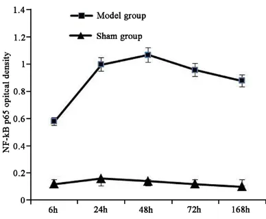
NF-κB p65 expression in venous endothelial cells
NF-κB p65 mainly locates in cytoplasm and nucleus. NF-κB p65 expression in model group
was obviously higher than that in sham group (P < 0.05). Its level increased at 6 h after model-ing, reached peak at 24 h, and declined after 72 h (Figure 2).
P-selectin, VCAM-1, and PSGL-1 mRNA expres-sion in blood
P-selectin, VCAM-1, PSGL-1 mRNA expression
increased significantly in blood after surgery
compared with sham group (P < 0.05). PSGL-1, VCAM 1, and P-selectin mRNA overexpressed at 6 h after DVT, reached peak at 24 h, and declined after 72 h (Figure 3).
P-selectin, VCAM-1, and PSGL-1 protein expres-sion in inferior vena cava wall
P-selectin, VCAM-1, and PSGL-1 protein expres-sion elevated obviously in venous wall after sur-gery. PSGL-1, VCAM-1, and P-selectin protein overexpressed at 6 h after DVT, achieved top at 48 h, and declined after 72 h (Figure 4). Discussion
DVT causes vein reflux obstruction, which usu -ally presents as lower extremity DVT. Slow
blood flow, blood vessel damage, and abnormal
blood components associated with DVT [14, 15]. Venous congestion leads to cell metabo-lism disorder, resulting in tissue hypoxia and local thrombin accumulation. Untimely correct-ing coagulation state may cause endothelial cells atrophy, basement membrane bareness, platelet adhesion to form thrombus core and activate coagulation material. For heparin exist-ed on the endothelial cell surface, integral
venous wall can prevent fibrin deposition.
Abnormal blood composition makes blood in high coagulation state, leading to venous thrombosis [16, 17]. Leukocytes involved
inflammation plays an important role in DVT
process. Leukocytes adhered to endothelial cells cause endothelial injury, further triggering coagulation system activation. Leukocyte adhe-sion function is related to cell adheadhe-sion mole-cules [18, 19]. Currently, there is still lack of investigation about the role and mechanism of cell adhesion factor in DVT process. Our study observed thrombosis at different time point in DVT model, and tested P-selectin, VCAM-1, and PSGL-1 mRNA and protein expression in blood and vena cava wall tissue. The results showed that obvious thrombus could be found in DVT model group at 6 h, 24 h, 48 h, 72 h, and 168 h. Under light microscope, red and mixed thrombus could be seen at 6 h and 24 h in model group. Vascular wall presented different
degree of inflammatory cell infiltration reached
peak at 24 h. Layered thrombus could be found
after 72 h, while thrombus and inflammatory cells infiltration could be found at 168 h, fur
[image:4.629.99.293.78.238.2]-ther confirming that inflammation was associ -ated with DVT.
DVT begins with leukocytes adhesion and
migration. P-selectin mainly mediates inflam -matory cells adhering to vascular endothelial cells, neutrophils activation, and monocytes adhering to platelet. E-selectin mainly mediates the starting process of vascular endothelial
cells adhering to leukocytes in inflammation
[20]. Integrin family plays an important role in the beginning of the leukocytes adhering to vascular endothelial cells. L-selectin overex-presses on the surface of leukocytes and falls off under proteolytic enzyme effect after leuko-cytes activation. E-selectin and P-selectin bind with PSGL-1 under histamine and thrombin
scription. Normally, NF-κB specific inhibitory protein I-κB binding with NF-κB dimers locates in cytoplasm. TNF-α or endotoxin stimulus acti
-vates I-κB kinase (IKK), leading to I-κB phos
-phorylation and NF-κB activation. NF-κB enters nucleus and binds with DNA to induce specific inflammatory factors gene transcription and protein synthesis, leading to inflammation [11, 12]. Generally, NF-κB in cytoplasm has no activ
-ity and presents as binding with IκB. NF-κB acti
[image:5.629.102.526.82.504.2]-vation promotes IκBα expression, and IκBα feedback regulates NF-κB expression [12]. IL-1 or LPS can degrade IκB, releasing NF-κB into nucleus to bind with specific κB sequence and
Figure 3. P-selectin, VCAM-1, and PSGL-1 mRNA ex-pression in leukocytes. ■P < 0.05, compared with
sham group.
Figure 4. P-selectin, VCAM-1, and PSGL-1 protein expression in inferior vena cava wall.
effect to mediate inflammato -ry cells adhering to endotheli-al cells. VCAM-1 overexpress-es in vascular endothelium
tran-P-selectin, VCAM-1, and PSGL-1 in DVT
cause inflammatory mediators and cytokines transcription. Large amount of proinflammatory factors and inflammatory mediators result in inflammation.
Inflammation is closely related to thrombosis, and thrombosis aggravates inflammatory inju
-ry. The key in early acute inflammation injury may be related to IκB phosphorylation and deg
-radation, and NF-κB release and translocation [11]. NF-κB binding site widely exists in the
upstream promoter and enhancer of ICAM-1,
VCAM-1, and IL-8, thus NF-κB activation ele -vates related gene expression level to promote endothelial cells and leukocyte adhesion func-tion [19]. Our study revealed that P-selectin,
VCAM-1, PSGL-1, and NF-κB expression in-creased significantly in blood and venous wall
after surgery. PSGL-1, VCAM-1, P-selectin and
NF-κB p65 overexpressed at 6 h after DVT,
reached peak at 24 h, and declined after 72 h. It suggested that P-selectin, VCAM-1, and PSGL-1 play important roles in regulating endothelial cells, leukocytes and platelet adhesion. DVT is a complex process with multiple system involve-ment. Endothelial cells adhere to leukocytes after activation, while activated leukocytes stimulate CD40/CD40L to enhance ROS re-
lease, activate NF-κB signaling pathway, and promote TF secretion, leading to inflammatory
factors and pro-coagulant release and throm- bosis.
To sum up, P-selectin, VCAM-1, and PSGL-1 mRNA and protein expression changes in DVT model are related to thrombosis. They can reg-ulate monocytes and endothelial cells
adhe-sion by activating NF-κB signaling pathway.
Acknowledgements
The Natural Science Foundation of China (NO. 81360291); The Natural Science Foundation
of Jiangxi Province, China (NO.
20122BAB-215011); Jiangxi province science and tech-
nology plan item (NO. 20132BBG70082); Jiangxi province health department of science and technology plan item (NO. 20121085); the science and technology plan item of Jiangxi province college (NO. KJLD14084).
Disclosure of conflict of interest
None.
Address correspondence to: Dr. Jianwen Mo, De-
partment of Orthopedic Surgery, The First Affiliated
Hospital of Gannan Medical University, 23 Qing- nian Road, Zhanggong District, Ganzhou 341000, Jiangxi, China. Tel: 797-8283927; Fax: +86-797-8283927; E-mail: mojianwen33@sina.com
References
[1] Magetsari R, Dewo P, Nugroho AS, Lanodiyu Z. Deep vein thrombosis in elderly patients fol-lowing surgery for fracture of the proximal fe-mur. Malays Orthop J 2014; 8: 7-10.
[2] Parunov LA, Soshitova NP, Ovanesov MV, Pan-teleev MA, Serebriyskiy II. Epidemiology of ve-nous thromboembolism (VTE) associated with
pregnancy. Birth Defects Res C Embryo Today
2015; 105: 167-184.
[3] Albayati MA, Grover SP, Saha P, Lwaleed BA, Modarai B, Smith A. Postsurgical inflammation
as a causative mechanism of venous thrombo-embolism. Semin Thromb Hemost 2015; 41: 615-620.
[4] Kirwan CC, McCollum CN, McDowell G, Byrne
GJ. Investigation of proposed mechanisms of chemotherapy-induced venous thromboembo-lism: endothelial cell activation and procoagu-lant release due to apoptosis. Clin Appl Thromb Hemost 2015; 21: 420-427.
[5] Obi AT, Diaz JA, Ballard-Lipka NL, Roelofs KJ, Farris DM, Lawrence DA, Wakefield TW, Henke
PK. Plasminogen activator-1 overexpression decreases experimental postthrombotic vein
wall fibrosis by a non-vitronectin-dependent
mechanism. J Thromb Haemost 2014; 12: 1353-1363.
[6] Bollen L, Wuyts J, Vermeire S, Gils A. Identifica
-tion of an inflammatory bowel disease patient
with a deep vein thrombosis and an altered
clot lysis profile. Blood Coagul Fibrinolysis
2016; 27; 223-5.
[7] Rabinovich A, Cohen JM, Kahn SR. Predictive
value of markers of inflammation in the post -thrombotic syndrome: a systematic review:
in-flammatory biomarkers and PTS. Thromb Res
2015; 136: 289-297.
[8] Rabinovich A, Cohen JM, Cushman M, Wells PS, Rodger MA, Kovacs MJ, Anderson DR, Ta-galakis V, Lazo-Langner A, Solymoss S, Miron MJ, Yeo E, Smith R, Schulman S, Kassis J, Ke-aron C, Chagnon I, Wong T, Demers C, Hanmi-ah R, Kaatz S, Selby R, Rathbun S, Desmarais S, Opatrny L, Ortel TL, Ginsberg JS, Kahn SR.
Inflammation markers and their trajectories
after deep vein thrombosis in relation to risk of post-thrombotic syndrome. J Thromb Haemost 2015; 13: 398-408.
[9] McEver RP. Selectins: initiators of leucocyte adhesion and signalling at the vascular wall. Cardiovasc Res 2015; 107: 331-339.
[10] Thomas GM, Brill A, Mezouar S, Crescence L,
expressed by circulating cancer cell-derived microparticles drastically increases the inci-dence of deep vein thrombosis in mice. J Thromb Haemost 2015; 13: 1310-1319. [11] Malaponte G, Signorelli SS, Bevelacqua V, Po
-lesel J, Taborelli M, Guarneri C, Fenga C, Umezawa K, Libra M. Increased Levels of
NF-kB-Dependent Markers in Cancer-Associated
Deep Venous Thrombosis. PLoS One 2015; 10: e0132496.
[12] Mosevoll KA, Lindas R, Wendelbo O, Bruserud
O, Reikvam H. Systemic levels of the endothe-lium-derived soluble adhesion molecules en-docan and E-selectin in patients with suspect-ed deep vein thrombosis. Springerplus 2014; 3: 571.
[13] Zhang YB, Li W, Yao LQ, Zhao XL, Wang B, Li
HK, Ning Y, Song E, Zhang XX. Expression changes and roles of matrix metalloproteinas-es in a rat model of traumatic deep vein throm-bosis. Chin J Traumatol 2010; 13: 188-192. [14] Hish GA Jr, Diaz JA, Hawley AE, Myers DD Jr,
Lester PA. Effects of analgesic use on inflam -mation and hematology in a murine model of venous thrombosis. J Am Assoc Lab Anim Sci 2014; 53: 485-493.
[15] Shi D, Xu X, Xu Z, Nakamura T, Pang Y, Yao C, Wang F, Chen D, Dai J, Jiang Q. P-selectin: an unpredicted factor for deep vein thrombosis
after total hip arthroplasty. Biomed Res Int
2014; 2014: 783967.
[16] Zolcinski M, Ciesla-Dul M, Potaczek DP, Undas
A. Atorvastatin favorably modulates proinflam
-matory cytokine profile in patients following
deep vein thrombosis. Thromb Res 2013; 132: e31-35.
[17] Coleman DM, Wakefield TW. Biomarkers for
the diagnosis of deep vein thrombosis. Expert Opin Med Diagn 2012; 6: 253-257.
[18] Siefert SA, Chabasse C, Mukhopadhyay S, Hoofnagle MH, Strickland DK, Sarkar R, Anta-lis TM. Enhanced venous thrombus resolution
in plasminogen activator inhibitor type-2 defi -cient mice. J Thromb Haemost 2014; 12: 1706-1716.
[19] Alias S, Lang IM. Coagulation and the vessel wall in pulmonary embolism. Pulm Circ 2013; 3: 728-738.
[20] Wang Y, Andrews M, Yang Y, Lang S, Jin JW, Cameron-Vendrig A, Zhu G, Reheman A, Ni H. Platelets in thrombosis and hemostasis: old topic with new mechanisms. Cardiovasc He-matol Disord Drug Targets 2012; 12: 126-132. [21] Li G, Han ZL, Dong HG, Zhang X, Kong XQ, Jin X.