Original Article
The balance between HGF and TGF-β1 acts as a switch
in the tissue remodeling of chronic rhinosinusitis
Jing Li1, Xiang Liu1, Min Sha2, Yong Li1
1Department of Otolaryngology, Affiliated Hangzhou First People’s Hospital, School of Medicine, Zhejiang Universi-ty, Hangzhou, Zhejiang Province, China; 2Department of Otolaryngology, Hangzhou Children’s Hospital, Hangzhou, Zhejiang Province, China
Received October 29, 2018; Accepted December 21, 2018; Epub March 1, 2019; Published March 15, 2019
Abstract: Objective: The remodeling patterns in different types of chronic rhinosinusitis (CRS) are controversial. This study aimed to investigate the roles of transforming growth factor-β1 (TGF-β1) and hepatocyte growth factor (HGF) in the CRS remodeling process. Methods: Surgical samples were obtained from CRSwNP patients (n=29), CRSsNP patients (n=34), and controls (n=21). Collagen deposition was detected via Masson trichrome (MT) staining. Im-munohistochemical staining was performed to examine the protein expression of α-smooth muscle actin (α-SMA). The expression of TGF-β1 and HGF was measured by ELISA. The relationship between the rate of TGF-β1/HGF and the expression of Collagen III and α-SMA was analyzed using a Pearson correlation test. Primary nasal epithelial cells (NECs) were cultured and divided into four groups. Collagen III secretion was measured in the supernatants by ELISA. The expression of α-SMA was studied by immunofluorescence. Results: Reduced collagen deposition and α-SMA expression were detected in the CRSwNP group (P=0.033). The expression of collagen deposition and α-SMA was increased in the CRSwNP group (P=0.001). The ELISA tests indicated that TGF-β1 levels were significantly in-creased in the CRSsNP compared with the controls. The expression of HGF was higher in the CRSwNP than in the other two groups. The ratio of TGF-β1/HGF was upregulated in CRSsNP and was correlated positively with collagen and α-SMA expression (P<0.05, R=0.762). TGF-β1 can increase collagen and α-SMA expression in NECs, and HGF can antagonize the remodeling action of TGF-β1. Conclusion: Distinct remodeling patterns are revealed for different types of CRS. The balance of TGF-β1 and HGF is important in the CRS remodeling process.
Keywords: Chronic rhinosinusitis, nasal polyp, tissue remodeling, transforming growth factor-β1, HGF
Introduction
Chronic rhinosinusitis (CRS) is one of the most common chronic diseases in adults. It is asso-ciated with impaired quality of life and substan-tial societal costs [1]. While it is sometimes associated with asthma, allergy, or nasal pol-yposis, many cases present without an appar-ent underlying cause. Despite a lack of etiology and the inflammatory nature of the condition, treatment protocols dictate intervention with antibiotics, anti-inflammatory drugs, or surgery [1, 2], often with poor long-term effects. CRS is typically classified into two types: chronic rhino-sinusitis with nasal polyps (CRSwNP) and chronic rhinosinusitis without nasal polyps (CRSsNP). The development of phenotype-ori-entated therapeutic strategies is critical for improving CRS treatment. Clinically, CRSwNP is
associated more closely with clinical com-plaints of nasal obstruction and olfactory loss, and more frequently linked to comorbidities such as asthma and aspirin hypersensitivity, than CRSsNP. Immunologically, CRSwNP tis-sues are characterized by more intense eosino-philic infiltration and a Th2-based cytokine pro-file, whereas sinonasal tissues from CRSsNP have been reported to display a predominant infiltration of leukocytes and Th1 cytokines [3]. However, there are still some controversies about the distinct role of Th1/Th2 profiles in CRS subtypes.
(ECM) protein deposition and tissue structure. Distinct disease entities can be distinguished within a group of chronic sinus diseases on the basis of different inflammation and remodeling patterns. CRSsNP is characterized by nasal epi-thelial injury, myofibroblast proliferation, and extracellular matrix remodeling with high levels of interferon receptor (IFN-r) and transforming growth factor-β1 (TGF-β1) [4]. In contrast, CRSwNP is characterized by predominant albu-min accumulation and edema formation within the ECM, and low levels of myofibroblast prolif-eration and TGF-β1-3 [5]. TGF-β1 is implicated as a key protein in the tissue remodeling pro-cess of CRSsNP. It stimulates fibrosis by attract-ing stromal cells and the transdifferentiation of primary nasal epithelial cells (NECs), angiogen-esis, and the accumulation of extracellular matrix [6]. Some studies have reported lower TGF-β1 expression in CRSwNP than in CRSsNP and healthy controls [5, 7]. There is still a con-troversy in the literature as to TGF-β1 expres-sion in nasal polyposis [8].
Hepatocyte growth factor (HGF), a pleiotropic growth factor, demonstrates antifibrotic prop-erties in experimental models of lung, kidney, heart, skin, and liver fibrosis. HGF protein atten-uates collagen accumulation and the extent of fibrosis. In addition, HGF affects fibroblast acti-vation and transdifferentiation indirectly by inhibiting the epithelial-to-mesenchymal cell transition (EMT) [9]. HGF acts as an antifibrotic agent that protects the host against TGF-β1-mediated profibrotic effects [10]. Some studies suggest that the HGF complex may play a role in the pathogenesis of nasal polyps [11]. Therefore, the aim of this study was to investi-gate the roles of TGF-β1/HGF in the remodeling preprocess in CRS.
Materials and methods
Patients and study experimental design
This study was approved by the medical ethics committee of Affiliated Hangzhou First People’s Hospital of Zhejiang University, and a written informed consent was obtained from each patient in the study. The work has been carried out in accordance with the declaration of Helsinki. Samples of the ethmoid bulla mucosa from patients without sinus disease who under-went endoscopic hypophysectomy or septo-plasty were used as controls. Samples from
patients with CRSwNP or CRSsNP were obtained during functional endoscopic sinus surgery (FESS) procedures. A total of 21 con-trols, 29 CRSwNP patients, and 34 CRSsNP patients were recruited into this study at the Department of Otorhinolaryngology of Hangzhou First People’s Hospital, Hangzhou, China, from September 2015 to December 2017. Sinus disease diagnosis was based on history, clinical examination, nasal endoscopy, and a computed tomography (CT) scan of the paranasal cavities according to the standard criteria issued in the European Position Paper on Rhinosinusitis and Nasal Polyps guidelines [1]. All subjects underwent a skin prick test for common inhalant allergens and all tests had to be negative for the subject to be included in the experiment. Other exclusion criteria were a diagnosis of asthma (based on a ventilatory test), aspirin intolerance (based on history), or cystic fibrosis. All patients stopped oral cortico-steroids for at least 1 month and topical appli-cation for at least 2 weeks before surgery. Patients did not take any other relevant medi-cations. Biopsy specimens were obtained for the assessment of nasal tissue remodeling.
Histological analysis
then incubated with a biotinylated goat anti-rabbit and mouse antibody (Thermo Scien- tific, USA) for 10 min. The sections were rinsed again with PBS. Finally, the sections were incu-bated with Streptavidin peroxidase (Thermo Scientific, USA). The slides were incubated for 10 min with 3,3’-diaminobenzidine tetrahydro-chloride (Sigma-Aldrich, USA) to visualize the reaction. The slides were counterstained with hematoxylin and then dehydrated and mount-ed. Primary antibodies were omitted and replaced with PBS for the negative controls. Ten random images from each sample were recorded on a digital camera (Olympus C-5050) without overlapping zones, and were assessed by two independent observers blinded to the tissue staining protocol. Images of the stained sections were analyzed by Image Pro Plus soft-ware in 10 microscopic fields.
TGF-β1 and HGF expression by ELISA
Samples in each group were homogenized in PBS and stored at -80°C. The levels of human TGF-β1 and HGF protein in the nasal mucosa were quantified using commercial enzyme-linked immunosorbent assay (ELISA) kits (eBio-science, USA) according to the manufacturer’s instructions. The values obtained by ELISA were corrected with a dilution factor and ulti-mately expressed in pg/mg pro. Correlations of the ratio of TGF-β1/HGF with collagen deposi-tion and α-SMA expression in nasal tissues were investigated. The Pearson correlation test was used, and R values indicate Pearson cor-relation coefficients.
Primary culture of NECs
The nasal specimens in the control group that did not require histopathologic examination were temporarily placed in a 50 mL conical tube containing 10 mL of DMEM/F-12 medium (Corning), 100 IU/mL penicillin, and 100 mg/ mL streptomycin. NECs were cultured accord-ing to previously published methods [10]. NECs were cultured to confluence as monolayers in 24-well plates containing BEGM under stan-dard culture conditions. At the end of culturing, the cells were harvested and seeded into 6-well plates at a concentration of 1×10⁵ cells / well. The NECs were divided into four groups. The culture media were then replenished with BEGM (control group), BEGM containing 5 ng/ mL TGF-β1 (R&D Systems Inc.), BEGM
contain-ing 100 ng/mL HGF (R&D Systems Inc.) (HGF group), BEGM containing 5 ng/mL TGF-β1 and
100 ng/mL HGF (TGF-β1+HGF group). The NECs were treated for 48 h at 37°C in a 5% CO2 in air atmosphere. At the end of culturing, superna-tants were collected, and the levels of collagen III were quantified using commercial ELISA kits (eBioscience, USA) according to the manufac-turer’s instructions. Each experimental condi-tion was tested in six replicate wells, and the mean was taken to represent an individual experiment.
Immunocytochemistry
NECs were fixed with 4% formaldehyde for 30 min and then incubated in 1% BSA/10% normal goat serum/0.3 M glycine in 0.1% PBS-Tween for 1 h to permeabilize the cells and block non-specific protein interactions. The cells were then incubated with the antibody to α-SMA (1:100; Santa Cruz Biotech, USA). Alexa 555-conjugated secondary antibodies (Mole- cular Probes/Invitrogen) were used. DAPI (Sigma-Aldrich, USA) was used for nuclear staining. Images were obtained via micro- scopy.
Statistical analysis
The data were statistically analyzed by SPSS version 16.0 statistical package. Data are expressed as the mean ± standard error (SE). Differences between the groups were assessed using one-way analysis of variance (ANOVA) fol-lowed by Tukey’s test. The Pearson correlation test was also used. Differences were consid-ered significant at P<0.05.
Results
Different collagen deposition and fibrotic marker in CRSwNP and CRSsNP patients
sections with bright-field microscopy, as shown in Figure 1A-C. The control group showed a nor-mal collagen distribution (Figure 1A). The CRSsNP group revealed extensive collagen deposition, bridging fibrosis (Figure 1B), and thicker and more numerous collagen fibers than collagen in the CRSwNP and control groups. Collagen fibers were reduced in the CRSwNP group compared to the control group (Figure 1C). The degree of collagen deposition is calculated in Figure 1G. It showed that there are significant differences in collagen deposi-tion among the three groups using ANOVA test (P<0.001). Reduced collagen deposition and α-SMA expression were detected in the CRS- wNP group (P=0.033). The expression of colla-gen deposition and α-SMA was increased in the CRSwNP group (P=0.001) (Figure 1H).
followed by Tukey’s test. TGF-β1 levels were sig-nificantly increased in the CRSsNP group com-pared with the controls (P<0.001). However, there was no difference in TGF-β1 between the control and CRSwNP groups (P=1.000). The expression of HGF was higher in the CRSwNP group, and no differences in HGF were found between the control and the CRSsNP groups.
Relation of TGF-β1/HGF with collagen deposi-tion and α-SMA
The ratios of TGF-β1/HGF were different in the three groups, where CRSsNP>control>CRSwNP, as presented in Figure 2C. Correlations of the ratio of TGF-β1/HGF with collagen deposition and α-SMA expression in nasal tissues were investigated. The Pearson correlation test was used, and R values indicate Pearson correla-Figure 1. Histopathological examination of nasal mucosa tissues from differ
-ent groups. (A-C) The MT staining showed that collagen fibers in the CRSwNP
groups were thinner and fewer in number than in the CRSsNP and control groups. The CRSsNP group revealed extensive collagen deposition, bridging
fibrosis and the collagen fibers were thicker and more. (G) The degree of collagen deposition in the different groups was calculated. (D-F) IHC staining
for α-SMA showed normal expression of α-SMA positive staining in control group and α-SMA expression in the CRSwNP group was significant reduced
compared to CRSsNP group. Positive staining is indicated by an arrow. The
semi-quantitative analysis of IHC is shown in (H). MT, Masson trichrome;
CRSsNP, chronic rhinosinusitis without nasal polyps; CRSwNP, chronic
rhino-sinusitis with nasal polyps; IHC, immunohistochemistry.
Representative sections of immunohistochemical stain-ing for α-SMA were performed in the controls, CRSsNP, and CRSwNP and are shown in Figure 1D-F. The expression of α-SMA was significantly higher in CRSsNP than in the controls and was significantly lower in CRSwNP than in the controls. α-SMA immunoreac-tivity was stained dark brown. The control group showed normal expression of α-SMA-positive staining. α-SMA ex- pression in the CRSwNP group showed a significant reduction compared to the CRSsNP group. Figure 1H summarizes the data of the immunohisto-chemical evaluation of α-SMA expression in the three grou- ps.
TGF-β1/HGF disbalance in CRS groups
tion coefficients. The result showed that the ratio of TGF-β1/HGF was positively related to the expression of collagen (P<0.05, R=0.889) and α-SMA (P<0.05, R=0.762) in the mucosa of patients.
HGF can antagonize the fibrotic effect caused by TGF-β1 in NECs
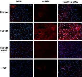
NECs were treated with different conditions to test whether HGF can antagonize the fibrotic effect of TGF-β1. ELISA was used to quantify collagen III expression in culture media of NECs under different culture conditions. As shown in Figure 2D, 100 ng/ml HGF significantly sup-pressed TGF-β1-induced collagen III release in the culture medium of NECs by ANOVA test (P=0.035). We also studied the release of α-SMA in NECs with immunocytochemistry, which plays an important role in the transdif-ferentiation of epithelial cells. The expression of α-SMA (red) was higher in the TGF-β1 group
polyps [13]. The aim of this study was to identi-fy the extracellular matrix factors that might play a role in the remodeling of CRS. Our study demonstrated that compared to CRSwNP patients and normal nasal mucosa, CRSsNP patients had a higher level of TGF-β1 and α-SMA expression in the nasal mucosa, which was paralleled by increased collagen deposi-tion. In the polyps, there was a lack of collagen deposition and low α-SMA expression.
TGF-β1 has been implicated as an important factor in the remodeling processes involved in chronic sinus disease and serves as a main switch for different remodeling patterns in CRS [14]. TGF-β1 also affects fibrosis by increasing ECM deposition, such as increasing collagen and fibronectin [15]. Van Bruaene et al. report-ed in a Belgian population that TGF-β1 and col-lagen were elevated in CRSsNP [16]. Li et al. reported that TGF-β1 and collagen were elevat-ed in CRSsNP in a Chinese population [17]. In
Figure 2. ELISA measurements for TGF-β1 (A), HGF (B), and collagen III, expressed as pg/mg protein. Tissue homogenates were prepared from the control, CRSsNP, and CRSwNP groups. In the CRSsNP group, expression of TGF-β1 was higher than in the other groups, and the HGF level was highest in the CRSwNP group. The ratio of TGF-β1 and HGF is shown in (C). There were significant differences between the three groups (P<0.05). The collagen III expression in the culture media of NECs is shown in (D). TGF-β1 can induce
the secretion of collagen III in NECs. However, HGF can antagonize this ef-fect and reduce the production of collagen III.
than in the other groups, which is shown in Figure 3. HGF treatment decreased the expression of α-SMA induced by TGF-β1 in NECs.
Discussion
[image:5.612.91.372.71.329.2]contrast, Shi et al. found in a Chinese popula-tion that TGF-β1 was reduced, but collagen was elevated in CRSsNP [12]. Although there are some discrepancies, our study suggests that TGF-β1 and collagen are elevated in CRSsNP and that they may contribute to the remodeling process in this form of the disease. As men-tioned above, the pattern of remodeling in CRSwNP is known to be different than that in CRSsNP. Indeed, collagen was reduced in CRSwNP and TGF-β1 was also reduced but showed no difference compared to the control group.
Histopathological analysis provided initial evi-dence of TGF-β1-induced fibrosis in CRS. Of note, α-SMA expression plays a key role in the pathophysiological mechanism of CRS remod-eling. α-SMA is a reliable unique marker of EMT. In this study, immunohistochemical analysis showed excessive α-SMA expression in CRSsNP compared to limited expression in controls, confirming that TGF-β1 stimulated the activa-tion of myofibroblasts in the CRSsN and that there was a marked reduction of α-SMA immu-noreactivity in CRSwNP.
tions [23]. These data suggest that polymor-phisms in the MET gene may play a role in susceptibility to developing CRS. The MET gene encodes the c-MET receptor of HGF, a trans-membrane protein receptor tyrosine kinase. The biological effects of HGF are mediated by binding the c-MET receptor and activating the tyrosine kinase signaling pathways. These results suggest that the MET/HGF pathway may have a role in the pathogenesis of nasal polyps [11].
In the current investigation, we found that the ratio of TGF-β1/HGF had a positive relationship with the expression of collagen and α-SMA in CRS patients. Higher TGF-β1/HGF ratios result-ed in greater collagen and α-SMA expression in the CRSsNP group; lower TGF-β1/HGF ratios reduced collagen deposition and α-SMA expres-sion, which may result in edema in the nasal mucosa of CRSwNP patients. In vitro studies have found that HGF specifically counteracts the profibrotic actions of TGF-β1 and reduces collagen secretion in NECs. The EMT maker α-SMA was also lower in the HGF-treated group than in the TGF-β1 group. The results
demon-Figure 3. Immunocytochemistry for α-SMA in NECs. The expression of α-SMA (Red) in the TGF-β1 group was significantly higher than in the HGF therapeu-tic group and controls. HGF treatment decreased the expression of α-SMA induced by TGF-β1 in NECs.
Importantly, we further found that HGF was significantly increased in CRSwNP patients compared with the control subjects and the CRSsNP group. HGF, a pleiotropic cyto
-kine demonstrates
antifibro-tic properties in experimental models of liver, kidney, heart,
skin, and lung fibrosis [18-20]. In addition, HGF acts as an antifibrotic agent that
prote-cts the host against
[image:6.612.92.376.72.339.2]popula-strated that the balance between HGF and TGF-β1 may play a decisive role in the patho-genesis of CRSsNP and CRSwNP. Hence, the balance of HGF and TGF-β1 acted as two sides of the same coin in the tissue fibrotic signals [24].
Above all, HGF effectively blocks TGF-β1-mediated cellular transdifferentiation into α-SMA-producing cells both in vivo and in vitro. The balance between HGF and TGF-β1 acts as a switch in the pathogenesis of CRSsNP and CRSwNP.
Acknowledgements
This paper was supported by projects of the Medical and Health Technology Development Program in Zhejiang Province (No. 2016KYB227 and 2019RC066) and the Medical and Health Technology Development Program in Hangzhou City (No. 2017A10).
Disclosure of conflict of interest
None.
Abbreviations
ANOVA, assessed using one-way analysis of variance; BSA, Bovine serum albumin; CRS, chronic rhinosinusitis; CRSwNP, chronic rhinosi-nusitis with nasal polyps; CRSsNP, chronic rhinosinusitis without nasal polyps; CT, com-puted tomography; ELISA, enzyme-linked immunosorbent assay; ECM, extracellular matrix; EMT, epithelial-to-mesenchymal cell transition; FESS, functional endoscopic sinus surgery; HGF, hepatocyte growth factor; IFN-r, interferon receptor; IOD, integrated OD; MT, Masson trichrome; NECs, nasal epithelial cells; TGF-β1, transforming growth factor-β1; SE, standard error; α-SMA, α-smooth muscle actin.
Address correspondence to: Yong Li, Department of Otolaryngology, Affiliated Hangzhou First People’s Hospital, School of Medicine, Zhejiang University, 261 Huansha Road, Shangcheng District, Hangzhou 310006, Zhejiang Province, China. Tel: +86-13806527172; Fax: +86-571-87914773; E-mail: leeyung828@163.com
References
[1] Fokkens WJ, Lund VJ, Mullol J, Bachert C, Alo-bid I, Baroody F, Cohen N, Cervin A, Douglas R,
Gevaert P, Georgalas C, Goossens H, Harvey R, Hellings P, Hopkins C, Jones N, Joos G, Kalogjera L, Kern B, Kowalski M, Price D, Riechelmann H, Schlosser R, Senior B, Thom-as M, Toskala E, Voegels R, Wang de Y and Wormald PJ. European position paper on rhino-sinusitis and nasal polyps 2012. Rhinol Suppl 2012; 23: 3 p preceding table of contents, 1-298.
[2] Cope EK and Lynch SV. Novel microbiome-based therapeutics for chronic rhinosinusitis. Curr Allergy Asthma Rep 2015; 15: 504. [3] Van Zele T, Claeys S, Gevaert P, Van Maele G,
Holtappels G, Van Cauwenberge P and Bachert C. Differentiation of chronic sinus diseases by measurement of inflammatory mediators. Al-lergy 2006; 61: 1280-1289.
[4] Van Bruaene N, Perez-Novo CA, Basinski TM, Van Zele T, Holtappels G, De Ruyck N, Schmidt-Weber C, Akdis C, Van Cauwenberge P, Bachert C and Gevaert P. T-cell regulation in chronic paranasal sinus disease. J Allergy Clin Immu-nol 2008; 121: 1435-1441.
[5] Bachert C, Gevaert P, Holtappels G, Cuvelier C and van Cauwenberge P. Nasal polyposis: from cytokines to growth. Am J Rhinol 2000; 14: 279-290.
[6] Little SC, Early SB, Woodard CR, Shonka DC Jr, Han JK, Borish L and Steinke JW. Dual action of TGF-beta1 on nasal-polyp derived fibro-blasts. Laryngoscope 2008; 118: 320-324. [7] Figueiredo CR, Santos RP, Silva ID and Weckx
LL. Microarray cDNA to identify inflammatory genes in nasal polyposis. Am J Rhinol 2007; 21: 231-235.
[8] Zaravinos A, Soufla G, Bizakis J and Spandidos DA. Expression analysis of VEGFA, FGF2, TGF-beta1, EGF and IGF1 in human nasal polypo-sis. Oncol Rep 2008; 19: 385-391.
[9] Lee YH, Suzuki YJ, Griffin AJ and Day RM. Hepa-tocyte growth factor regulates cyclooxygen-ase-2 expression via beta-catenin, Akt, and p42/p44 MAPK in human bronchial epithelial cells. Am J Physiol Lung Cell Mol Physiol 2008; 294: L778-786.
[10] Li J, Zheng CQ, Li Y, Yang C, Lin H and Duan HG. Hepatocyte growth factor gene-modified mes-enchymal stem cells augment sinonasal wound healing. Stem Cells Dev 2015; 24: 1817-1830.
[11] Castano R, Bosse Y, Endam LM, Filali-Mouhim A and Desrosiers M. c-MET pathway involve-ment in chronic rhinosinusitis: a genetic asso-ciation analysis. Otolaryngol Head Neck Surg 2010; 142: 665-671.e661-662.
remodel-ing in different types of Chinese chronic rhino-sinusitis are associated with inflammation pat-terns. Allergy 2013; 68: 101-109.
[13] Meng J, Zhou P, Liu Y, Liu F, Yi X, Liu S, Holtap-pels G, Bachert C and Zhang N. The develop-ment of nasal polyp disease involves early na-sal mucona-sal inflammation and remodelling. PLoS One 2013; 8: e82373.
[14] Van Bruaene N and Bachert C. Tissue remod-eling in chronic rhinosinusitis. Curr Opin Aller-gy Clin Immunol 2011; 11: 8-11.
[15] Al-Alawi M, Hassan T and Chotirmall SH. Trans-forming growth factor beta and severe asthma: a perfect storm. Respir Med 2014; 108: 1409-1423.
[16] Van Bruaene N1, Derycke L, Perez-Novo CA, Gevaert P, Holtappels G, De Ruyck N, Cuvelier C, Van Cauwenberge P, Bachert C. TGF-beta signaling and collagen deposition in chronic rhinosinusitis. J Allergy Clin Immunol 2009; 124: 253-259.
[17] Li X, Meng J, Qiao X, Liu Y, Liu F, Zhang N, Zhang J, Holtappels G, Luo B, Zhou P, Zheng Y, Lin P, Liu S and Bachert C. Expression of TGF, matrix metalloproteinases, and tissue inhibi-tors in Chinese chronic rhinosinusitis. J Allergy Clin Immunol 2010; 125: 1061-1068. [18] Ogaly HA, Eltablawy NA, El-Behairy AM, El-Hindi
H and Abd-Elsalam RM. Hepatocyte growth factor mediates the antifibrogenic action of ocimum bacilicum essential oil against CCl4-Induced liver fibrosis in rats. Molecules 2015; 20: 13518-13535.
[19] Gazdhar A, Temuri A, Knudsen L, Gugger M, Schmid RA, Ochs M and Geiser T. Targeted gene transfer of hepatocyte growth factor to alveolar type II epithelial cells reduces lung fi-brosis in rats. Hum Gene Ther 2013; 24: 105-116.
[20] Kajihara I, Jinnin M, Makino T, Masuguchi S, Sakai K, Fukushima S, Maruo K, Inoue Y and Ihn H. Overexpression of hepatocyte growth factor receptor in scleroderma dermal fibro-blasts is caused by autocrine transforming growth factor beta signaling. Biosci Trends 2012; 6: 136-142.
[21] Chakraborty S, Chopra P, Hak A, Dastidar SG and Ray A. Hepatocyte growth factor is an at-tractive target for the treatment of pulmonary fibrosis. Expert Opin Investig Drugs 2013; 22: 499-515.
[22] Rho HS, Lee SH, Lee HM, Lee SH, Jung HH, Choi J, Park MK and Kang SM. Overexpression of hepatocyte growth factor and its receptor c-Met in nasal polyps. Arch Otolaryngol Head Neck Surg 2006; 132: 985-989.
[23] Stankovic KM, Goldsztein H, Reh DD, Platt MP and Metson R. Gene expression profiling of na-sal polyps associated with chronic sinusitis and aspirin-sensitive asthma. Laryngoscope 2008; 118: 881-889.