Original Article
Epidermal growth factor promotes mesenchymal
stem cell-mediated wound healing and hair
follicle regeneration
Ying Xia1*, Xiao-En You2*, Hong Chen1, Yong-Jun Yan3, Yu-Cheng He3, Shi-Zhang Ding3
1Department of Plastic Surgery, Shaoxing People’s Hospital, Zhejiang, China; 2Department of Burn and Plastic Surgery, People’s Hospital of Lishui City, Zhejiang, China; 3Department of Surgery, The Seventh Hospital of Shaoxing City, Zhejing, China. *Equal contributors.
Received November 18, 2016; Accepted January 7, 2017; Epub July 1, 2017; Published July 15, 2017
Abstract: Background and Aim: Bone marrow mesenchymal stem cells (MSC) are receiving increasing attention for
skin wound repair. However, the specific mechanisms underlying MSC-mediated improvement in wound healing
have not been fully elucidated. This study aims at testing whether epidermal growth factor (EGF) can promote MSC-mediated wound healing and hair follicle regeneration. Methods: Excisional wounds in rats were transplanted with different collagen-chitosan scaffolds: control, MSC, and MSC + EGF. Regenerated tissues were harvested 1, 3, or 5 weeks following transplantation, stained with hematoxylin and eosin and evaluated microscopically. The forma-tion of sebaceous glands was examined by Oil red staining and the regeneraforma-tion of hair follicles by
immunohisto-chemical staining and Western blot to test the expression of hair follicle-specific factors. Results: Gross observations
showed that the wounds were much smaller and the hairs grew faster in the MSC + EGF group. Histological analysis demonstrated that there were more hair follicles, sebaceous glands, and newly formed blood vessels in the MSC + EGF group compared with that in the MSC group. In addition, oil red staining showed that MSCs + EGF induced seba-ceous gland regeneration. Finally, immunohistochemistry and western blot revealed that MSCs + EGF increased the
expression of hair follicle-specific factors. Conclusion: MSCs alone cannot achieve the regeneration of hair follicles
and EGF can promote MSC-mediated wound healing and hair follicle regeneration.
Keywords: EGF, hair follicles, MSC, wound healing
Introduction
As skin is the largest organ of the body, skin loss caused by trauma, burning, and chronic diseases becomes one of the most serious clinical problems. Millions of patients suffering from skin loss require treatment with skin sub-stitutes such as wound dressings, allografts, and autografts. However, traditional autografts are limited by their availability in terms of both time and donor sites [1, 2]. Recent advances in regenerative medicine and tissue engineering have expanded our understanding of wound healing and led to the development of diverse methods for skin repair and regeneration. In full-thickness skin defects, the absence of der-mis prevents the operation of autonomous repair mechanisms. In such cases, a proper dermal equivalent, which can function as a regenerating template for wound healing, is
Many studies have focused on wound healing and hair regeneration [8-11]. The use of mes-enchymal stem cells (MSCs) is one of the most promising discoveries, which can contribute to wound closure acceleration, improvement of neovascularization, and reduction of scar for-mation. In addition, Rustad et al. suggested that MSC induced hair regeneration during wound healing [1]. While the specific mecha-nisms by which MSCs improve wound healing have not been fully elucidated, it has been pro-posed that MSC differentiation plays a signifi-cant role. A suitable environment is necessary for MSCs to differentiate into specific cells. However, in a wound following skin damage, the physiological environment of the skin is com-promised, interfering with the differentiation pathways of transplanted MSCs. Consequently, hair and sebaceous gland regeneration is impeded. Epidermal growth factor (EGF) has been shown to induce differentiation of MSCs into dermal papilla cells, and to promote hair follicle regeneration during wound healing [12]. In this study, we generated three
collagen-chi-tosan scaffolds coated with PBS, MSCs, or MSCs + EGF respectively, and tested whether the combination of MSCs and EGF could accel-erate wound healing, improve neovasculariza-tion, and promote hair regeneration.
Materials and methods
MSC isolation
[image:2.612.89.524.70.389.2]Preparation of collagen-chitosan scaffolds
A porous collagen-chitosan scaffold was fabri-cated as previously described [13-16]. Briefly, collagen and chitosan were dissolved in 0.5 M acetic acid to form a 0.5% (w/v) solution, in a mass ratio of 9:1. The collagen-chitosan com-posite was injected into homemade molds, fro-zen at -20°C for 2 h, and lyophilized for 24 h to produce a porous collagen-chitosan scaffold. EGF, MSC, and MSC + EGF scaffolds were pre-pared according to a multi-point injection meth-od. MSC scaffolds were prepared by injecting 1.0 × 10⁶-1.0 × 10⁷ MSCs into the previously
prepared collagen-chitosan scaffold. MSC + EGF scaffolds were prepared by injecting both 1.0 × 10⁶-1.0 × 10⁷ MSCs and ~40 μg EGF into
the collagen-chitosan scaffold.
Excisional wound healing model
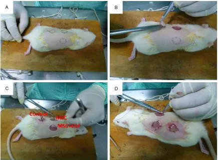
All animal studies conformed to the Guide for the Care and Use of Laboratory Animals (NIH Publication No. 85-27, revised 1996) and were approved by the Institutional Animal Care and Use Committee of Zhejiang University. Twenty-seven male Sprague-Dawley (SD) rats (2 mon- ths old, 250 ± 12 g) were randomized into three treatment groups: Control, MSC, and MSC + EGF. Prior to the operation, the rats were anes-thetized by intraperitoneal administration of 3% pentobarbital sodium solution (Sigma) at a dose of 1.0 ml/kg. An additional dose (0.5 ml/ kg) was administered as required to maintain deep anesthesia. The back hair of the rat was removed with 8% Na2S aqueous solution (Fig- ure 1A). Three standardized full-thickness exci-sional skin wounds (diameter = 2.0 cm) were created at the rat dorsum with a distance of no less than 2.0 cm from each other (Figure 1B). The excised skin was trimmed into thin split-thickness skin. The scaffolds were then extend-ed on the wound bextend-ed and suturextend-ed to the adja-cent skin (Figure 1C, 1D). The operative inci-sions were sterilized with 2.5% povidone iodine, the rats received a pressure dressing (petrola-tum gauze and elastoplast) and were housed in single cages. The rats were inspected every day to ensure that the pressure dressings were intact. One week post-surgery, the wounds healed and the dressings were removed. The animals were killed in batches 1, 3, and 5 weeks post-surgery. Following imaging of the wound sites, tissue specimens were harvested
and were sectioned along the uniform direc-tion, and maintained in 10% buffered formalin, liquid nitrogen, and physiological saline solu-tion for histological investigasolu-tion and molecular biology analysis respectively.
Macroscopic observation
Images of the wounds were acquired at differ-ent time points and analyzed using the image processing software package Photoshop (ver. 11.0.1; Adobe, San Jose, CA, USA). The wound area was calculated from the images according to a method previously described [10, 11].
Histology and oil red staining
Five-millimeter-thick sections were prepared from paraffin-embedded tissue samples, and were stained with hematoxylin and eosin (H&E) according to standard protocols [6, 17, 18]. Sebaceous glands budding from the follicle epi-thelial root are an accessory for hair follicle for-mation. Therefore, the regeneration of seba-ceous glands indirectly indicates hair follicle regeneration. We investigated the regeneration of sebaceous glands during wound healing by oil red staining. Five-millimeter-thick sections were prepared from paraffin-embedded tissue samples, and were stained using oil red accord-ing to standard protocols.
Immunohistochemistry
The expression of factors related to hair follicle formation, such as alkaline phosphatase (ALP), was investigated by immunohistochemistry. ALP immunohistochemical staining was carried out to identify newly hair follicle formation in the transplanted scaffolds. Briefly, following deparaffinization and hydration, the paraffin sections were blocked in 5% goat serum for 30 min, exposed to rabbit ALP primary anti-body (1:100, Abcam, Cambridge, UK) at 4°C overnight, incubated with goat anti-rabbit sec-ondary antibody (1:200, Dako, Ely, UK) at 37°C for 30 min, developed with 3,30-diaminobenzi-dine tetrahydrochloride solution and counter-stained using haematoxylin. Successful stain-ing was ensured if the outline of the lumen of blood vessels appeared brown.
Western blot analysis
buffer (RIPA Lysis Buffer, Beyotime, China),
[image:4.612.90.524.72.218.2]con-taining protease and phosphatase inhibitors (1 mM phenylmethanesulfonyl fluoride (PMSF), Beyotime, China). The homogenates were cen-Figure 2. Images of regenerated skin following scaffold transplantation.
[image:4.612.92.525.258.656.2]trifuged at 13,000×g for 5 min at 4°C and the protein concentration in the supernatant was determined by the bicinchoninic acid (BCA) assay (BCA Protein assay kit, Beyotime, China). The supernatant was mixed with 5× concen-trated SDS sample buffer (Beyotime, China), boiled for 5 min to ensure protein denaturation, and subjected to sodium dodecyl sulfate poly-acrylamide gel electrophoresis (SDS-PAGE).
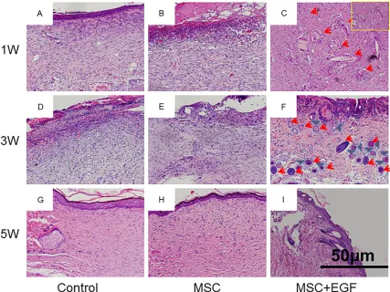
[image:5.612.94.521.71.391.2]The separated proteins were electrotransfer- red to a polyvinylidene fluoride (PVDF) mem-brane. The membrane was incubated consecu-tively with blocking buffer for 30 min at room temperature, with the primary antibody over-night at 4°C and with a horseradish peroxidase-linked secondary antibody for 1 h at room tem-perature. Between the incubations in primary and secondary antibody, the membrane was Figure 4. H&E staining of wound sections. Wounds were transplanted with empty scaffold (A, D, G), MSC scaffold (B, E, H) and MSC + EGF (C, F, I).
[image:5.612.93.523.442.575.2]washed three times, 10 min each, with Tris-buffered saline containing 0.1% Tween 20 (TBS-T). The membrane was treated with a standard chemical luminescence reagent (Beyotime, China). For signal detection, Kodak X-Omat AR films were exposed to the mem-brane, were scanned on a gel imaging and sys-tem, and analyzed by the Quantity One 4.4 soft-ware (Bio-Rad, Hercules, CA, USA).
Results
Gross observations of wound healing and hair growth
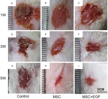
Images were taken to record and analyze wound size changes and healing (Figure 2). Repaired wounds could be readily distinguished from normal skin, owing to the naked appear-ance of the skin on the wound site, displaying little hair growth and attachment. At day 7, the appearance of the skin on the wound sites of the rat in the MSC + EGF group was more red and fresh, and with more scales, compared to
that of the rat in the control and MSC groups (Figure 3A-C). These results suggest that MSC + EGF improved wound healing and promoted vascularization. At day 21, the wound area in the rat in the MSC + EGF group was much smaller than in the other two groups (Figure 3D-F). At day 35, we found that the skin in the wound area was nearly completely regenerated in the MSC + EGF group whereas there were still a few wounds unhealed in the control and MSC groups (Figure 3G-I).
Furthermore, since hair growth indicates hair follicle regeneration, we monitored hair regrow- th in the regenerated wound area at day 35 and we found more hair growth in the animals in the MSC + EGF group compared to the those in the control and MSC groups (Figure 3G-I).
Histology
[image:6.612.91.522.71.395.2]cles began to appear in the MSC + EGF group (Figure 4A-C). On day 21, we clearly observed hair follicles wrapped by sebaceous glands in the MSC + EGF group, but no hair follicle or sebaceous gland was found in the control and MSC groups (Figure 4D-F). On day 35, mature hair follicles and sebaceous glands could be clearly identified in the MSC + EGF group, whereas still no hair follicle or sebaceous gland was observed in the control and MSC groups (Figure 4G-I).
Newly formed blood vessels are necessary for skin regeneration. At day 7, newly formed blood vessels could be visualized in all three groups (Figure 5). Compared with that in the control group, blood vessel density in both the MSC and MSC + EGF groups were much higher (P<0.01).
Oil red staining
[image:7.612.92.523.71.391.2]Oil red staining was performed to further inves-tigate the ability of MSCs + EGF to induce seba-Figure 7. ALP immunohistochemistry of wound sections. Wounds were transplanted with empty scaffold (A, D, G), MSC scaffold (B, E, H) and MSC + EGF (C, F, I). ALP positive areas were stained yellow or brown.
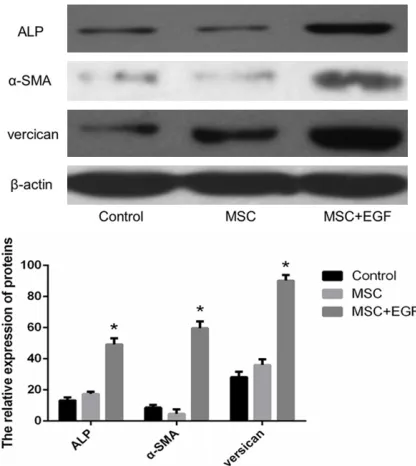
Figure 8. Western blot analysis of ALP, α-SMAs and versican expression. *denotes statistical significance
[image:7.612.92.300.441.674.2]follicle regeneration.
Immunohistochemistry
In normal skin, ALP is a hair follicle-specific fac-tor in dermal papilla cells, which is continuously expressed throughout the development of the hair follicle. ALP can be used as an important indicator of hair follicle regeneration. Therefore, in order to further identify hair follicle regenera-tion during wound healing, we tested ALP expression by immunohistochemistry in the regenerated skin from the three experimental groups. ALP positive cells were stained yellow-brown. As show in Figure 7, few ALP-positive areas could be found in the control and MSC groups, while a large number of ALP-positive areas could be observed in the MSC + EGF group. These results indicated that MSC + EGF promoted hair follicle regeneration.
Western blot analysis
The immunohistochemistry results were con-firmed by Western blot analysis. As shown in
Figure 8, compared to the control and MSC groups, the expression of ALP, α-SMA, and ver-sican increased in the MSC + EGF group (P<0.01).
Discussion
In this study, we utilized biomimetic hydrogel scaffolds to effectively deliver MSC or MSC + EGF into cutaneous wounds. Our results dem-onstrate that delivery of MSCs + EGF acceler-ated wound healing. Moreover, we found that MSCs + EGF could promote hair growth, angio-genesis, and sebaceous gland recovery. Hair follicles are necessary accessory organs of the skin. Hairs have physiological functions, such as providing thermoregulation and
protec-maintain cell homology and differentiate into various different cell types. Asakawa et al. iso-lated epithelial and dermal cells from embry-onic skin tissue and found that their transplan-tation to the wound could facilitate hair follicle regeneration [19]. Higgins et al. showed that dermal hair follicle cells could induce human hair-follicle growth [2]. In addition, Huang et al. found that transplantation of cultured sweat gland cells into the wound in biomimetic hydro-gel scaffolds was a promising method for sweat gland regeneration [20]. These studies suggest that regeneration of the accessory organs could be achieved by transplantation of acces-sory organ-derived cells. However, it is not straightforward to isolate these seed cells because skin appendages are very small, and the separation and purification processes are complex and need to be performed under a microscope [21]. Moreover, cells isolated from an accessory organ often have low viability dur-ing in vitro culture, and lose the ability to differ-entiate into specific cells that are needed for organ regeneration. Therefore, transplantation of accessory organ-derived cells has not been widely applied in the clinic.
received the most attention for hair follicle regeneration. As seed cells, MSCs can differen-tiate into multiple cell types and participate in cutaneous wound healing. Transplantation of MSCs into full-thickness burn wounds induces epidermal thickening, increases the number of dermal nerve fibers, accelerates wound heal-ing, and greatly improves healing quality [27]. Moreover, MSCs participate in wound reepithe-lialization and the regeneration of accessory organs like hair follicles and sweat glands [10, 27]. However, there are studies reporting that although transplantation of MSCs can promote skin recovery, they have little effect on hair fol-licle regeneration [28]. Our results are consis-tent with those of these studies. We found that MSCs could accelerate wound healing and blood vessel formation, but had little effect on hair follicle or sebaceous glands regeneration. Hair follicle regeneration requires a variety of different cell types and a suitable external envi-ronment that is essential for MSC differentia-tion [29]. When the skin is wounded, the physi-ological environment under the skin is often compromised. This might be the reason why MSC transplantation fails to regenerate hair fol-licle or sebaceous glands. Recent studies sug-gest that MSCs must migrate to wound sites before they can be involved in the repair pro-cess, and the ability of MSCs to secrete biologi-cally active substances, such as cytokines and growth factors, play key roles in wound recov-ery [30, 31]. Thus, to regenerate hair follicles during skin wound recovery, it is very important to both promote MSC migration and induce MSC differentiation into dermal papilla cells. A wide variety of growth factors and cytokines are involved in the regulation of all phases of wound healing. Yamazaki et al. and Jindo et al. found that pHGF has a stimulatory effect on hair growth in vivo and in vitro [32, 33]. Zhang et al. showed that Activin B could promote MSC-mediated cutaneous wound healing by regulat-ing cell migration via the JNK-ERK signalregulat-ing pathway [27]. EGF is a multi-functional cell growth factor, affecting physiological process-es, such as epidermization, cell proliferation, and migration [12, 34, 35]. Therefore, EGF may play an important role in MSC-mediated skin repair. In this study, we examined the impact of combined administration of MSCs and EGF on wound healing in a rat model. We found that EGF did indeed promote MSC-mediated wound
healing and hair follicle regeneration in vivo. These results indicated that combined admin-istration of MSCs and EGF might be a promising therapeutic strategy for wound management.
Acknowledgements
This study was supported by the Plan Project of Zhejiang Provincial Department of Health, China (No. 2016RCA027).
Disclosure of conflict of interest
None.
Address correspondence to: Hong Chen, Depart- ment of Plastic Surgery, Shaoxing People’s Hospital, Shaoxing 312000, Zhejiang Province, China. E-mail: dingshizhang7@163.com
References
[1] Rustad KC, Wong VW, Sorkin M, Glotzbach JP,
Major MR, Rajadas J, Longaker MT and Gurt-ner GC. Enhancement of mesenchymal stem cell angiogenic capacity and stemness by a biomimetic hydrogel scaffold. Biomaterials 2012; 33: 80-90.
[2] Higgins CA, Chen JC, Cerise JE, Jahoda CA and Christiano AM. Microenvironmental repro-gramming by three-dimensional culture en-ables dermal papilla cells to induce de novo human hair-follicle growth. Proc Natl Acad Sci U S A 2013; 110: 19679-19688.
[3] Garcin CL and Ansell DM. The battle of the bulge: Re-evaluating hair follicle stem cells in wound repair. Exp Dermatol 2016; 26: 101-104.
[4] Garcin CL, Ansell DM, Headon DJ, Paus R and Hardman MJ. Hair follicle bulge stem cells ap-pear dispensable for the acute phase of wound re-epithelialization. Stem Cells 2016; 34: 1377-1385.
[5] Wang X, Wang X, Liu J, Cai T, Guo L, Wang S, Wang J, Cao Y, Ge J, Jiang Y, Tredget EE, Cao M and Wu Y. Hair follicle and sebaceous gland de novo regeneration with cultured epidermal stem cells and skin-derived precursors. Stem Cells Transl Med 2016; 5: 1695-1706. [6] Guo R, Xu S, Ma L, Huang A and Gao C.
En-hanced angiogenesis of gene-activated dermal equivalent for treatment of full thickness inci-sional wounds in a porcine model. Biomateri-als 2010; 31: 7308-7320.
scarring. Biomaterials 2013; 34: 2038-2048. [10] Liu H, Peng H, Wu Y, Zhang C, Cai Y, Xu G, Li Q,
Chen X, Ji J, Zhang Y and OuYang HW. The
pro-motion of bone regeneration by nanofibrous
hydroxyapatite/chitosan scaffolds by effects on integrin-BMP/Smad signaling pathway in BMSCs. Biomaterials 2013; 34: 4404-4417. [11] Wang W, Li B, Li Y, Jiang Y, Ouyang H and Gao
C. In vivo restoration of full-thickness cartilage defects by poly(lactide-co-glycolide) sponges
filled with fibrin gel, bone marrow mesenchy -mal stem cells and DNA complexes. Biomateri-als 2010; 31: 5953-5965.
[12] Doma E, Rupp C and Baccarini M. EGFR-ras-raf signaling in epidermal stem cells: roles in hair follicle development, regeneration, tissue remodeling and epidermal cancers. Int J Mol Sci 2013; 14: 19361-19384.
[13] Hong Y, Gong Y, Gao C and Shen J. Collagen-coated polylactide microcarriers/chitosan hy-drogel composite: injectable scaffold for carti-lage regeneration. J Biomed Mater Res A 2008; 85: 628-637.
[14] Wang X, You C, Hu X, Zheng Y, Li Q, Feng Z, Sun H, Gao C and Han C. The roles of knitted mesh-reinforced collagen-chitosan hybrid scaffold in the one-step repair of full-thickness skin de-fects in rats. Acta Biomater 2013; 9: 7822-7832.
[15] Shi H, Han C, Mao Z, Ma L and Gao C. En-hanced angiogenesis in porous collagen-chito-san scaffolds loaded with angiogenin. Tissue Eng Part A 2008; 14: 1775-1785.
[16] Wang X, Wu P, Hu X, You C, Guo R, Shi H, Guo S, Zhou H, Yu C, Zhang Y and Han C. Polyure-thane membrane/knitted mesh-reinforced collagen-chitosan bilayer dermal substitute for the repair of full-thickness skin defects via a two-step procedure. J Mech Behav Biomed Mater 2016; 56: 120-133.
[17] Haifei S, Xingang W, Shoucheng W, Zhengwei M, Chuangang Y and Chunmao H. The effect of collagen-chitosan porous scaffold thickness on dermal regeneration in a one-stage grafting procedure. J Mech Behav Biomed Mater 2014; 29: 114-125.
constitution and in vivo implantation of engi-neered skin constructs with sweat glands. Bio-materials 2010; 31: 5520-5525.
[21] Aoi N, Inoue K, Kato H, Suga H, Higashino T, Eto H, Doi K, Araki J, Iida T, Katsuta T and Yo-shimura K. Clinically applicable transplanta-tion procedure of dermal papilla cells for hair follicle regeneration. J Tissue Eng Regen Med 2012; 6: 85-95.
[22] Kamstrup M, Faurschou A, Gniadecki R and Wulf HC. Epidermal stem cells - role in normal, wounded and pathological psoriatic and can-cer skin. Curr Stem Cell Res Ther 2008; 3: 146-150.
[23] Li H and Fu X. Mechanisms of action of mesen-chymal stem cells in cutaneous wound repair and regeneration. Cell Tissue Res 2012; 348: 371-377.
[24] Peng LH, Mao ZY, Qi XT, Chen X, Li N, Tabata Y and Gao JQ. Transplantation of bone-marrow-derived mesenchymal and epidermal stem cells contribute to wound healing with different regenerative features. Cell Tissue Res 2013; 352: 573-583.
[25] Festa E, Fretz J, Berry R, Schmidt B,
Rodehef-fer M, Horowitz M and Horsley V. Adipocyte lin -eage cells contribute to the skin stem cell niche to drive hair cycling. Cell 2011; 146: 761-771.
[26] Li H, Fu X, Ouyang Y, Cai C, Wang J and Sun T. Adult bone-marrow-derived mesenchymal stem cells contribute to wound healing of skin appendages. Cell Tissue Res 2006; 326: 725-736.
[27] Zhang M, Sun L, Wang X, Chen S, Kong Y, Liu N, Chen Y, Jia Q, Zhang L and Zhang L. Activin B promotes BMSC-mediated cutaneous wound healing by regulating cell migration via the JNK-ERK signaling pathway. Cell Transplant 2014; 23: 1061-1073.
and epidermal cells: biotinylated galectins as
tool to detect specific binding sites. Biol Cell
2003; 95: 535-545.
[29] Badiavas EV, Abedi M, Butmarc J, Falanga V
and Quesenberry P. Participation of bone mar-row derived cells in cutaneous wound healing. J Cell Physiol 2003; 196: 245-250.
[30] Fu X, Qu Z and Sheng Z. Potentiality of mesen-chymal stem cells in regeneration of sweat glands. J Surg Res 2006; 136: 204-208. [31] Wu Y, Chen L, Scott PG and Tredget EE.
Mesen-chymal stem cells enhance wound healing through differentiation and angiogenesis. Stem Cells 2007; 25: 2648-2659.
[32] Yamazaki M, Tsuboi R, Lee YR, Ishidoh K, Mit-sui S and Ogawa H. Hair cycle-dependent ex-pression of hepatocyte growth factor (HGF) ac-tivator, other proteinases, and proteinase inhibitors correlates with the expression of HGF in rat hair follicles. J Investig Dermatol Symp Proc 1999; 4: 312-315.
[33] Jindo T, Tsuboi R, Takamori K and Ogawa H. Local injection of hepatocyte growth factor/ scatter factor (HGF/SF) alters cyclic growth of murine hair follicles. J Invest Dermatol 1998; 110: 338-342.
[34] Myers SR, Leigh IM and Navsaria H. Epidermal repair results from activation of follicular and epidermal progenitor keratinocytes mediated by a growth factor cascade. Wound Repair Re-gen 2007; 15: 693-701.