Original Article
Genetic heterogeneity in hepatocellular carcinoma
and paired bone metastasis revealed by
next-generation sequencing
Ketao Jin1*, Huanrong Lan2*, Xuanwei Wang3, Jieqing Lv1
Departments of 1Gastrointestinal Surgery, 2Breast and Thyroid Surgery, Shaoxing People’s Hospital, Shaoxing Hospital of Zhejiang University, Shaoxing, Zhejiang Province, P. R. China; 3Department of Orthopedics, The First Affiliated Hospital, School of Medicine, Zhejiang University, Hangzhou, Zhejiang, P. R. China. *Equal contributors.
Received June 21, 2017; Accepted August 3, 2017; Epub October 1, 2017; Published October 15, 2017
Abstract: Although bone involvement is reported as uncommon in hepatocellular carcinoma (HCC), its incidence
has significantly increased in the last decade due to the longer survival of HCC patients related to recent progresses made both in the diagnosis and treatment of the disease. A better understanding of the pathogenic mechanisms underlying the spread of bone metastases in HCC is important. The primary tumor and its corresponding metasta
-ses are different at the molecular marker expression or gene status levels and that these differences may affect the clinical outcome of anticancer therapy, particularly in molecularly targeted therapies for the treatment of cancer. No study has investigated the genetic heterogeneity between HCC and paired bone metastasis. In this study, we inves
-tigated the genetic heterogeneity in HCC and paired metastasis using a next generation sequencing (NGS) platform to illustrate the molecularly targeted therapy related genes mutations.
Keywords: Hepatocellular carcinoma, bone metastasis, genetic heterogeneity, next generation sequencing
Introduction
Liver cancer in men is the fifth most frequently
diagnosed cancer worldwide but the second
most frequent cause of cancer death. In wo-men, it is the seventh most commonly diag
-nosed cancer and the sixth leading cause of cancer death. Half of these cases and deaths
were estimated to occur in China [1]. Among
pri-mary liver cancers, hepatocellular carcinoma
(HCC) represents the major histological
sub-type, accounting for 70% to 85% of the total liver cancer burden worldwide [2]. The primary risk factor for HCC is liver injury from diverse
causes that leads to hepatic cirrhosis in most
patients. An estimated 78% of HCC cases and 57% of cases of liver cirrhosis are caused by chronic infection with hepatitis B virus (HBV) or hepatitis C virus (HCV) [2, 3]. HCC is the sixth
most prevalent cancer worldwide and the third
leading cause of cancer-related death, although
its geographical distribution is heterogeneous with the highest incidence in sub-Saharan
Africa and Eastern Asia [4]. Bone is an uncom
-mon site of metastasis in HCC, with the inci
-dence ranging from 3% to 20% [5]. Although
bone involvement is reported as uncommon in
HCC, its incidence has significantly increased
in the last decade due to the longer survival
of HCC patients related to recent progresses made both in the diagnosis and treatment of
the disease [5-7].
A better understanding of the pathogenic mechanisms underlying the spread of bone
metastases in HCC is important. Some retro-spective studies have described the
character-istics of bone metastasis from HCC [5, 8-10]. However, few data are yet available about bone
involvement in patients with HCC, and no
agree-ment has yet been reached about the treat
-ment strategy for extrahepatic HCC metasta
-ses. The nature and the characteristics of bone metastases in HCC have not been fully explored in literature, presumably because HCC skeletal involvement was rarely diagnosed.
Intratumor heterogeneity is a recognized char
-acteristic of human tumors, and occurs on mul
the method of mutation analysis), protein (de-tected by the method of immunohistochemical analysis) and macroscopic level in a wide range of tumors, such as breast, colorectal (CRC),
non-small cell lung (NSCLC), prostate, ovarian, pancreatic, gastric, and brain cancer and renal
clear cell carcinoma. In recent years, many studies have focused on the heterogeneity found in primary tumors and related metasta
-ses with the consideration that evaluation of metastatic rather than primary sites could be of clinical relevance. Numerous reports have evaluated the genetic heterogeneity in primary
tumors and corresponding metastases in a
study investigated the role of PTEN loss, AKT phosphorylation and KRAS mutations in prima
-ry colorectal tumors and their corresponding metastases on the activity of cetuximab plus irinotecan [17]. This study gave us direct evi
-dence to reveal that the genetic heterogeneity in primary colorectal tumors and their corre
-sponding metastases have different responses to molecularly targeted therapy.
However, no study has investigated the genetic heterogeneity between HCC and paired bone metastasis. Here, we hypothesize that there is genetic heterogeneity between HCC and paired
bone metastasis, which might result in the
therapeutic failure of molecularly targeted ther
-apy for treatment of HCC bone metastasis. In this study, we investigated the genetic hetero
-geneity in HCC and paired metastasis using a next generation sequencing (NGS) platform to illustrate the molecularly targeted therapy
related genes mutations. Patients and methods
Patients and tumor tissue samples
[image:2.612.91.392.98.335.2]The institutional ethical committee approved the current retrospective study. A written in- Table 1. Clinical characteristics of two hepatocellular carcinoma pa
-tients with metachronous bone metastasis received surgery
Patient 1 Patient 2
Gender (Male/Female) Male Male
Age (Years) 39 58
AFP level (ng/ml) 8.4 27.1
ALT (IU/L) Not available Not available
AST (IU/L) Not available Not available
HBV Positive Positive
HCV Negative Negative
BCLC staging (A/B/C) Not available A
Child’s score (A/B/C) Not available A
Hepatocellular carcinoma (number of masses) 2 2
Bone metastases Metachronous Metachronous
Overall survival (months) 57.5 42.3
Chemotherapy. (Yes/No) Yes Not available
Radiotherapy (Yes/No) Yes No
Molecularly targeted therapy (Yes/No) Yes No
Primary tumor ID 10-13656-1 10-42240-1
Metastatic tumor ID 12-60901-3 13-39592-3
Abbreviations: HBV, hepatitis B virus infection. HCV, hepatitis C virus infection. AST, aspartate aminotransferase. ALT, aspartate aminotransferase. AFP, alpha-fetoprotein. BCLC staging, Barcelona clinic liver cancer staging.
Table 2. Fifty known cancer genes Gene name
ABL1 EGFR GNAQ KRAS PTPN11
AKT1 ERBB2 GNAS MET RB1
ALK ERBB4 HNF1A MLH1 RET
APC EZH2 HRAS MPL SMAD4
ATM FBXW7 IDH1 NOTCH1 SMARCB1
BRAF FGFR1 IDH2 NPM1 SMO
CDH1 FGFR2 JAK2 NRAS SRC
CDKN2A FGFR3 JAK3 PDGFRA STK11
CSF1R FLT3 KDR PIK3CA TP53
CTNNB1 GNA11 KIT PTEN VHL
range of solid tumors such
as breast cancer [11], CRC [12] and NSCLC [13]. As discussed earlier, the
pri-mary tumor and its corre -sponding metastases are
different at the molecular marker expression or
ge-ne status levels and that
these differences may af-fect the clinical outcome of anticancer therapy, par
-ticularly in molecularly tar
-geted therapies for the treatment of cancer [14, 15]. Monaco et al. sug
-gested that the EGFR and KRAS status of primary
lung carcinomas might not predict the status in the corresponding metastas-
es. Their observation may
have important implicati-
[image:2.612.91.289.400.548.2]10497 Int J Clin Exp Pathol 2017;10(10):10495-10504
formed consent was obtained for both two
patients. We reviewed the electronic medical
records of consecutive patients in whom HCC and metachronous bone metastasis was newly diagnosed from January 2009 to October 2014 at the Department of Orthopedics, the First Affiliated Hospital, Zhejiang University School of Medicine (Table 1). The diagnosis of HCC was mainly based on recommendations of the American Association for the Study of Liver Diseases [18]. Both patients underwent blood
investigations, which included complete blood
count, liver function tests, and tests for viral markers of hepatitis B and C infection. Serum alpha-fetoprotein (AFP) was estimated using a particle enzyme immunoassay (Axsym System; Abbott Laboratories, Abbot Park, Illinois, USA; normal value <20 ng/ml). Upper gastrointesti
-nal endoscopy was done in each case to detect the presence of esophageal varices. Patients with underlying cirrhosis were classified into Child’s A, B or C based on the Child-Pugh clas
-sification [19]. Staging of HCC was done based on the Barcelona Clinic Liver Cancer (BCLC) staging protocol [20]. Both patients were evalu
-ated pre-operatively using abdominal comput
-ed tomographic (CT) scan, magnetic resonance imaging (MRI), or fluorine-13 fluorodeoxyglu
-cose positron emission tomography/computed
tomographic scan (18F-FDG PET/CT).
DNA extraction
Formalin-fixed paraffin-embedded (FFPE) tis
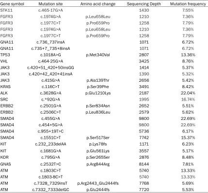
[image:3.612.92.521.96.495.2]-sues were sectioned, and hematoxylin and eosin (H&E) stained slides were reviewed by
Table 3. Hepatocellular carcinoma primary tumor (sample number: 10-13656-1) detected gene muta -tions
Gene symbol Mutation site Amino acid change Sequencing Depth Mutation frequency
STK11 c.465-17G>A 1430 7.55%
FGFR3 c.1974G>A p.Leu658Leu 1210 7.36%
FGFR3 c.1977C>T p.Pro659Pro 1258 7.79%
FGFR3 c.1974G>A p.Leu658Leu 1210 7.36%
FGFR3 c.1977C>T p.Pro659Pro 1258 7.79%
GNA11 c.736_737insA 1071 6.72%
GNA11 c.735+7_735+8insA 1071 6.72%
TP53 c.1018A>G p.Met340Val 2807 13.36%
VHL c.464-25G>A 3425 8.76%
JAK3 c.420+51_420+50insGG 1414 5.37%
JAK3 c.420+42_420+41insA 1390 5.32%
JAK3 c.415G>A p.Ala139Thr 2656 5.42%
KRAS c.116C>T p.Ser39Phe 3491 8.42%
ALK c.3628G>A p.Glu1210Lys 2187 22.04%
SRC c.*92G>A 1995 16.74%
ERBB2 c.2501G>A p.Ser834Asn 2652 5.51%
ERBB2 c.2506C>T p.Leu836Leu 2579 5.62%
SMAD4 c.455G>A 9800 22.69%
SMAD4 c.454+5G>A 9800 22.69%
SMAD4 c.955+19T>C 5736 6.17%
SMAD4 c.1551C>T p.Ser517Ser 7742 15.37%
KIT c.232_233delAA p.Lys78fs 1171 6.23%
KIT c.1681G>A p.Glu561Lys 3557 5.17%
KDR c.795G>A p.Ser265Ser 2876 8.48%
GNAS c.2532T>C p.Arg844Arg 8144 7.81%
ATM c.1803C>T 5740 13.33%
ATM c.1803-8C>T 5740 13.33%
ATM c.7328_7329insT p.Arg2443_Glu2444fs 7768 5.69%
one surgical pathologist (FL) to confirm the tumor content in each section. Ten serial sec
-tions (4 μm) were cut from selected tissue areas with tumor tissue were microdissected
fr-om those slides using the H&E slides as
tem-plates. The pathologic diagnosis of each case was confirmed on routine H&E slides. All sam
-ples sent for DNA extraction contained a mini
-mum of 20% DNA derived from tumor cells. The tissues were deparaffinized with 1 mL xylene at 56°C for 10 min, washed with 1 mL 100% etha
-nol for 5 min at RT, and then dried at 37°C for 10 min. QIAamp DSP DNA FFPE tissue kit (Qiagen, Valencia, CA, USA) was used to extract the genomic DNA from FFPE samples. DNA concentration was determined by Qubit dsDNA HS assay kit on the Qubit Fluorometer accord
-ing to the manufactur-ing protocol (Life Tec-hnologies, Carlsbad, CA, USA). DNA quality (A260/280 and A260/230) was measured by
Hybrid capture and ultra-deep next generation sequencing
The 5’-biotinylated probe solution is provided
as capture probes, the baits target 416
cancer-related genes. 1 μg of each DNA-fragment se-quencing library is mixed with 5 μg of human Cot-1 DNA, 5 μg of salmon sperm DNA, and 1 unit adaptor-specific blocker DNA in hybridiza-tion buffer, heated for 10 minutes at 95°C, and held for 5 minutes at 65°C in the thermocycler.
Within 5 minutes, the capture probes are added
[image:4.612.91.383.95.479.2]to the mixture, and the solution hybridization is performed for 16-18 hours at 65°C. After hybridization is complete, the captured targets are selected by pulling down the biotinylated probe/target hybrids using streptavidin-coated magnetic beads, and off-target library is removed by washing with wash buffer. The PCR master mix is added to directly amplify (6-8
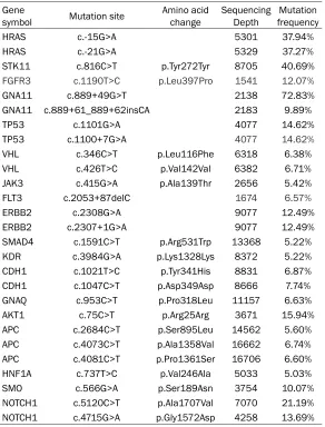
Table 4.Hepatocellular carcinoma bone metastasis (sample num-ber: 12-60901-3) detected gene mutations
Gene
symbol Mutation site Amino acid change Sequencing Depth frequencyMutation
HRAS c.-15G>A 5301 37.94%
HRAS c.-21G>A 5329 37.27%
STK11 c.816C>T p.Tyr272Tyr 8705 40.69%
FGFR3 c.1190T>C p.Leu397Pro 1541 12.07%
GNA11 c.889+49G>T 2138 72.83%
GNA11 c.889+61_889+62insCA 2183 9.89%
TP53 c.1101G>A 4077 14.62%
TP53 c.1100+7G>A 4077 14.62%
VHL c.346C>T p.Leu116Phe 6318 6.38%
VHL c.426T>C p.Val142Val 6382 6.71%
JAK3 c.415G>A p.Ala139Thr 2656 5.42%
FLT3 c.2053+87delC 1674 6.57%
ERBB2 c.2308G>A 9077 12.49%
ERBB2 c.2307+1G>A 9077 12.49%
SMAD4 c.1591C>T p.Arg531Trp 13368 5.22%
KDR c.3984G>A p.Lys1328Lys 8372 5.22%
CDH1 c.1021T>C p.Tyr341His 8831 6.87%
CDH1 c.1047C>T p.Asp349Asp 8666 7.74%
GNAQ c.953C>T p.Pro318Leu 11157 6.63%
AKT1 c.75C>T p.Arg25Arg 3671 15.94%
APC c.2684C>T p.Ser895Leu 14562 5.60%
APC c.4073C>T p.Ala1358Val 16662 6.74%
APC c.4081C>T p.Pro1361Ser 16706 6.60%
HNF1A c.737T>C p.Val246Ala 5033 5.03%
SMO c.566G>A p.Ser189Asn 3754 10.07%
NOTCH1 c.5120C>T p.Ala1707Val 7070 21.19%
NOTCH1 c.4715G>A p.Gly1572Asp 4258 13.69%
Nanodrop 2000 (Thermo Fi-sher Scientific, Waltham, MA, USA).
Library construction
Sequencing library was pr-epared by Illumina TruSeq DNA PCR-Free Sample
Pre-paration Kit (Illumina, San
Diego, CA, USA) according to the manufacturing protocol. In brief, genomic DNA sam
-ple was fragmented into 350 or 550 bp in AFA fiber snap-cap microTUBE using Covaris M220 (Covaris, Woburn, MA, USA). End repair and size selection were performed according to the fragment size, followed by 30 end ad-enylation. Finally, multiple
in-dexing adapters were ligated
to the ends of the DNA frag
10499 Int J Clin Exp Pathol 2017;10(10):10495-10504
cycles) the captured library from the washed beads. After amplification, the samples are purified by AMPure XP beads, quantified by qPCR (KAPA Biosystems, Boston, MA, USA) and sized on bioanalyzer 2100 (Agilent Technolo-gies, Santa Clara, CA, USA). Libraries are nor
-malized to 2.5 nM and pooled. Deep Sequencing is performed on Illumina HiSeq 4000 using PE75 V1 Kit. Cluster generation and sequenc
-ing is performed accord-ing to manufacturer’s
protocol.
Sequence alignment and processing
Quality control (QC) was applied with Trimmo-matic 1. High quality reads were mapped to the human genome (hg19, GRCh37 Genome Reference Consortium Human Reference 37) using modified BWA aligner 0.7.12 2 with BWA-MEM algorithm and default parameters to create SAM files. Picard 1.119 (http://picard. sourceforge.net/) was used to convert SAM files to compressed BAM files which were then
sorted according to chromosome coordinates.
The Genome Analysis Toolkit 3 (GATK, version 3.4-0) was modified and used to locally realign the BAMs files at intervals with indel mismatch
-es and recalibrate base quality scor-es of reads in BAM files.
month after treatment. Further treatments were based on clinical evaluation, laboratory
values and imaging response. Patients were
followed-up every 3 months. The patients were followed up until death or until the date of last follow-up. Follow-up was finished on February 28, 2015.
Results
HCC primary tumor (sample number: 10-13656-1) detected gene mutations
We analyzed fifty molecularly targeted therapy
related genes (Table 2) to illustrate the genetic
heterogeneity in HCC and paired metastasis using a next generation sequencing (NGS) plat
-form as described in Materials and Methods. HCC primary tumor (sample number:
10-13656-1) involves 15 mutant genes, 30 independent
mutation sites, and an average mutation fre
-quency of 11.1% (Table 3).
HCC bone metastasis (sample number: 12-60901-3) detected gene mutations
HCC bone metastasis (sample number: 1260-
901-3) involves 18 mutant genes, 27 indepen -dent mutation sites, and an average mutation
[image:5.612.91.332.121.353.2]frequency of 12.34% (Table 4). Table 5. Genes mutations detected in hepatocellular
carcinoma primary tumor (sample number:
10-13656-1) and its paired bone metastasis (sample number: 12-60901-3)
Number Mutant genes in HCC primary tumor and
bone metastasis
Mutant genes in HCC primary
tumor
Mutant genes
in HCC bone metastasis
1 ABL1 ALK AKT1
2 ATM APC
3 CTNNB1 FGFR3 CDH1
4 EGFR GNAS HNF1A
5 ERBB2 KDR NOTCH1
6 ERBB4 KIT SMAD4*
7 FLT3 KRAS VHL*
8 GNA11 SMAD4
9 GNAQ SRC
10 HRAS STK11
11 JAK3 VHL
12 NPM1
13 NRAS
14 SMO
15 TP53
*, The same mutant genes with different mutation sites in hepatocel-lular carcinoma primary tumor and bone metastasis.
Validation of SNPs/Indels and CNVs
Single nucleotide variants (SNVs) and
short insertions/deletions (indels) were
identified using VarScan 2 2.3.9 4 with minimum variant allele frequency thresh -old set at 0.01 and p-value threshold for
calling variants set at 0.05 to generate
Variant Call Format (VCF) files. All SNVs/ indels were annotated with ANNOVAR, and each SNV/indel was manually che-cked with the Integrative Genomics Viewer (IGV) 5. Copy number variations (CNVs) were identified using ADTEx 1.0.4
6.
Data collection and follow-up
The clinical, laboratory, and radiologic records of the two patients were retro
-spectively reviewed (Table 1). Liver func
-tion tests were checked in the patients every three months in order to evaluate hepatic functional reserve. The results of two HCC patients with metachronous bone metastasis were analyzed.
Follow-up cross-sectional imaging
Genes mutations detected in HCC primary tumor (sample number: 10-13656-1) and its paired bone metastasis (sample number: 12-60901-3)
HCC primary tumor (sample number:
10-13656-1) and paired bone metastasis (sample
num-ber: 12-60901-3) identified 15 common mutant
genes. Ten unique mutations in the primary cancer were identified. We found 7 unique
mutations in metastatic cancer, including 2
genes, which are different from mutations in the primary mutation. Five new mutations were only found in metastatic site (Table 5).
new mutations were only found in metastatic
site (Table 8).
Genes mutations detected in HCC primary tu-mor (sample number: 10-13656-1; 10-42240-1) and its paired bone metastasis (sample number: 12-60901-3; 13-39592-3)
Of the two HCC primary tumor (sample number 1013656-1; 10-42240-1) identified 18 non
common genes; the sample 10-13656-1 has 6 non common genes, and the sample 10-42240-1 number has 10-42240-12 non common genes (Table 9).
[image:6.612.93.372.98.548.2]After analyzing two pairs of HCC primary tumor
Table 6.Hepatocellular carcinoma primary tumor (sample number:
10-42240-1) detected gene mutations
Gene
symbol Mutation site Amino acid change Sequencing Depth frequencyMutation
TP53 c.504C>A p.His168Gln 2566 5.85
TP53 c.501_502insG p.Gln167_His168fs 8.38
VHL c.484T>C p.Cys162Arg 3256 5.04
ERBB2 c.2310A>G 2302 8.95
ERBB2 c.2310A>G p.Glu770Glu 2302 8.95
ERBB2 c.2498T>C p.Met833Thr 2937 11.68
FGFR1 c.839-15T>C 1877 9.59
PDGFRA c.2003G>A p.Gly668Asp 2752 6.29
PDGFRA c.2003G>A 2752 6.29
EGFR c.286G>A p.Val96Met 4311 11.53
EGFR c.2184+63delC 1260 14.29
KIT c.2597T>C 2263 9.68
KIT c.2596+2T>C 2263 9.68
KDR c.3496G>A p.Ala1166Thr 2746 10.31
KDR c.2962G>A p.Glu988Lys 2143 27.2
GNAQ c.650T>C p.Ile217Thr 10582 7.4
GNAQ c.496C>T p.Arg166Cys 2698 13.86
ATM c.5918+37T>C 2371 18.52
ATM c.7328G>A p.Arg2443Gln 7559 14.14
APC c.2674G>A p.Glu892Lys 8094 10.49
PTPN11 c.1448C>T 4161 12.62
PTPN11 c.1448-8C>T 4161 12.62
MET c.2986C>T p.Pro996Ser 28255 17.48
FGFR2 c.880G>A p.Val294Met 4164 17.65
SMO c.654G>A p.Gln218Gln 4469 6.58
SMO c.1264+41A>G 2321 69.19
SMO c.1599C>T p.Ser533Ser 4612 22.61
ABL1 c.1010T>C p.Met337Thr 5156 6.92
ABL1 c.1143-44A>T 1067 6.28
PIK3CA c.2719G>A p.Ala907Thr 5771 8.14
ERBB4 c.884-23A>T 1163 5.93
ERBB4 c.764G>A p.Ser255Asn 4417 28.59
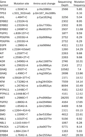
HCC primary tumor (sample number: 10-42240-1) de-tected gene mutations
HCC primary tumor (sample
number: 10-42240-1) involv- es 19 mutant genes, 32 inde-pendent mutation sites, and
an average mutation frequen
-cy of 13.81% (Table 6).
HCC bone metastasis (sample number: 13-39592-3) detected gene mutations
HCC bone metastasis (sample number: 1339592-3) involves 10 mutant genes, 32 inde-pendent mutation sites, and
an average mutation frequen
-cy of 9.19% (Table 7).
Genes mutations detected in HCC primary tumor (sample number: 10-42240-1) and its paired bone metastasis (sample number: 13-39592-3)
HCC primary tumor (sample
number: 10-42240-1) and pa- ired bone metastasis (sample number: 13-39592-3)
identi-fied 21 common mutant ge-nes. Twelve unique mutations in the primary cancer were identified. We found 6 unique
mutations in metastatic can-cer, including 4 genes, which
10501 Int J Clin Exp Pathol 2017;10(10):10495-10504 (sample number 10-13656-1; 1042240-1) and
bone metastases (sample number:
12-60901-3; 13-39592-3), we found 13 unique mutations
in bone metastases, including 7 genes, which
were only different from the mutation sites of primary mutations. Five new mutations were only found in metastatic sites (Table 9). This suggests that the importance of genetic muta
-tions after metastasis is declining. If additional
range of solid tumors such as breast cancer, CRC and NSCLC. However, no study has investi
-gated the genetic heterogeneity between HCC and paired bone metastasis. We hypothesize that there would be genetic heterogeneity
between HCC and paired bone metastasis. Recent advances in genomics technologies are
now providing new opportunities for the analy -Table 7. Hepatocellular carcinoma bone metastasis (sample number:
13-39592-3) detected gene mutations
Gene
symbol Mutation site Amino acid change Sequencing Depth frequencyMutation
PDGFRA c.1958C>T p.Pro653Leu 1415 9.47
PDGFRA c.1960_1961insT p.His654_Leu655fs 10.22
PDGFRA c.1962T>C p.His654His 1432 10.27
PDGFRA c.1966A>C p.Asn656His 1433 9.84
EGFR c.391G>A p.Gly131Arg 3743 7.59
EGFR c.2539A>G p.Thr847Ala 2582 6.51
KIT c.222_223insA p.Thr74_Asn75fs 7.08
KIT c.223A>T p.Asn75Tyr 1080 9.35
KIT c.226_227insA p.Glu76_Asn77fs 8.87
KIT c.229A>C p.Asn77His 1108 8.66
KIT c.230A>G p.Asn77Ser 1103 9.16
KIT c.1740C>T p.His580His 7062 9.8
KDR c.3405G>A 2008 17.53
KDR c.3405-1G>A 17.53
KDR c.795G>A p.Ser265Ser 1445 8.37
GNAQ c.477-30G>A 8.26
GNAQ c.477-42G>A 8.58
PTEN c.926_927insCC p.Ala309_Asp310fs 7.36
PTEN c.928G>C p.Asp310His 8209 7.53
PTEN c.931A>C p.Asn311His 12188 5.03
APC c.2697C>T p.Thr899Thr 6481 7.36
MET c.3757T>C p.Tyr1253His 15747 10.48
MET c.3790C>T p.Pro1264Ser 15146 7.16
MET c.3793G>A p.Val1265Met 15509 12.41
FBXW7 c.1443C>T p.Ala481Ala 3967 5.14
FBXW7 c.843_844insA p.Ile281_Ser282fs 8.29
FBXW7 c.828_832delTCAAC p.Phe276fs 1710 6.08
FBXW7 c.826T>G p.Phe276Val 1718 8.56
ERBB4 c.2825C>T p.Pro942Leu 1901 6
ERBB4 c.1091G>A p.Gly364Glu 6467 21.97
ERBB4 c.884-20T>C 5.51
ERBB4 c.831C>T p.His277His 7092 12.11
163787 9.19
sample analysis is added, it
will increase the persuasi- veness.
Discussion
HCC is the fifth most com -mon cancer in men
world-wide [4]. The bone is well known to be the third most frequent site of metastases by all tumors, after the lu-ngs and lymph nodes, and HCC bone colonization has
been reported in
approxi-mately 20% of patients aff-ected by this tumor [5, 21, 22]. Recently, the progress
in both diagnostic modali-ties and therapeutic proce-dures, such as surgical res-
ection, radiofrequency abla -tion, and transcatheter art-
erial chemoembolization in
association with treatments using small molecules as
multikinase inhibitors, has
prolonged the survival in HCC patients which led to a
concurrent worsening of the
tumor progression within the skeleton and the forma
-tion of bone metastases [5-7, 21]. To date, few data are yet available about
bo-ne involvement in patients with HCC, and the nature
and the characteristics of
bone metastases in HCC
have not been fully explo-red. In recent years, numer -ous reports have evaluated
[image:7.612.93.386.100.592.2]sis of tumor DNA and circulating tumor DNA (ctDNA). The technique of next-generation se-quencing (NGS) has been directly applied to tumor DNA and ctDNA analysis, to provide an unprecedented, genome-wide view of somatic chromosomal alterations and copy number
aberrations [23, 24]. NGS of ctDNA has been demonstrated to be an effective non-invasive tool for monitoring tumor burden, evolution,
therapeutic responses and resistance to
thera-py [25-30].
This is the first study to investigate the genetic heterogeneity between HCC and paired bone metastasis. In this study, we investigated the genetic heterogeneity in HCC and paired metas
-tasis using a next generation sequencing (NGS) platform to illustrate the molecularly targeted therapy related genes mutations. In this stu-dy, after analyzing two pairs of HCC and pair-ed bone metastases, we found a total of 18
Projects (grants no. 2015C33264, 2017C33- 212 and 2017C33213), and Zhejiang Provincial
Medical and Healthy Science and Technology Projects (Grants No. 2013KYA228 and 2016-KYA180).
Disclosure of conflict of interest
None.
Address correspondence to: Drs. Ketao Jin and Jieqing Lv, Department of Gastrointestinal Surgery, Shaoxing People’s Hospital, Shaoxing Hospital of Zhejiang University, 568 Zhongxing North Road,
Shaoxing 312000, Zhejiang Province, P. R. China.
E-mail: [email protected] (KTJ); dr.lvjieqing@ aliyun.com (JQL); Dr. Xuanwei Wang, Department of Orthopedics, The First Affiliated Hospital, School of Medicine, Zhejiang University, 79 Qingchun Road, Hangzhou 310003, Zhejiang, P. R. China. E-mail: jin
[image:8.612.91.332.119.434.2]Table 8. Genes mutations detected in hepatocellular carcinoma primary tumor (sample number:
10-42240-1) and its paired bone metastasis (sample number: 13-39592-3)
Number Mutant genes in HCC primary tumor and
bone metastasis
Mutant genes in HCC primary
tumor
Mutant genes
in HCC bone metastasis
1 HRAS GNA11 EGFR*
2 SMARCB1 VHL KIT*
3 KRAS ERBB2 KDR*
4 FLT3 FGFR1 MET*
5 CTNNB1 RET FBXW7
6 GNAQ EGFR DEAR
7 PTEN KIT
8 ATM KDR
9 APC PTPN11
10 NRAS MET
11 SMO FGFR2
12 NPM1 ABL1
13 PIK3CA
14 ERBB4
15 TP53
16 JAK3
17 FLT3
18 PDGFRA
19 ATM
20 APC
21 NOTCH1
*, The same mutant genes with different mutation sites in
hepatocel-lular carcinoma primary tumor and bone metastasis.
mutants involved, mainly due to frame
-shift and missense mutations. These
include important driver mutations in
cancer cells, known as cancer driven genes such as EGFR, ALK, KRAS, and PI3KCA. If further samples are added, there will be more evidence for the dif
-ferential analysis of gene mutation sta
-tus in hepatocellular carcinoma primary tumor and bone metastases. The differ
-ent mutations of the functional genes in
HCC and paired bone metastasis might
result in the therapeutic failure of molec
-ularly targeted therapy for treatment of
HCC bone metastasis, which should be
demonstrated in our following work.
Conclusion
Our study revealed the different gene
mutation status in HCC and paired bone
metastases. However, whether the dif
-ferent mutations of the functional genes
in HCC and paired metastasis would
result in the therapeutic failure of molec
-ularly targeted therapy for treatment of
HCC bone metastasis should be
demon-strated further.
Acknowledgements
10503 Int J Clin Exp Pathol 2017;10(10):10495-10504 References
[1] Torre LA, Bray F, Siegel RL, Ferlay J, Lortet-Tieu
-lent J and Jemal A. Global cancer statistics, 2012. CA Cancer J Clin 2015; 65: 87-108.
[2] Perz JF, Armstrong GL, Farrington LA, Hutin YJ and Bell BP. The contributions of hepatitis B virus and hepatitis C virus infections to cirrho
-sis and primary liver cancer worldwide. J Hepa
-tol 2006; 45: 529-538.
[3] Chu CM and Liaw YF. Hepatitis B virus-related cirrhosis: natural history and treatment. Semin Liver Dis 2006; 26: 142-152.
[4] El-Serag HB. Epidemiology of viral hepatitis and hepatocellular carcinoma. Gastroenterol
-ogy 2012; 142: 1264-1273, e1261.
[5] Fukutomi M, Yokota M, Chuman H, Harada H, Zaitsu Y, Funakoshi A, Wakasugi H and Iguchi H. Increased incidence of bone metastases in hepatocellular carcinoma. Eur J Gastroenterol Hepatol 2001; 13: 1083-1088.
[6] Si MS, Amersi F, Golish SR, Ortiz JA, Zaky J, Finklestein D, Busuttil RW and Imagawa DK. Prevalence of metastases in hepatocellular carcinoma: risk factors and impact on survival. Am Surg 2003; 69: 879-885.
[11] Gong Y, Booser DJ and Sneige N. Comparison of HER-2 status determined by fluorescence in situ hybridization in primary and metastatic
breast carcinoma. Cancer 2005; 103: 1763-1769.
[12] Scartozzi M, Bearzi I, Berardi R, Mandolesi A, Fabris G and Cascinu S. Epidermal growth fac
-tor recep-tor (EGFR) status in primary colorec
-tal tumors does not correlate with EGFR ex -pression in related metastatic sites: implica-
tions for treatment with EGFR-targeted mono -clonal antibodies. J Clin Oncol 2004; 22:
4772-4778.
[13] Sasatomi E, Finkelstein SD, Woods JD, Bakker A, Swalsky PA, Luketich JD, Fernando HC and Yousem SA. Comparison of accumulated allele loss between primary tumor and lymph node
metastasis in stage II non-small cell lung car-
cinoma: implications for the timing of lymph
node metastasis and prognostic value. Cancer
Res 2002; 62: 2681-2689.
[image:9.612.91.338.119.394.2][14] Lan H, Jin K, Xie B, Han N, Cui B, Cao F and Teng L. Heterogeneity between primary colon carcinoma and paired lymphatic and hepatic metastases. Mol Med Rep 2012; 6: 1057-1068.
Table 9. Genes mutations detected in hepatocellular carcinoma primary tumor (sample number:
10-13656-1; 10-42240-1) and its paired bone metastasis (sample number: 12-60901-3; 13-39592-3)
Number Mutant genes in HCC primary tumor and
bone metastasis
Mutant genes in HCC primary
tumor
Mutant genes
in HCC bone metastasis
1 ABL1: del ALK DEAR
2 ATM: del or ins APC EGFR*
3 CTNNB1: mis ATM FBXW7
4 EGFR: mis or del FGFR1 KDR*
5 ERBB2: mis FGFR2 KIT*
6 ERBB4: del or ins FGFR3 MET*
7 FLT3: del GNAS AKT1
8 GNA11: sillent MET APC
9 GNAQ: del or ins NOTCH1 CDH1
10 HRAS: mis PDGFRA HNF1A
11 JAK3: mis PIK3CA NOTCH1*
12 KDR: ins or mis PTEN SMAD4*
13 KIT: del PTPN11 VHL*
14 KRAS: mis or ins or del RET
15 NPM1: del or ins SMAD4
16 NRAS: sillent SMARCB1
17 SMO: mis SRC
18 TP53: mis or ins STK11
*, The same mutant genes with different mutation sites in
hepatocellu-lar carcinoma (HCC) primary tumor and bone metastasis.
[7] Cho HS, Oh JH, Han I and Kim HS.
Survival of patients with skeletal me
-tastases from hepatocellular carcino
-ma after surgical -management. J Bone Joint Surg Br 2009; 91:
1505-1512.
[8] Ahmad Z, Nisa AU, Uddin Z and Azad NS. Unusual metastases of hepato -cellular carcinoma (hcc) to bone and
soft tissues of lower limb. J Coll Physi
-cians Surg Pak 2007; 17: 222-223.
[9] Kim S, Chun M, Wang H, Cho S, Oh YT, Kang SH and Yang J. Bone metas
-tasis from primary hepatocellular car
-cinoma: characteristics of soft tissue formation. Cancer Res Treat 2007; 39: 104-108.
[10] Santini D, Pantano F, Riccardi F, Di Costanzo GG, Addeo R, Guida FM, Ce
-ruso MS, Barni S, Bertocchi P, Mari
-nelli S, Marchetti P, Russo A, Scar
-tozzi M, Faloppi L, Santoni M, Cascinu S, Maiello E, Silvestris F, Tucci M, Ibra
-him T, Masi G, Gnoni A, Comandone A, Fazio N, Conti A, Imarisio I, Pisconti S, Giommoni E, Cinieri S, Catalano V, Palmieri VO, Infante G, Aieta M, Trogu A, Gadaleta CD, Brunetti AE, Lorusso V and Silvestris N. Natural history of
malignant bone disease in
hepatocel-lular carcinoma: final results of a mul
[15] Jin K, Lan H, Cao F, Han N, Xu Z, Li G, He K and Teng L. Differential response to EGFR- and VEGF-targeted therapies in patient-derived tu
-mor tissue xenograft models of colon carcino -ma and related metastases. Int J Oncol 2012;
41: 583-588.
[16] Monaco SE, Nikiforova MN, Cieply K, Teot LA, Khalbuss WE and Dacic S. A comparison of EGFR and KRAS status in primary lung carci -noma and matched metastases. Hum Pathol 2010; 41: 94-102.
[17] Loupakis F, Pollina L, Stasi I, Ruzzo A, Scartozzi M, Santini D, Masi G, Graziano F, Cremolini C, Rulli E, Canestrari E, Funel N, Schiavon G, Pe
-trini I, Magnani M, Tonini G, Campani D, Flori
-ani I, Cascinu S and Falcone A. PTEN expres
-sion and KRAS mutations on primary tumors and metastases in the prediction of benefit from cetuximab plus irinotecan for patients
with metastatic colorectal cancer. J Clin Oncol 2009; 27: 2622-2629.
[18] Bruix J, Sherman M; Practice Guidelines Com
-mittee AAftSoLD. Management of hepatocel
-lular carcinoma. Hepatology 2005; 42:
1208-1236.
[19] Pugh RN, Murray-Lyon IM, Dawson JL, Pietroni MC and Williams R. Transection of the oesoph
-agus for bleeding oesophageal varices. Br J
Surg 1973; 60: 646-649.
[20] Cillo U, Vitale A, Grigoletto F, Farinati F, Brolese A, Zanus G, Neri D, Boccagni P, Srsen N, D’Amico F, Ciarleglio FA, Bridda A and D’Amico DF. Prospective validation of the Barcelona Clinic Liver Cancer staging system. J Hepatol
2006; 44: 723-731.
[21] Uchino K, Tateishi R, Shiina S, Kanda M, Masu
-zaki R, Kondo Y, Goto T, Omata M, Yoshida H and Koike K. Hepatocellular carcinoma with extrahepatic metastasis: clinical features and prognostic factors. Cancer 2011; 117: 4475-4483.
[22] Natsuizaka M, Omura T, Akaike T, Kuwata Y, Yamazaki K, Sato T, Karino Y, Toyota J, Suga T and Asaka M. Clinical features of hepatocellu -lar carcinoma with extrahepatic metastases. J
Gastroenterol Hepatol 2005; 20: 1781-1787.
[23] Leary RJ, Sausen M, Kinde I, Papadopoulos N, Carpten JD, Craig D, O’Shaughnessy J, Kinzler KW, Parmigiani G, Vogelstein B, Diaz LA Jr and Velculescu VE. Detection of chromosomal al
-terations in the circulation of cancer patients with whole-genome sequencing. Sci Transl Med 2012; 4: 162ra154.
[24] Heitzer E, Ulz P, Belic J, Gutschi S, Quehen
-berger F, Fischereder K, Benezeder T, Auer M, Pischler C, Mannweiler S, Pichler M, Eisner F, Haeusler M, Riethdorf S, Pantel K, Samonigg H, Hoefler G, Augustin H, Geigl JB and Speicher MR. Tumor-associated copy number changes in the circulation of patients with prostate can
-cer identified through whole-genome sequenc
-ing. Genome Med 2013; 5: 30.
[25] Diehl F, Li M, Dressman D, He Y, Shen D, Szabo S, Diaz LA Jr, Goodman SN, David KA, Juhl H, Kinzler KW and Vogelstein B. Detection and quantification of mutations in the plasma of
patients with colorectal tumors. Proc Natl Acad
Sci U S A 2005; 102: 16368-16373.
[26] Diehl F, Schmidt K, Choti MA, Romans K, Good
-man S, Li M, Thornton K, Agrawal N, Sokoll L, Szabo SA, Kinzler KW, Vogelstein B and Diaz LA Jr. Circulating mutant DNA to assess tumor dynamics. Nat Med 2008; 14: 985-990.
[27] Forshew T, Murtaza M, Parkinson C, Gale D, Tsui DW, Kaper F, Dawson SJ, Piskorz AM, Jimenez-Linan M, Bentley D, Hadfield J, May AP, Caldas C, Brenton JD and Rosenfeld N. Noninvasive identification and monitoring of cancer mutations by targeted deep sequenc
-ing of plasma DNA. Sci Transl Med 2012; 4: 136ra168.
[28] Misale S, Yaeger R, Hobor S, Scala E, Janakira
-man M, Liska D, Valtorta E, Schiavo R, Busca
-rino M, Siravegna G, Bencardino K, Cercek A, Chen CT, Veronese S, Zanon C, Sartore-Bianchi A, Gambacorta M, Gallicchio M, Vakiani E, Bos
-caro V, Medico E, Weiser M, Siena S, Di Nicol
-antonio F, Solit D and Bardelli A. Emergence of
KRAS mutations and acquired resistance to
anti-EGFR therapy in colorectal cancer. Nature 2012; 486: 532-536.
[29] Dawson SJ, Tsui DW, Murtaza M, Biggs H, Rue
-da OM, Chin SF, Dunning MJ, Gale D, Forshew T, Mahler-Araujo B, Rajan S, Humphray S, Becq J, Halsall D, Wallis M, Bentley D, Caldas C and Rosenfeld N. Analysis of circulating tumor DNA
to monitor metastatic breast cancer. N Engl J
Med 2013; 368: 1199-1209.
[30] Murtaza M, Dawson SJ, Tsui DW, Gale D, For
-shew T, Piskorz AM, Parkinson C, Chin SF, Kingsbury Z, Wong AS, Marass F, Humphray S, Hadfield J, Bentley D, Chin TM, Brenton JD, Cal