Original Article
Expression of CX43 and SOCS-1
in patients with sudden cardiac death
Li’e Zhang, Suilong Zhang
Department of Geriatric Cardiovascular Diseases, Second Affiliated Hospital of Xi’an Jiaotong University, Xi’an, Shaanxi, China
Received December 28, 2016; Accepted March 6, 2017; Epub May 1, 2017; Published May 15, 2017
Abstract: Background: This research was aimed to study the expression of CX43 and SOCS-1 in cardiac tissues of patients with sudden cardiac death (SCD). Method: From March 2013 to March 2015, 21 cardiac tissue specimens from patients with SCD as well as cardiac tissue specimens from non-SCD patients were collected. RT-PCR and immunohistochemical staining were applied to detect mRNA and protein expression of CX43 and SOCS-1 in the cardiac tissues, respectively. Result: Relative expression of CX43 and SOCS-1 in the cardiac tissues from patients with SCD was 0.395+0.014 and 0.823+0.021, respectively, vs. 0.892+0.103 and 0.429+0.037 in the non-SCD pa-tients (P<0.05). The positive area (S value) of immunohistochemical staining and OD value of CX43 in SCD papa-tients
were 1.538+0.328 and 0.42+0.003, respectively, with a significant decline as compared with those in non-SCD
patients, which were 2.402+0.928 and 1.421+0.007, respectively (P<0.05). The S value of immunohistochemical
staining and OD value of SOCS-1 in the SCD patients were 2.231+0.515 and 1.129+0.002, respectively, signifi -cantly higher as compared with those in non-SCD patients (1.369+0.257 and 0.201+0.005) (P<0.05). Conclusion: Downregulation of CX43 and upregulation of SOCS-1 were the risk factors of SCD.
Keywords: Sudden cardiac death, suppressor of cytokine signaling-1 (SOCS-1), connexin-43, immunohistochemis-try
Introduction
Sudden cardiac death (SCD) is an unexpected death due to cardiac causes occurring in a short time period (generally within 1 h of sy- mptom onset) [1]. Epidemiological study has shown that the incidence of SCD is increasing as more and more people are suffered from cardiovascular diseases [2]. Since SCD is as- sociated with multiple factors and the mole- cular mechanism of SCD remains unclear, the diagnosis and prevention of SCD are generally based on the criteria for primary diagnosis. No independent diagnostic plan is yet available for SCD [3, 4]. Suppressor of cytokine signaling-1 (SOCS-1) is a novel therapeutic target for car- diac injury. It is produced through the induct- ion of cytokines and regulates cytokine expres-sion via negative feedback. SOCS-1 is upregu-lated in a variety of cancers [5]. CX43 is an im- portant gap junction protein [6, 7]. This study compared the mRNA and protein expression of CX43 and SOCS-1 in cardiac tissues from 21
SCD patients and 21 non-SCD patients. We aimed to reveal the molecular mechanism for the prevention and diagnosis of SCD.
Materials and methods
General data
Cardiac tissues from 21 SCD patients and 21 non-SCD patients were collected by autopsies at the pathology laboratory of Second Affili-ated Hospital of Xi’an Jiaotong University from March 2013 to March 2015. The study was approved by the ethics committee of Second Affiliated Hospital of Xi’an Jiaotong University. Reagents
Pufei Biotech Co., Ltd.), and immunohistochem-ical staining kit (Shanghai Jiyan Biotech Co., Ltd.).
RT-PCR detection of mRNA expression of CX43 and SOCS-1 in the cardiac tissues
Total RNA was extracted from the cardiac tissues using the tissue RNA extraction kit. RNA concentration was determined. 1000 ng of cDNA was generated by using the reverse transcription kit (20 µl). Then 20 µl MilliQ water was added and RT-PCR was performed using cDNA as the template. The primers were de- signed for SOCS-1 and CX43 according to the sequences downloaded from NCBI (Table 1). After 1.5% agarose gel electrophoresis (120 V, 15 min) was performed using 10 µl PCR prod-uct, the gel was scanned by Bio-Rad imaging system, and the gel images were analyzed us- ing Quantity One. The relative expression of the target genes was determined with β-actin as an internal reference marker.
Immunohistochemical detection of CX43 and SOCS-1 protein expressions in cardiac tissues: The tissues were incubated with primary anti-bodies and subjected to immunohistochemi- cal staining using the immunohistochemistry kit. For negative control, PBS was added in- stead. Ten fields of view were selected rando-mly for each slice (400 × magnification), and the positive area (S value) and mean optical densities (OD value) of CX43 and SOCS-1 were determined.
Protein expression of CX43 and SOCS-1
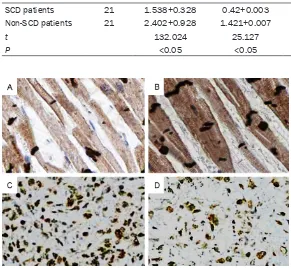
S and OD value of CX43 in the left ventricle of SCD patients showed a significant decrease as compared with that of the non-SCD patients (P<0.05) (Table 3; Figure 2A and 2B); however, the S and OD value of SOCS-1 in the left ven- tricle of SCD patients were significantly increa-sed as compared with that of the non-SCD pa- tients (P<0.05) (Table 3; Figure 2C and 2D).
Discussion
The S and OD value of CX43 mRNA expression were significantly lower in the SCD patients as compared with that of the non-SCD patients (P<0.05). This indicated the downregulation of CX43 in SCD. Gap junction (GJ) channel plays an important role in function execution in car-diac cells by transmitting the electric coupling signals and intercellular chemical/metabolic signals. As a key component of the GJ channel, CX43 is highly expressed in the ventricular myo-cytes [8]. Severs NJ [9] and Tribulova N et al. [10] pointed out that the expression, localiza-tion and phosphorylalocaliza-tion of CX43 in the ven-tricular myocytes all had an impact on GJ chan-nel in the myocytes, thus contributing to car- diac diseases. Moreover, Rhett JM et al. [11] showed that CX43 was involved in the patho-genesis of cardiac diseases through GJ chan-nel, but also facilitated the onset through he- michannels.
Deficiency of blood and oxygen supply to the myocytes is among the major cases of myocyte
Table 1. Primer design
Gene Primer sequence (5’-3’) Length (bp) SOCS-1 F: AGCCTTTCTCCGGCCCTA 243
R: GTTCAGCCTCAGTGGACACA
CX43 F: GCAGCCATTGGCGTTGATAG 380 R: ATCGCCAAGGTCTGTTCTGG
β-actin F: AAGTACTCCGTGTGGATCGG 615 R: TCAAGTTGGGGGACAAAAAG
Table 2. Comparison of relative mRNA expression of CX43 and SOCS-1 between the two groups
Groups N/case CX43 SOCS-1
SCD patients 21 0.395+0.014 0.892+0.103 Non-SCD patients 21 0.823+0.021 0.429+0.037
t --- 19.336 18.267
P --- <0.05 <0.05
Statistical analysis
SPSS 19.0 was used for statistical analy-sis. The mRNA and protein expressions of CX43 and SOCS-1 were compared betw- een the two groups by using paired t-test. P<0.05 indicated significant difference.
Results
injury. Wang et al. [12] showed that myocyte injury was associated with a downregulation of CX43 and a change in the distribution pat-tern of CX43 in the myocytes. CX43 is an impor-tant molecule associated with myocyte injury. Saez JC et al. [13] demonstrated the molecu- lar mechanism involving CX43 in the mediation of myocyte injury. As CX43 is downregulated, there will be a considerable influx of Na+ and
Ca2+ into the myocytes, leading to a high loss
of intracellular metabolites and hence ischemic injury caused by ATP leakage. We deduce that
[image:3.612.89.521.70.159.2] [image:3.612.91.521.227.303.2]nized C-terminal motif [14]. SOCS-1 is an im- portant SOCS protein and is generated by the induction of IL-6, IL-4, IL-3 and (IFN)-γ [15]. Be-sides, SOCS-1 plays a negative feedback regu-lation role in the cytokines through the inter- action with the JAK signaling pathway. Clinical studies [16] have shown that IL-6 expressed by the myocytes can activate the JAK/STAT signal-ing pathway and MAPK signalsignal-ing pathway by binding with the receptors. The pathogenesis of myocardial hypertrophy [17-19] involves the working of JAK/STAT pathway, MAPK pathway Figure 1. MRNA expression of CX43 and SOCS-1 and in the two groups. 1 stands non-SCD patients group; 2 stands SCD patients group.
Table 3. S values and OD values of CX43 and SOCS-1 in the left ventricle in the two groups
Groups N/case CX43 SOCS-1
S OD S OD
SCD patients 21 1.538+0.328 0.42+0.003 2.231+0.515 1.129+0.002
Non-SCD patients 21 2.402+0.928 1.421+0.007 1.369+0.257 0.201+0.005
t 132.024 25.127 105.371 15.664
P <0.05 <0.05 <0.05 <0.05
Figure 2. Immunohistochemical staining of CX43 and SOCS-1 in the cardiac tissues (400 ×). A: CX43 expression in the cardiac tissues of the SCD pa-tients; B: CX43 expression in the cardiac tissues of the non-SCD papa-tients; C: SOCS-1 expression in the cardiac tissues of the SCD patients; D: SOCS-1 expression in the cardiac tissues of the non-SCD patients.
CX43 is involved in the patho-genesis of cardiac diseases by being part of the GJ chan-nels and hemichanchan-nels. Along with myocyte injury in car- diac disease, CX43 is usually downregulated in the cytes, thus aggravating myo-cyte injury.
[image:3.612.88.379.246.514.2]unrecog-and CaN pathway, unrecog-and myocardial hypertrophy may further contribute to SCD. Therefore SOCS-1 participates in the pathogenesis of cardiac diseases by interacting with the JAK/STAT sig-naling pathway and its regulation of the cyto-kines. According to the study of Ottani A et al. [20], the JAK/STAT signaling pathway fulfills a crucial role in the early stage and second win-dow of protection by ischemic preconditioning of myocytes. The upregulation of SOCS-1 may inhibit the expression of various cytokines in the myocytes, thus contribute to myocardial hypertrophy via the JAK/STAT signaling path-way and MAPK signaling pathpath-way. Moreover, by inhibiting the JAK/STAT signaling pathway, up- regulation of SOCS-1 can weaken the protec-tive effect against the ischemic injury of the myocytes, which further increases the risk of SCD.
SOCS-1 can regulate the expression of cyto-kines expressed by the myocytes and partici-pate in the pathogenesis of myocardial hyper-trophy via the JAK/STAT signaling pathway and MAPK signaling pathway. Its upregulation is one of the major risk factors of SCD.
Conclusions
Abnormal expression of SOCS-1 and CX43 in cardiac tissues is closely related to SCD. Down- regulation of CX43 will aggravate myocyte inju-ry, thus increasing the risk of SCD. In contrast, upregulation of SOCS-1 will inhibit the expres-sion of cytokines and further aggravate myo-cyte injury via the JAK/STAT signaling pathway and MAPK signaling pathway, thus contributing to SCD. Therefore, downregulation of CX43 and upregulation of SOCS-1 are the risk factors of SCD.
Disclosure of conflict of interest
None.
Address correspondence to: Dr. Suilong Zhang, Department of Geriatric Cardiovascular Diseases,
Second Affiliated Hospital of Xi’an Jiaotong
Uni-versity, 157 Xiwu Road and Beida Street, Xi’an 710004, Shaanxi, China. Tel: +86-29-87679522; E-mail: zhangslsahxju@163.com
References
[1] Kuch M, Janiszewski M, Mamcarz A,
Cudnoch-Jedrzejewska A, Dłuzniewski M. Major adverse
cardiac event predictors in survivors of myo-cardial infarction with asymptomatic left ven-tricular dysfunction or chronic heart failure. Med Sci Monit 2009; 15: PH40-48.
[2] Fan G, Zhang L. [Sudden cardiac death: prog-ress in epidemiological research]. Zhonghua Liu Xing Bing Xue Za Zhi 2015; 36: 87-89. [3] John RM, Tedrow UB, Koplan BA, Albert CM,
Epstein LM, Sweeney MO, Miller AL, Michaud GF, Stevenson WG. Ventricular arrhythmias and sudden cardiac death. Lancet 2012; 380: 1520-1529.
[4] Lam CS, Anand I, Zhang S, Shimizu W, Narasi- mhan C, Park SW, Yu CM, Ngarmukos T, Omar R, Reyes EB, Siswanto B, Ling LH, Richards AM. Asian sudden cardiac death in heart fail-ure (ASIAN-HF) registry. Eur J Heart Fail 2013; 15: 928-936.
[5] Tagami-Nagata N, Serada S, Fujimoto M, Tane- mura A, Nakatsuka R, Ohkawara T, Murota H, Kishimoto T, Katayama I, Naka T. Suppressor
of cytokine signalling-1 induces significant pre -clinical antitumor effect in malignant melano-ma cells. Exp Dermelano-matol 2015; 24: 864-871. [6] Forster T, Rausch V, Zhang Y, Isayev O, Heil-
mann K, Schoensiegel F, Liu L, Nessling M, Richter K, Labsch S, Nwaeburu CC, Mattern J, Gladkich J, Giese N, Werner J, Schemmer P, Gross W, Gebhard MM, Gerhauser C, Schaefer M, Herr I. Sulforaphane counteracts aggres-siveness of pancreatic cancer driven by dys-regulated Cx43-mediated gap junctional inter-cellular communication. Oncotarget 2014; 5: 1621-1634.
[7] Solan JL, Lampe PD. Specific Cx43 phosphory -lation events regulate gap junction turnover in vivo. FEBS Lett 2014; 588: 1423-1429. [8] de Wit C, Griffith TM. Connexins and gap junc
-tions in the EDHF phenomenon and conduct-
ed vasomotor responses. Pflugers Arch 2010;
459: 897-914.
[9] Severs NJ, Bruce AF, Dupont E, Rothery S. Re- modelling of gap junctions and connexin ex-pression in diseased myocardium. Cardiovasc Res 2008; 80: 9-19.
[10] Tribulova N, Seki S, Radosinska J, Kaplan P, Babusikova E, Knezl V, Mochizuki S. Myocardial Ca2+ handling and cell-to-cell coupling, key factors in prevention of sudden cardiac death. Can J Physiol Pharmacol 2009; 87: 1120-1129.
[11] Rhett JM, Jourdan J, Gourdie RG. Connexin 43 connexon to gap junction transition is regulat-ed by zonula occludens-1. Mol Biol Cell 2011; 22: 1516-1528.
ex-pression in rat hearts. Asian Pac J Trop Med 2015; 8: 658-663.
[13] Sáez JC, Schalper KA, Retamal MA, Orellana JA, Shoji KF, Bennett MV. Cell membrane per-meabilization via connexin hemichannels in living and dying cells. Exp Cell Res 2010; 316: 2377-2389.
[14] Feng X, Tang H, Leng J, Jiang Q. Suppressors of cytokine signaling (SOCS) and type 2 dia- betes. Mol Biol Rep 2014; 41: 2265-2274. [15] Galic S, Sachithanandan N, Kay TW, Steinberg
GR. Suppressor of cytokine signalling (SOCS)
proteins as guardians of inflammatory re -sponses critical for regulating insulin sensitivi-ty. Biochem J 2014; 461: 177-188.
[16] Čokić VP, Mitrović-Ajtić O, Beleslin-Čokić BB, Marković D, Buač M, Diklić M, Kraguljac-Kurtović N, Damjanović S, Milenković P, Gotić M, Raj PK. Proinflammatory cytokine IL-6 and
JAK-STAT signaling pathway in
myeloprolifera-tive neoplasms. Mediators Inflamm 2015;
2015: 453020.
[17] Liu J, Shen Q, Wu Y. Simvastatin prevents car-diac hypertrophy in vitro and in vivo via JAK/ STAT pathway. Life Sci 2008; 82: 991-996. [18] Streicher JM, Ren S, Herschman H, Wang Y.
MAPK-activated protein kinase-2 in cardiac hy-pertrophy and cyclooxygenase-2 regulation in heart. Circ Res 2010; 106: 1434-1443. [19] Jeong D, Kim JM, Cha H, Oh JG, Park J, Yun
SH, Ju ES, Jeon ES, Hajjar RJ, Park WJ. PICOT attenuates cardiac hypertrophy by disrupting calcineurin-NFAT signaling. Circ Res 2008; 102: 711-719.