Original Article
MAP4 and MAP6 expression in breast cancer cell lines
Eduardo Gómez-Conde
1,2, María Alicia Díaz-Orea
2, Aarón Pimentel-Morales
2, María Elena Cárdenas-Perea
2,
Ana Luisa Galicia-Zamalloa
2, Tayde Guerrero-González
3, Juan Antonio González-Barrios
4, Modesto
Gómez-López
5, Eleazar Lara-Padilla
51División de Investigación en Salud, Unidad Médica de Alta Especialidad (UMAE), Hospital de Especialidades, Centro Médico Nacional General de División “Manuel Ávila Camacho”, Instituto Mexicano del Seguro Social (IMSS), Puebla, México; 2Laboratorio de Investigación en Inmunobiología, Facultad de Medicina, Benemérita Universidad Autónoma de Puebla (BUAP), Puebla, México; 3Servicio de Traumatología y Ortopedia, Hospital Regional 1o de Octubre, Instituto de Seguridad y Servicios Sociales de los Trabajadores del Estado (ISSSTE), Ciudad de México (CDMX), México; 4Laboratorio de Medicina Genómica, Hospital Regional 1 de Octubre, ISSSTE, CDMX, México; 5Laboratorio de Obesidad, Sección de Posgrado e Investigación de la Escuela Superior de Medicina, Instituto Politécnico Nacional (IPN), CDMX, México
Received January 13, 2017; Accepted April 8, 2017; Epub June 1, 2017; Published June 15, 2017
Abstract: Breast Cancer (BCa) remains one of the most prevalent forms of cancer and is the most common cancer found in women around the world. Multiple steps are involved in the process of tumorigenesis and metastasis, but one of the most important is motility and reorganization. In this process, cytoskeletal elements such as actin, microtubules, and other proteins are important. MAP4 and MAP6 are microtubule-associated proteins that have been related with different mechanisms in the cytoskeletal process; we analyzed the gene expression of MAP4 and MAP6 in cultured cell lines MCF-10A, MDA-MB-231, SKBR3, and T47D by real-time PCR and protein interactions with STRING network analysis. We found mRNA expression of MAP4, but not of MAP6. Gene expression of MAP4 was higher in the MDA-MB-231 than in the MCF-10A cell line. With respect to protein interactions, MAP4 were related with different proteins that are involved in the process, such as tumorigenesis, cell cycle progress, apoptosis and autophagy, ubiquitination, platelet activation and vascular development, formation and elongation of filopodia, and the dynamic process of intracellular movements. In conclusion, regulation of MAP4 could be related with different proteins in different important molecular mechanisms in BCa and could comprise an important anticancer drug target.
Keywords: MAP4, MAP6, patients with breast cancer, microtubule
Introduction
MicroTubules (MT) play important roles in
fun-damental cellular processes, such as
chromo-some segregation, intracellular transport, direc-
tional migration, and cell morphogenesis [1].
Microtubule-targeting agents have been used
for treatment of different types of aggressive
cancer [2]. MAP4 and MAP6 are
microtubule-associated proteins [3-6]. MAP4 was
recog-nized as a cytosolic MT-binding protein that is
ubiquitously expressed in non-neural cells and
it possesses an important role in microtubu-
le dynamics [7, 8]. Once it is phosphorylated,
MAP4 dissociates from tubulin, resulting in MT
instability [9, 10]. Thus, MAP4 may play a major
role in the maintenance of vascular integrity
[7]; this could be an important role in
tumori-teins is not yet fully understood, but phenotypic
and cellular analyses of MAP6-null mice
indi-cated that MAP6 proteins are involved in a
number of neuronal functions. MAP6-null mice
present defects in synaptic plasticity and
neu-rotransmission associated with severe
behav-ioral disorders [11, 12]. In our study, we showed
the genetic expression of MAP4 and MAP6 in
Breast Cancer (BCa) cell lines, suggesting that
these proteins may be related in different tumor
mechanisms, in addition to their potential as a
drug target in different tumor types.
Materials and methods
Cell culture
USA). MCF-10A (ATCC
®CRL-10317™) were
cul-tured in DMEM F12 (GIBCO) supplemented with
4.18 µf/ml insulin, 10 µg/ml HEGF, and 0.4 µg/
ml Hydrocortisone. T47D (ATCC
®HTB-133™)
were cultured in RPMI-1640 (GIBCO). MDA-MB-
231 (ATCC
®HTB-26™) was cultured in DMEM
high glucose (Hyclon, Logan, UT, USA). SKBR
(ATCC
®HTB-30™) was cultured in McCoy’s 5a
Modified Medium (Thermo Fisher Scientific). All
cell lines were supplemented with 10% FBS
(GIBCO) and 1% Penicillin/Streptomycin (SIG-
MA). Cells were grown as monolayers under
standard conditions at 37°C in a humidified
atmosphere containing 5% CO
2. The cells were
cultured in BD Falcon 250-ml, 75-cm
2Cell
Culture Flasks for gene expression analysis.
RNA extraction and RT-PCR assays
Total RNA from the BCa cell lines and tissue
human brain was isolated using TRIzol Reagent
according to the manufacturer’s protocol (Life
Technologies, USA). We utilized 0.5 μg of the
total simple RNA for reverse-transcription with
random hexamers for 10 min at 65°C, for 50
min at 35°C, and for 5 min at 75°C in a 20-μl
reaction volume employing Transcriptor First
Strand cDNA Synthesis kit (Roche Diagnostics).
Reactions were performed in an Eppendorf
Mastercycler
®Thermal Cycler (Eppendorf, Mé-
xico). PCRq reactions were carried out utilizing
the Human Universal Probe Library (Roche
Diagnostics). Specific oligonucleotide primers
for MAP4 and MAP6 were originally generat-
ed by employing online assay design softwa-
re (ProbeFinder:
http://www.universal-probeli-brary.com) and the primer sequence for each
gene that is depicted in
Table 1
. The reaction
mixture was prepared according to the
manu-facturer’s instructions (Roche Diagnostics,
GmbH Mannheim, Germany). Amplification was
performed in borosilicate glass capillaries (Ro-
che Diagnostics) with a LightCycler 2.0
instru-ment. Amplification conditions for UPL-based
were calculated using the comparative
param-eter threshold Cycle (Ct) method
and
normal-ized to the endogenous control: 18S rRNA.
Results were calculated as a percentage of the
mean level found in the control sample utilizing
the ΔCT method.
Protein network analysis
STRING network analysis of protein-protein in-
teractions was performed to identify
function-ally linked proteins and to determine the po-
tentially affected biological processes [PMID:
12519996]. The network is presented under
confidence view, whereby stronger associa
-tions are represented by thicker lines or edges
and vice versa, whereas proteins are
represent-ed as nodes. All gene symbols were derivrepresent-ed
from the HUGO Gene Nomenclature Committee
(HGNC) (http://www.genenames.org).
Statistics
The percentage of cells with different
morphol-ogy was determined for observed field and was
plotted. R
esults were expressed as the mean ±
Standard Deviation (SD). Data were analyzed
with one-way ANOVA and the Dunnett test for
multiple comparisons, using SigmaPlot ver.
12.0 software (San Jose, CA, USA), and
differ-ences were considered statistically significant
with P≤0.05.
Results
MAP gene expression in breast cancer and
non-tumor cell lines
[image:2.612.91.396.100.177.2]We analyzed the MAP4 and MAP6 mRNA
tran-script in three cancer cell lines compared with
MCF-10A (non-transformed epithelial cell lines,
derived from human fibrocystic mammary tis
-sue (
Figure 1
). MAP4 (
Figure 1A
) exhibited
mRNA expression in all cell lines. In all cases,
we found differences between MCF-10A and
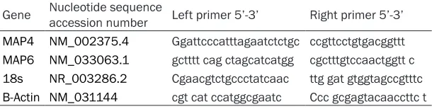
Table 1.
Primer sets for MAP4 and MAP6 used in real-time PCR. The
de-sign was based on ensemble transcript ID of the Human Probe Library
Gene Nucleotide sequence accession number Left primer 5’-3’ Right primer 5’-3’ MAP4 NM_002375.4 Ggattcccatttagaatctctgc ccgttcctgtgacggttt MAP6 NM_033063.1 gctttt cag ctagcatcatgg cgctttgtccaactggtt c 18s NR_003286.2 Cgaacgtctgccctatcaac ttg gat gtggtagccgtttc B-Actin NM_031144 cgt cat ccatggcgaatc Ccc gcgagtacaaccttc tMDA-MB-231 cell lines (P<0.01), but did not
find MAP6 mRNA expression in MCF-10A or in
BCa cell lines (
Figure 1B
).
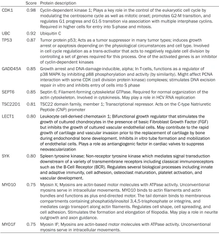
[image:3.612.92.514.74.263.2]Protein network analysis
Figure 2
illustrates the interaction between 10
identified proteins and the additional interac
-tions related with MAP4 proteins. We found
that MAP4 protein was related with proteins
that are involved in ubiquitination, modulation
of centrosome cycle, apoptosis, cytokinesis,
angiogenesis, cell adhesion, osteoclast
matu-ration, platelet activation, and vascular
devel-opment and elongation of filopodia, and the
dynamical process of intracellular movements
(
Table 2
).
Discussion
MAP4 is primarily recognized as a cytosolic
MT-binding protein that is ubiquitously expres-
[image:3.612.94.522.322.529.2]Figure 1. The relative expression level of genes MAP4 (A) and MAP6 (B) was determined after normalization against the 18S internal control for each simple. Data represent the mean ± Standard Deviation (SD). *P<0.01. Human Brain was used as positive control.
sed in non-neural cells and that is possesses
an important role in microtubule dynamics [7].
Therefore, it has been mentioned that MAP4
may play a major role in the maintenance of
vascular integrity, and that it could be involved
in the modulation of inflammation [7] and in
invasion through microtubule dynamics. In
bladder cancer, it has been described that
overexpression of MAP4 appears to be
posi-tively correlated with tumor stage and degree
of malignancy [13]. With respect to with STRING
protein analysis, MAP4 were related with the
Ubiquitin C protein. Ubiquitin Carboxyl-terminal
[image:4.612.89.521.85.545.2]Hydrolase L1 (UCH-L1) is an abundant neuronal
protein; it was overexpressed in wide-type tu-
mors and was related with invasion in some
solid tumors, including BCa [14-16]; Goto et al.
demonstrated that Ubiquitin C-terminal Hydro-
lase-L1 (UCHL1) abrogates von
Hippel-Lindau-mediated ubiquitination of HIF-1α, the regula
-tory subunit of HIF-1, and consequently
pro-motes metastasis, and that overexpression of
UCHL1 increases tumor metastases in an
HIF-1-dependent manner in murine models of
pul-monary tumor. Meanwhile, blockade of the
UCHL1-HIF-1 axis could suppress the formation
Table 2.
MAP4 interaction protein (http://string-db.org/cgi/network.pl?taskId=IAcDMtF1EKwX)
Score Protein description
CDK1 0.98 Cyclin-dependent kinase 1; Plays a key role in the control of the eukaryotic cell cycle by modulating the centrosome cycle as well as mitotic onset; promotes G2-M transition, and regulates G1 progress and G1-S transition via association with multiple interphase cyclins. Required in higher cells for entry into S-phase and mitosis.
UBC 0.92 Ubiquitin C
TP53 0.87 Tumor protein p53; Acts as a tumor suppressor in many tumor types; induces growth arrest or apoptosis depending on the physiological circumstances and cell type. Involved in cell cycle regulation as a trans-activator that acts to negatively regulate cell division by controlling a set of genes required for this process. One of the activated genes is an inhibitor of cyclin-dependent kinases
GADD45A 0.85 Growth arrest and DNA-damage-inducible, alpha; In T-cells, functions as a regulator of p38 MAPK by inhibiting p88 phosphorylation and activity (by similarity). Might affect PCNA interaction with some CDK (cell division protein kinase) complexes; stimulates DNA excision repair in vitro and inhibits entry of cells into S phase
SEPT6 0.85 Septin 6; Filament-forming cytoskeletal GTPase. Required for normal organization of the actin cytoskeleton. Involved in cytokinesis. May play a role in HCV RNA replication TSC22D1 0.81 TSC22 domain family, member 1; Transcriptional repressor. Acts on the C-type Natriuretic
Peptide (CNP) promoter
LECT1 0.80 Leukocyte cell-derived chemotaxin 1; Bifunctional growth regulator that stimulates the growth of cultured chondrocytes in the presence of basic Fibroblast Growth Factor (FGF) but inhibits the growth of cultured vascular endothelial cells. May contribute to the rapid growth of cartilage and vascular invasion prior to the replacement of cartilage by bone during endochondral bone development. Inhibits in vitro tube formation and mobilization of endothelial cells. Plays a role as antiangiogenic factor in cardiac valves to suppress neovascularization
SYK 0.80 Spleen tyrosine kinase; Non-receptor tyrosine kinase which mediates signal transduction downstream of a variety of transmembrane receptors including classical immunoreceptors such as the B-Cell Receptor (BCR). Regulates several biological processes including innate and adaptive immunity, cell adhesion, osteoclast maturation, platelet activation, and vascular development.
MYO10 0.75 Myosin X; Myosins are actin-based motor molecules with ATPase activity. Unconventional myosins serve in intracellular movements. MYO10 binds to actin filaments and actin bundles and functions as plus end-directed motor. The tail domain binds to membranous compartments containing phosphatidylinositol 3,4,5-trisphosphate or integrins, and mediates cargo transport along actin filaments. Regulates cell shape, cell spreading, and cell adhesion. Stimulates the formation and elongation of filopodia. May play a role in neurite outgrowth and axon guidance.
of metastatic tumors. Goto et al. also found
that the expression levels of UCHL1 correlate
positively with HIF-1α, and that they were relat
-ed with poor prognosis of patients with breast
and lung cancer [15]. In addition, these authors’
findings are similar to those reported by others.
In colorectal and pancreatic tumors,
overex-pression of UCHL1 was associated with higher
incidence of tumor recurrence and shorter
sur-vival time [17, 18]. Thus, UCHL1 could be a
can-cer drug target in wide-type tumors [19-21].
Another important protein related with MAP4
was Tumor Protein p53 (TP53). TP53, the
guardian of the genome, possesses a tumor
suppressor function through the maintenance
of genetic integrity, cell-cycle machinery,
apop-tosis, and DNA repair [22, 23]. In order to check
genetic errors, p53 accumulates in the nucleus
in response to cellular stress, such as DNA
damage, hypoxia, and nucleotide deprivation
[23, 24]. Once p53 is transported into the
nucleus, it transactivates its target genes,
involved either in cell-cycle arrest or in
apopto-sis [25]. Some studies have demonstrated that
G-actin is guides through p53 transport toward
the nucleus; the p53 cargo reaches the
peri-nuclear region and interacts importantly in
receptors [23, 26]. Microtubules have been
tar-geting several drugs that are important in the
treatment of a wide variety of tumor types [27,
28], but proteins associated with microtubules
are acquiring importance within this context in
different cancer types [29, 30].
Conclusions
In conclusion, this study shows the gene
expression of MAP4, but not of MAP6, in BCa
cell lines and its in silico relationship among
proteins involved in different cancer processes.
However, further studies are needed to clarify
our results.
Disclosure of conflict of interest
None.
Address correspondence to: Eleazar Lara-Padilla, Escuela Superior de Medicina, Instituto Politécnico Nacional, Calles Plan de San Luis y Salvador Díaz Mirón s/n, Col. Casco de Santo Tomás, 11340, CDMX, México. E-mail: [email protected]
References
[1] Toya M, Takeichi M. Organization of non-cen-trosomal microtubules in epithelial cells. Cell Struct Funct 2016; 41: 127-135.
[2] Taranejoo S, Janmaleki M, Pachenari M, Seyedpour SM, Chandrasekaran R, Cheng W, Hourigan K. Dual effect of F-actin targeted car-rier combined with antimitotic drug on aggres-sive colorectal cancer cytoskeleton: allying dis-similar cell cytoskeleton disrupting mechanis- ms. Int J Pharm 2016; 513: 464-472.
[3] Riederer BM. Microtubule-associated protein 1B, a growth-associated and phosphorylated scaffold protein. Brain Res Bull 2007; 71: 541-558.
[4] Ankam S, Lim CK, Yim EK. Actomiosin contrac-tility plays a role in MAP2 expression during naotopography-directed neuronal differentia-tion of human embryonic stem cells. Biomate-rials 2015; 47: 20-28.
[5] Holmfeldt P, Brattsand G, Gullberg M. MAP4 counteracts microtubule catastrophe promo-tion but not tubulsequestering activity in in-tact cells. Curr Biol 2002; 12: 1034-1039. [6] Shimizu H, Iwayama Y, Yamada K, Toyota T,
Mi-nabe Y, Nakamura K, Nakajima M, Hattori E, Mori N, Osumi N, Yoshikawa T. Genetic and ex-pression analyses of the STOP (MAP6) gene in schizophrenia. Schizophr Res 2006; 84: 244-252.
[7] Li L, Hu J, He T, Zhang Q, Yang X, Lan X, Zhang D, Mei H, Chen B, Huang Y. P38/MAPK contrib-utes to endothelial barrier dysfunction via MAP4 phosphorylation-dependent microtu-bule disassembly in inflammation-induced acute lung injury. Sci Rep 2015; 5: 8895. [8] Illenberger S, Drewes G, Trinczek B, Biernat J,
Meyer HE, Olmsted JB, Mandelkow EM, Man-delkow E. Phosphorylation of microtubule-as-sociated proteins MAP2 and MAP4 by the pro-tein kinase p110mark. Phosphorylation sites and regulation of microtubule dynamics. J Biol Chem 1996; 271: 10834-10843.
[9] Ebneth A, Drewes G, Mandelkow EM, Mandel-kow E. Phosphorylation of MAP2c and MAP4 by MARK kinases leads to the destabilization of microtubules in cells. Cell Motil Cytoskeleton 1999; 44: 209-224.
[10] Hu J, Chu Z, Han J, Zhang Q, Zhang D, Dang Y, Ren J, Chan HC, Zhang J, Huang Y. Phosphory-lation-dependent mitochondrial translocation of MAP4 is an early step in hypoxia-induced apoptosis in cardiomyocytes. Cell Death Dis 2014; 5: 1424.
[11] Lefèvre J, Savarin P, Gans P, Hamon L, Clément MJ, David MO, Bosc C, Andrieux A, Curmi PA. Structural basis for the association of map6 protein with microtubules and its regulation by calmodulin. J Biol Chem 2013; 288: 24910-24922.
Job D. The suppression of brain cold-stable mi-crotubules in mice induces synaptic defects associated with neuroleptic-sensitive behav-ioral disorders. Genes Dev 2002; 16: 2350-2364.
[13] Ou Y, Zheng X, Gao Y, Shu M, Leng T, Li Y, Yin W, Zhu W, Huang Y, Zhou Y, Tang J, Qiu P, Yan G, Hu J, Ruan H, Hu H. Activation of cyclic AMP/ PKA pathway inhibits bladder cancer cell inva-sion by targeting MAP4-dependent microtu-bule dynamics. Urol Oncol 2014; 32: 47, e21-28.
[14] Schröder C, Milde-Langosch K, Gebauer F, Schmid K, Mueller V, Wirtz RM, Meyer-Schwesinger C, Schlüter H, Sauter G, Schum-acher U. Prognostic relevance of ubiquitin C-terminal hydrolase L1 (UCH-L1) mRNA and protein expression in breast cancer patients. J Cancer Res Clin Oncol 2013; 139: 1745-1755. [15] Goto Y, Zeng L, Yeom CJ, Zhu Y, Morinibu A,
Shi-nomiya K, Kobayashi M, Hirota K, Itasaka S, Yoshimura M, Tanimoto K, Torii M, Sowa T, Menju T, Sonobe M, Kakeya H, Toi M, Date H, Hammond EM, Hiraoka M, Harada H. UCHL1 provides diagnostic and antimetastatic strate-gies due to its deubiquitinating effect on HIF-1α. Nat Commun 2015; 6: 6153.
[16] Hurst-Kennedy J, Chin LS, Li L. Ubiquitin C-ter-minal hydrolase l1 in tumorigenesis. Biochem Res Int 2012; 2012: 123706.
[17] Akishima-Fukasawa Y, Ino Y, Nakanishi Y, Miu-ra A, Moriya Y, Kondo T, Kanai Y, Hirohashi S. Significance of PGP9.5 expression in cancer-associated fibroblasts for prognosis of colorec-tal carcinoma. Am J Clin Pathol 2010; 134: 71-79.
[18] Tezel E, Hibi K, Nagasaka T, Nakao A. PGP9.5 as a prognostic factor in pancreatic cancer. Clin Cancer Res 2000; 6: 4764-4767.
[19] Miyoshi Y, Nakayama S, Torikoshi Y, Tanaka S, Ishihara H, Taguchi T, Tamaki Y, Noguchi S. High expression of ubiquitin caboxy-terminal hydrolase-L1 and -L3 mRNA predicts early re-currence in patients with invasive breast can-cer. Cancer Sci 2006; 97: 523-529.
[20] D’Arcy P, Brnjic S, Olofsson MH, Fryknäs M, Lindsten K, De Cesare M, Perego P, Sadeghi B, Hassan M, Larsson R, Linder S. Inhibition of proteasome deubiquitinating activity as a new cancer therapy. Nat Med 2011; 17: 1636-1640.
[21] Qu X, Wang Y. Effect of liposomal transfection of UCH-L1 siRNA on proliferation and apopto-sis of lung cancer cell line H157. Zhongguo Fei Ai Za Zhi 2010; 13: 292-296.
[22] Lane DP. Cancer. p53, guardian of the ge-nome. Nature 1992; 358: 15-16.
[23] Saha T, Guha D, Manna A, Panda AK, Bhat J, Chatterjee S, Sa G. G-actin guides p53 nuclear transport: potential contribution of monomeric actin in altered localization of mutant p53. Sci Rep 2016; 6: 32626.
[24] Meek DW. The p53 response to DNA damage. DNA Repair (Amst) 2004; 3: 1049-1056. [25] Beckerman R, Prives C. Transcriptional
regula-tion by p53. Cold Spring Harb Perspect Biol 2010; 2: 000935.
[26] Liang SH, Clarke MF. A bipartite nuclear local-ization signal is required for p53 nuclear im-port regulated by a carboxyl-terminaldomain. J Biol Chem 1999; 274: 32699-32703.
[27] Florian S, Mitchison TJ. Anti-microtubule drugs. Methods Mol Biol 2016; 1413: 403-421. [28] Nepali K, Ojha R, Lee HY, Liou JP. Early
investi-gational tubulin inhibitors as novel cancer therapeutics. Expert Opin Investig Drugs 2016; 25: 917-936.
[29] Shao C, Duan C, Wang J, Luan S, Gao Y, Jin D, Wang D, Li Y, Xu L. Expression of microtubule-associated protein TPX2 in human gastric car-cinoma and its prognostic significance. Cancer Cell Int 2016; 16: 79.