Original Article
Synergistic effects of AKAP95, Cyclin D1, Cyclin E1,
and Cx43 in the development of rectal cancer
Fengjie Qi1*, Yangyang Yuan2*, Xuehong Zhi2, Qian Huang2, Yuexin Chen2, Wenxin Zhuang2, Dengcheng Zhang2, Bogang Teng2, Xiangyu Kong3, Yongxing Zhang2
1Department of Pathology of Liaoning Medical College, Jinzhou, China; 2State Key Laboratory of Molecular
Vaccin-ology and Molecular Diagnostics, School of Public Health, Xiamen University, Xiamen 361102, Fujian, PR China;
3Affiliated Zhongshan Hospital of Dalian University, Dalian 116001, Liaoning, PR China. *Equal contributors.
Received November 25, 2014; Accepted January 28, 2015; Epub February 1, 2015; Published February 15, 2015
Abstract: Objective: To explore the expression of A-kinase anchor protein 95 (AKAP95), Cyclin D1, Cyclin E1, and Connexin43 (Cx43) in rectal cancer tissues and assess the associations between each of the proteins and patho-logical parameters, as well as their inter-relationships. Methods: AKAP95, Cyclin D1, Cyclin E1, and Cx43 protein expression rates were evaluated by immunohistochemistry in 50 rectal cancer specimens and 16 pericarcinoma tissues. Results: The positive rates of AKAP95, Cyclin E1, and Cyclin D1 proteins were 54.00 vs. 18.75%, 62.00 vs. 6.25%, and 72.00 vs. 31.25% in rectal cancer specimens and pericarcinoma tissues, respectively, representing statistically significant differences (P < 0.05). The positive rate of Cx43 protein expression in rectal cancer tissues was 44.00% and 62.50% in pericarcinoma tissues, and the difference between them was not significant (P > 0.05). No significant associations were found between protein expression of AKAP95, Cyclin E1, Cyclin D1, and Cx43, and the degree of differentiation, histological type, and lymph node metastasis of rectal cancer (P > 0.05). However, significant correlations were obtained between the expression rates of AKAP95 and Cyclin E1, Cyclin E1 and Cyclin D1, Cyclin E1 and Cx43 protein, and Cyclin D1 and Cx43, respectively (P < 0.05). Conclusion: AKAP95, Cyclin E1, and Cyclin D1 protein expression rates were significantly higher in rectal cancer tissues compared with pericarci -noma samples, suggesting an association between these proteins and the development and progression of rectal cancer. In addition, the significant correlations between the proteins (AKAP95 and Cyclin E1, Cyclin E1 and Cyclin D1, Cyclin E1 and Cx43 protein, and Cyclin D1 and Cx43) indicate the possible synergistic effects of these factors in the development and progression of rectal cancer.
Keywords: Rectal cancer, AKAP95, Cyclin E1, Cyclin D1, Cx43
Introduction
Cell cycle signaling pathway assessment has provided new insights for understanding the mechanisms involved in the development and progression of rectal cancer, a common diges-tive tract malignancy. AKAP95 was shown to anchor to the RII subunit of PKA; the anchored PKA catalyzes the phosphorylation of target proteins, thus ensuring signal transduction in the cAMP pathway [1, 2]. Cyclin D and Cyclin E are known to promote mitosis of mammalian cells in the G1 phase. Previous studies have demonstrated that the Cyclin D1 protein is over-expressed in tissues from rectal and esophageal cancers [3, 4]; meanwhile, Cyclin E1 is also over-expressed in liver cancer and serous cystadenocarcinoma of the ovary [5, 6].
AKAP95, Cyclin D1, Cyclin E1, and Cx43 in rectal cancer
Data assessment
Brown-yellow staining was considered as posi-tive protein expression. For each slice, 10
dif-ferent visual fields were randomly chosen under
microscope (BA310 Digital, Motic China Group Co., Ltd), and 200 rectal cancer cells were
counted in each visual field. The percentage of positive cells in the field was used for result
determination. “-” indicated no brown-yellow
staining to <10%; “+-” indicated ≥10% but <25%; “+” indicated ≥25% but <50%; “++” indi
-cated ≥50% but <75%; and “+++” indi-cated ≥75%. “-” and “+-” were considered as negative
protein expression, while “+”, “++”, and “+++” were considered to be positive protein expres-sion [17].
Statistical analysis
The SPSS13.0 software (SPSS Inc., Chicago, IL, USA) was used for statistical analysis. Chi-square test was used for ratio comparisons. Spearman rank correlation was used to assess the associations among the proteins expres-sion. P < 0.05 was considered statistically
significant.
Results
AKAP95, Cyclin E1, Cyclin D1, and Cx43 pro-tein expression in rectal cancer and pericarci-noma tissues
The positive rates of AKAP95 expression in rec-tal cancer tissues and pericarcinoma speci-mens were 54.00 and 18.75%, respectively (Table 1). The AKAP95 protein in pericarcinoma these 4 proteins in rectal cancer tissues and
their associations with pathological parame-ters, as well as their inter-relationships. Materials and methods
Tissue collection
Fifty rectal cancer tissues were obtained from rectal cancer patients that received surgical
resection in the First Affiliated Hospital of
Liaoning Medical University. In all patients,
diagnosis was confirmed by pathological exami -nation. The patients were 64±10 (ranging from 39 to 83) years old, including 32 males and 18 females. Forty-four patients had tubular or pap-illary adenocarcinoma, while 4 presented with mucinous adenocarcinoma and 2 had signet-ring cell carcinoma. Cancer cells were highly differentiated in 4 patients, moderately differ-entiated in 42 individuals, and poorly differenti-ated in 4 subjects. Twenty-six patients had lymph node metastasis, 18 were free from lymph node metastasis, and lymph node metastasis status was unclear for the remain-ing 6 individuals. In addition, pericarcinoma tis-sues were obtained in normal rectal tistis-sues at least 3 cm away from cancerous tissues in 16 of the 50 patients. Pathological examination was also performed with pericarcinoma tissues
to confirm the absence of cancer cells.
Reagents and methods
All specimens were fixed with 10% neutral for
-malin, embedded with paraffin, and sectioned
at 4 µm. The S-P immunohistochemical method
[image:2.612.91.345.97.227.2]was performed to evaluate the ex- pression of various proteins. In brief, high pressure antigen retrieval was carried out with citrate buffer (pH6.0) for 1.5 min, followed by DAB stai- ning; cell nuclei were stained with hematoxylin. The SP kit and DAB sub-strate were purchased from Maixin biotechnology co., LTD (Fuzhou, Fujian, China); mouse anti-human AKAP95, Cyclin D1, and Cx43 monoclonal an- tibodies were purchased from Santa Cruz (Dallas, Texas, USA); mouse an- ti-human Cyclin E1 antibody was pur-chased from Epitomics (Burlingame, California, USA). PBS was used in- stead of primary antibody in negative controls.
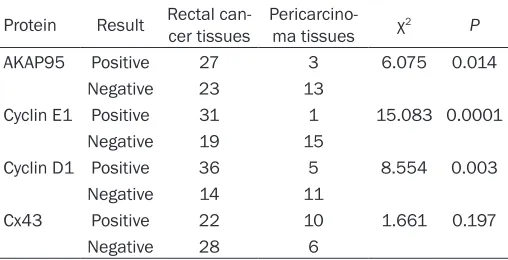
Table 1. AKAP95, Cyclin E1, Cyclin D1, and Cx43 protein expression in rectal cancer tissues
Protein Result Rectal can-cer tissues Pericarcino-ma tissues χ2 P
AKAP95 Positive 27 3 6.075 0.014
Negative 23 13
Cyclin E1 Positive 31 1 15.083 0.0001
Negative 19 15
Cyclin D1 Positive 36 5 8.554 0.003
Negative 14 11
Cx43 Positive 22 10 1.661 0.197
Negative 28 6
The positive rates for AKAP95, Cyclin E1, and Cyclin D1 protein expression
were significantly higher in rectal cancer tissues compared with pericar -cinoma specimens; the positive rate of Cx43 protein expression was not
AKAP95, Cyclin D1, Cyclin E1, and Cx43 in rectal cancer
and rectal cancer tissues was mainly located in the cell nucleus, while a marginal portion was located in the cytoplasm (Figure 1). The positive rate of Cyclin E1 expression was 62.00% in rectal cancer tissues (31/50), but only 6.25% in pericarcinoma specimens (1/16). The Cyclin E1 protein in rectal cancer tissues was mainly in the cytoplasm and less repre-sented in the cell nucleus (Figure 2). The posi-tive rates of Cyclin D1 expression were 72.00% (36/50) and 31.25% (5/16) in rectal cancer tis-sues and pericarcinoma tistis-sues, respectively. The Cyclin D1 protein in rectal cancer and
peri-carcinoma tissues was mainly confined to the
cytoplasm (Figure 3). The positive rates of AKAP95, Cyclin E1, and Cyclin D1 protein
expression were all significantly higher in can -cer tissues compared with pericarcinoma spec-imens (P < 0.01 or 0.05); in contrast, Cx43 pro-tein expression was lower in cancer tissues compared with pericarcinoma samples, alth-
ough the difference was not statistically signifi -cant (P > 0.05); the Cx43 protein was also mainly located in the cytoplasm of rectal can-cer cells (Figure 4).
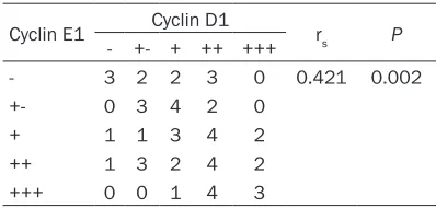
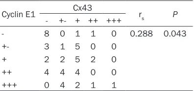
Correlations among the expression of the AKAP95, Cyclin E1, Cyclin D1, and Cx43 pro-teins in rectal cancer tissues
As shown in Tables 2-7, significant
correla-tions were obtained between the protein expression rates of AKAP95 and Cyclin E1 (Table 2), Cyclin E1 and Cyclin D1 (Table 3), Cyclin E1 and Cx43 (Table 4), and Cyclin D1 and Cx43 (Table 5) (P < 0.05) in rectal cancer
speci-mens; no significant correlation was found
between the expression rates of AKAP95 and Cyclin D1 (Table 6) as well as AKAP95 and Cx43 (Table 7) (P > 0.05).
Correlations between pathological parameters and protein expression rates of AKAP95, Cyclin E1, Cyclin D1, and Cx43 in rectal cancer tis-sues
No significant association was found
be-tween the protein expression rates of AKAP95, Cyclin E1, Cyclin D1, and Cx43 and the degree of differentiation, histological type, and lymph node metastasis in rectal cancer tissues (P > 0.05).
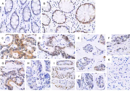
Figure 2. Expression of Cyclin E1 in pericarcinoma and rectal cancer tissues (× 400). A. Low expression of Cyclin E1 in the cytoplasm in pericarcinoma rectal tissues; B. Negative expression in pericarcinoma rectal tissues; C and D. Positive expression in the cytoplasm in poorly and moderately differentiated rectal adenocarcinoma tissues, respec-tively; E. Positive expression in highly differentiated rectal adenocarcinoma tissue; the protein was mainly expressed in the cytoplasm; a marginal proportion was found in the cell nucleus; F. Low expression the cytoplasm in highly differentiated rectal adenocarcinoma tissues; G. Positive expression in the cytoplasm in poorly differentiated rectal mucinous adenocarcinoma tissues; H. Negative expression in rectal signet-ring cell carcinoma tissues.
Discussion
The AKAP95 protein is mainly expressed in the cell nucleus; it participates in signal
transduc-tion, specifically in the cAMP pathway by
[image:5.612.91.519.73.405.2]anchoring the RII subunit of PKA, which phos-phorylates target proteins [1]; in addition, AKAP59 also plays a role in chromosome con-Figure 4. Expression of the Cx43 protein in pericarcinoma and rectal cancer tissues (× 400). A. Positive cytoplasmic expression of Cx43 in pericarcinoma rectal tissues; B. Negative expression in pericarcinoma rectal tissues; C. Low expression in the cytoplasm in poorly differentiated rectal adenocarcinoma tissues; D and E. Negative expression in poorly and moderately differentiated rectal adenocarcinoma tissues, respectively; F. Low expression in the cyto-plasm in poorly differentiated rectal mucinous adenocarcinoma tissues; G. Positive expression in the cytocyto-plasm in moderately differentiated rectal adenocarcinoma tissues; H and I. Negative expression in moderately differentiated rectal adenocarcinoma and signet-ring cell carcinoma tissues.
Table 2. Correlation between the protein expression rates of AKAP95 and Cyclin E1 in rectal cancer tissues
AKAP95 Cyclin E1 rs P
- +- + ++ +++
- 7 2 2 2 1 0.353 0.012
+- 0 2 4 3 0
+ 2 3 4 4 4
++ 1 1 1 2 3
+++ 0 1 0 1 0
[image:5.612.323.522.540.635.2]rs: Spearman’s rank correlation coefficient.
Table 3. Correlation between the protein expression rates of Cyclin E1 and Cyclin D1 in rectal cancer tissues
Cyclin E1 Cyclin D1 rs P
- +- + ++ +++
- 3 2 2 3 0 0.421 0.002
+- 0 3 4 2 0
+ 1 1 3 4 2
++ 1 3 2 4 2
+++ 0 0 1 4 3
[image:5.612.90.288.541.634.2]AKAP95, Cyclin D1, Cyclin E1, and Cx43 in rectal cancer
Table 7. Correlation between the protein ex-pression rates of AKAP95 and Cx43 in rectal cancer tissues
AKAP95 Cx43 rs P
- +- + ++ +++
- 7 2 5 0 0 0.071 0.623
+- 1 3 4 1 0
+ 4 5 6 2 0
++ 4 0 2 1 1
+++ 1 1 0 0 0
rs: Spearman’s rank correlation coefficient.
densation and DNA replication during mitosis, and cell apoptosis [16-18]. Cyclin D1 and Cyclin E1 are two proteins that regulate the G1 to S phase progression in mitotic cells; these two proteins are mainly over-expressed in cancer tissues [4-6]; in addition, over-expression of Cyclin D1 [19] and Cyclin E1 [6] was shown to predict poor prognosis. In a study performed by Arsenijevic [11], the investigators found that the AKAP95 protein could combine to Cyclin D and Cyclin E to form a complex in CHO cells; in addition, AKAP95 competitively combined with Cyclin D3 instead of CKD4, or with Cyclin E1 instead of CKD2, suggesting that the AKAP95 protein could participate in cell cycle regulation by affecting Cyclin D and Cyclin E. In the
pres-ent study, we found a significant correlation
between the expression of AKAP95 and Cyclin E1 but not between AKAP95 and Cyclin D1 expression rates in rectal cancer tissues, which
was inconsistent with our previous findings in
lung cancer tissues (in which AKAP95 and
Cyclin D1 expression rates were significantly
correlated, as well as those of AKAP95 and Cyclin E1 [20]), suggesting that the cell cycle regulatory role of AKAP95 could differ with tis-sue type. Lodén M et al. [21] found that in
estrogen receptor (ER) positive breast cancer tissues, over-expressed Cyclin D1 increased the phosphorylation of the pRB protein, thus elevating cell proliferation; while in ER negative breast cancer tissues, over-expression of Cyclin E was accompanied with a down-regulation of the Cyclin D1 protein, which also increased cell proliferation in the absence of regulatory
effects of pRB [21]. These findings suggested
that Cyclin D1 and Cyclin E promote cell prolif-eration via different mechanisms or through different pathways, which could vary in differ-ent tissues. Harbour et al. [22] found that pRB phosphorylation by Cyclin D is the precondition for the transcription factor E2F to activate Cyclin E transcription. In the present study, the association between the expression rates of Cyclin D1 and Cyclin E1 suggested that these two proteins play synergistic roles in promoting tumor development, providing histological
evi-dence to support Harbour’s findings.
Cx43 is known as a tumor suppressor; howev-er, Cx43 protein expression is decreased in many tumors [8-10]. In our previous work, we found that AKAP95 and Cx43 protein
[image:6.612.325.522.107.202.2]expres-sion rates were significantly associated in lung
Table 5. Correlation between the protein ex-pression rates of Cyclin D1 and Cx43 in rectal cancer tissues
Cyclin D1 Cx43 rs P
- +- + ++ +++
- 4 0 1 0 0 0.481 0.0004
+- 7 2 0 0 0
+ 3 2 7 0 0
++ 2 4 7 3 1
+++ 1 3 2 1 0
[image:6.612.91.289.108.201.2]rs: Spearman’s rank correlation coefficient.
Table 4. Correlation between the protein ex-pression rates of Cyclin E1 and Cx43 in rectal cancer tissues
Cyclin E1 Cx43 rs P
- +- + ++ +++
- 8 0 1 1 0 0.288 0.043
+- 3 1 5 0 0
+ 2 2 5 2 0
++ 4 4 4 0 0
+++ 0 4 2 1 1
rs: Spearman’s rank correlation coefficient.
Table 6. Correlation between the protein expression rates of AKAP95 and Cyclin D1 in rectal cancer tissues
AKAP95 Cyclin D1 rs P
- +- + ++ +++
- 3 2 6 2 1 0.211 0.141
+- 1 2 1 3 2
+ 1 2 4 7 3
++ 0 1 1 5 1
+++ 0 2 0 0 0
[image:6.612.91.288.275.368.2]tissues [23]; cytological experiments in our laboratory also showed the dynamic combina-tion of AKAP95 and Cx43 in the whole cell cycle
(data not shown); however, no significant asso -ciation between AKAP95 and Cx43 protein expression rates was found in rectal cancer tis-sues, suggesting that the mechanisms underly-ing the interactions between AKAP95 and Cx43 that regulate the cell cycle could be different in various tissues.
The PTEN protein is known to reduce Akt activ-ity [24], thus regulating the phosphorylation of the Cx43 protein [25]; in addition, PTEN pro-motes p27kip1 to combine with CyclinE-CDK
2 and form a complex [26]. MAPKs (p38, p42/44, and JNK) were shown to activate Cyclin D1 via
transcription factors, including NF-κB and AP-1
[27]; meanwhile, phosphorylated MAPKs (p38, p42/44, and JNK) increased AP-1 and CREB levels, thus enhancing Cx43 protein synthesis [28]. Therefore, we speculated that Cx43 could regulate the cell cycle through the complex formed by AKAP95 and Cyclin D/E-CDKs; this
was supported by the present findings that Cx43 expression is significantly associated
with that of Cyclin D1 and Cyclin E1.
Of note, no significant correlations were
obtai-ned between the protein expression of AKAP95, Cyclin E1, Cyclin D1, and Cx43, and the degree of differentiation, histological type, and lymph node metastasis in rectal cancer tissues.
Acknowledgements
This work is supported by National Natural Sci- ence Foundation (No.81071927), Fujian Cre- ative Project (No.2012-CXB-25), Graduate Creative Project (No.ZX11A1) and Xiamen University Creative Grant (No.CXB2013024). Disclosure of conflict of interest
None.
Address correspondence to: Dr. Yongxing Zhang, State Key Laboratory of Molecular Vaccinology and Molecular Diagnostics, School of Public Health, Xia- men University, Xiamen 361102, Fujian, PR China. Tel: +86-18959244508; E-mail: [email protected]. cn; Dr. Xiangyu Kong, Affiliated Zhongshan Hospital of Dalian University, Dalian 116001, Liaoning, PR China. E-mail: [email protected]
References
[1] Eide T, Coghlan V, Orstavik S, Holsve C, Solberg R, Skâlhegg BS, Lamb NJ, Langeberg L, Fer-nandez A, Scott JD, Jahnsen T, Taskén K. Mo-lecular cloning, chromosomal localization, and cell cycle-dependent sub cellular distribution of the A-kinase anchoring protein, AKAP95. Exp Cell Res 1998; 238: 305-316.
[2] Wall EA, Zavzavadjian JR, Chang MS, Randha-wa B, Zhu X, Hsueh RC, Liu J, Driver A, Bao XR, Sternweis PC, Simon MI, Fraser ID. Suppres-sion of LPS-induced TNF-alpha production in macrophages by cAMP is mediated by PKA-AKAP95-p105. Sci Signal 2009; 2: ra28. [3] Schwandner O, Bruch HP, Broll R. P21, p27,
cyclin D1, and p53 in rectal cancer:
immuno-histology with prognostic significance. Int J
Colorectal Dis 2002; 17: 11-19.
[4] Nagasawa S, Onda M, Sasajima K, Makino H, Yamashita K, Takubo K, Miyashita M. Cyclin D1 overexpression as a prognostic factor in pa-tients with esophageal carcinoma. J Surg On-col 2001; 78: 208-214.
[5] Farra R, Dapas B, Pozzato G, Scaggiante B, Agostini F, Zennaro C, Grassi M, Rosso N, Giansante C, Fiotti N, Grassi G. Effects of E2F1-cyclin E1-E2 circuit down regulation in hepatocellular carcinoma cells. Dig Liver Dis 2011; 43: 1006-1014.
[6] Guo H, Lu Y, Wang J, Liu X, Keller ET, Liu Q, Zhou Q, Zhang J. Targeting the Notch signaling pathway in cancer therapeutics. Thoracic Can-cer 2014; 5: 473-486.
[7] Moorby C, Patel M. Dual functions for connex-ins: Cx43 regulates growth independently of gap junction formation. Exp Cell Res 2001; 271: 238-248.
[8] Yi ZC, Wang H, Zhang GY, Xia B. Downregula-tion of connexin 43 in nasopharyngeal carci-noma cells is related to promoter methylation. Oral Oncol 2007; 43: 898-904.
[9] Tang B, Peng ZH, Yu PW, Yu G, Qian F.
Expres-sion and significance of Cx43 and E-cadherin
in gastric cancer and metastatic lymph nodes. Med Oncol 2011; 28: 502-508.
[10] Sirnes S, Bruun J, Kolberg M, Kjenseth A, Lind GE, Svindland A, Brech A, Nesbakken A, Lothe RA, Leithe E, Rivedal E. Connexin43 acts as a colorectal cancer tumor suppressor and pre-dicts disease outcome. Int J Cancer 2012; 131: 570-581.
[11] Arsenijevic T, Degraef C, Dumont JE, Roger PP, Pirson I. G1/S Cyclins interact with regulatory subunit of PKA via A-kinase anchoring protein, AKAP95. Cell Cycle 2006; 5: 1217-1222. [12] Lampe PD, Lau AF. The effects of connexin
AKAP95, Cyclin D1, Cyclin E1, and Cx43 in rectal cancer
[13] Yun SP, Ryu JM, Park JH, Kim MO, Lee JH, Han HJ. Prostaglandin E2 maintains mouse ESC undifferentiated state through regulation of connexin31, connexin43 and connexin45 ex-pression: Involvement of glycogen synthase
ki-nase 3β/β-catenin. Biol Cell 2012; 104:
378-396.
[14] Solan JL, Lampe PD. Connexin phosphoryla-tion as a regulatoy event link to gap juncphosphoryla-tion chanel assembly. Biochim Biophys Acta 2005; 1711: 154-163.
[15] Decrock E, De Vuyst E, Vinken M, Van Moor-hem M, Vranckx K, Wang N, Van Laeken L, De Bock M, D’Herde K, Lai CP, Rogiers V, Evans WH, Naus CC, Leybaert L. Connexin43 hemi-channels contribute to the propagation of apoptotic cell death in a rat C6 glioma cellmod-el. Cell Death Differ 2009; 16: 151-163. [16] Eide T, Carlson C, Taskén KA, Hirano T, Taskén
K, Collas P. Distinct but overlapping domains of AKAP95 are implicated in chromosome con-densation and condensin targeting. EMBO Rep 2002; 3: 426-432.
[17] Eide T, Taskén KA, Carlson C, Williams G, Jahn-sen T, Taskén K, Collas P. Protein kinase A-an-choring protein AKAP95 interacts with MCM2, a regulator of DNA replication. J Biol Chem 2003; 278: 26750-26756.
[18] Kamada S, Kikkawa U, Tsujimoto Y, Hunter T. A-kinase-anchoring protein 95 functions as a potential carrier for the nuclear translocation of active caspase 3 through an enzyme-sub-strate- like association. Mol Cell Biol 2005; 25: 9469-9477.
[19] Weinstein IB. Disorders in cell circuitry during multistage carcinogenesis: the role of homeo-stasis. Carcinogenesis 2000; 21: 857-864. [20] Hu SX, Kong XY, Yuan YY, Teng BG, Zhi XH,
Zhuang WX, Yu XY, Liu WZ, Zhang YX. Relation-ship between AKAP95, cyclin E1, cyclin D1, and clinicopathological parameters in lung cancer tissue. Zhonghua Lao Dong Wei Sheng Zhi Ye Bing Za Zhi 2013; 31: 890-894. [21] Lodén M, Stighall M, Nielsen NH, Roos G,
Em-din SO, Ostlund H, Landberg G. The cyclin D1 high and cyclinE high subgroups of breast can-cer: separate pathways in tumorogenesis based on pattern of genetic aberrations and inactivation of the pRb node. Oncogene 2002; 21: 4680-4690.
[22] Harbour JW, Luo RX, Dei Santi A, Postigo AA, Dean DC. Cdk phosphorylation triggers se-quential intramolecular interactions that pro-gressively block Rb functions as cells move through G1. Cell 1999; 98: 859-869.
[23] Chen YD, Chen XX, Shen LN, Liang FC, Ding Y, Yu XY, Xue MQ, Zhang YX. Expression of A-ki-nase anchor protein 95, cyclinE2, and
connex-in43 in lung cancer tissue, clinical significance
of their expression, and their expression cor-relation. Zhonghua Lao Dong Wei Sheng Zhi Ye Bing Za Zhi 2012; 30: 725-729.
[24] Kenneth MY and Masaru A. Tumor suppressor PTEN: modulator of cell signaling, growth, mi-gration and apoptosis. J Cell Sci 2001; 114: 2375-2382.
[25] Dunn CA, Su V, Lau AF, Lampe PD. Activation of Akt, not connexin43 protein ubiquitination, regulates gap junction stability. J Biol Chem 2012; 287: 2600-2607.
[26] Cheney IW, Neuteboom ST, Vaillancourt MT, Ramachandra M, Bookstein R. Adenovirus-mediated gene transfer of MMAC1/PTEN to glioblastoma cells Inhibits S phase entry by the recruitment of p27Kip1 into Cyclin E/CDK2 complexes. Cancer Res 1999; 59: 2318-2323. [27] Mishra S, Tripathi A, Chaudhari BP, Dwivedi PD,
Pandey HP, Das M. Deoxynivalenol induced
mouse skin cell proliferation and inflammation
via MAPK pathway. Toxicol Appl Pharmacol 2014; 279: 186-197.
[28] Salameh A, Krautblatter S, Karl S, Blanke K, Gomez DR, Dhein S, Pfeiffer D, Janousek J. The signal transduction cascade regulating the ex-pression of the gap junction protein
connex-in43 by β-adrenoceptors. Br J Pharmacol