Int J Clin Exp Pathol 2015;8(3):2274-2287 www.ijcep.com /ISSN:1936-2625/IJCEP0005347
Original Article
Silent information regulator 1 (
SIRT1
) promotes the
migration and proliferation of endothelial progenitor
cells through the PI3K/Akt/eNOS signaling pathway
Wei Li1*, Dayong Du1*, Hang Wang2, Yang Liu1, Xiaohui Lai1, Feng Jiang1, Dong Chen1, Yanbin Zhang1, Jiaxin Zong1, Yuntian Li1
1Department of Cardiology, 305 Hospital of PLA, Beijing 100017, China; 2Cadre Ward Two, Wuhan General
Hospital of Guangzhou Military Command, Wuhan 430070, China. *Equal contributors.
Received December 29, 2014; Accepted February 25, 2015; Epub March 1, 2015; Published March 15, 2015
Abstract: Silent information regulator 1 (SIRT1) mediates many effects of caloric restriction (CR) on an organism’s lifespan and metabolic pathways. Recent reports have also emphasized its role in vascular function. The pres-ent study was designed to investigate the effects of SIRT1 on the properties of mouse spleen derived endothelial progenitor cells (EPCs). SIRT1 in EPCs was significantly increased by serum and by vascular endothelial growth
factor (VEGF). Moreover, an adenovirus (Ad) vector expressing SIRT1 (Ad-SIRT1)-mediated overexpression of SIRT1
directly enhanced migration and proliferation of EPCs, whereas silencing of endogenous SIRT1 in EPCs inhibited cell functions. In addition, LY294002 (a PI3K inhibitor), sc-221226 (an Akt inhibitor), and L-NAME (an NOS inhibi-tor) abolished Ad-SIRT1-induced migration and proliferation of EPCs, and prevented nitric oxide (NO) production. Phosphorylation of Akt, PI3K, and endothelial nitricoxide synthase (eNOS) were up-regulated by Ad-SIRT1, which was attenuated by LY294002, sc-221226, and L-NAME. Together, the results suggested that through the PI3K/Akt/ eNOS signaling pathway, SIRT1 plays an important role in the biological properties of EPCs.
Keywords: Silent information regulator 1, endothelial progenitor cells, migration, proliferation
Introduction
Enhancement of re-endothelialization plays an important role in the repair of injured blood ves-sels. Endothelial progenitor cells (EPCs) have the capacity to proliferate and differentiate into mature endothelial cells, which facilitate repair of injured blood vessels [1, 2]. EPCs can migrate to sites of injury and differentiate into endothe-lial cells (ECs), to eventually participate in the re-endothelialization after vascular injury [3-5]. However, the regulatory mechanisms of the migration and proliferation of EPCs in re-endo-thelialization after vascular injury remain un- clear. It was reported that one of the sirtuins, silent information regulator 1 (SIRT1), is respon-sible for maintenance of vascular endothelial cell homoeostasis [6-8], and was shown to exert anti-atherosclerotic effects against EPC dysfunction [9, 10].
The sirtuins are a highly conserved family of NAD+-dependent histone deacetylases that help regulate the lifespan of diverse organisms.
SIRT1
and
endothelial progenitor cells
remodeling and angiogenesis [13-15]. PI3K/ Akt activation induced by pro-angiogenic fac-tors has been shown to participate in the prolif-eration and migration of EPCs [16]. NO plays critical roles in EPC migration and proliferation. It is mainly produced by endothelial nitricoxide synthase (eNOS), and endothelial dysfunction is characterized by a loss of NO bioavailability. Furthermore, VEGF increased EPC survival and angiogenesis by promoting Akt-dependent eNOS phosphorylation and NO production [14]. In this study, we identified the effects of SIRT1
on the migration and proliferation of EPCs, in cultured mouse spleen-derived EPCs. In addi-tion, we provided evidence that the biological properties of EPCs are mediated through the PI3K/Akt/eNOS pathway.
Materials and methods
Ethics statement
All experimental procedures were approved by the Ethics Committee of the 305 Hospital of PLA (Beijing, P. R. China).
Isolation and characterization of EPCs
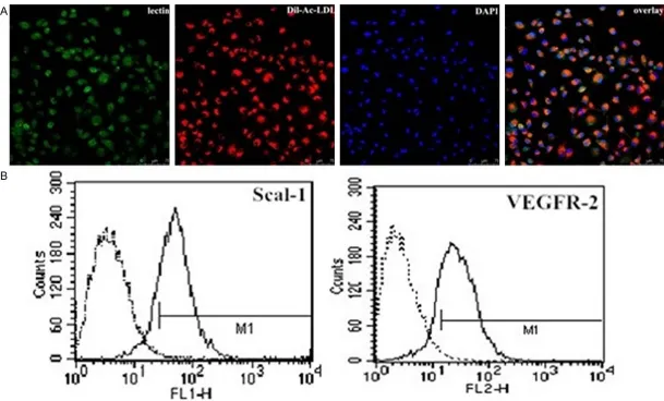
Culture and characterization of EPCs were done as previously described by Werner N et al [5]. Spleens were explanted from C57BL/6 mice (6 to 8 weeks of age, 20 to 25 g of weight, Beijing, P. R. China). Total spleen-derived mononuclear cells were isolated using a Ficoll gradient (Lympholite-M, Cedarlane). After three washing steps, 4 × 10⁶ spleen-derived mononuclear cells were seeded on fibronectin-coated cell culture flasks and re-suspended in 6 ml endo-thelial basal medium (Cell Systems) supple-mented with 1 μg/ml hydrocortisone, 3 μg/ml bovine brain extract, 30 μg/ml gentamicin, 50 μg/ml amphotericin B, 10 μg/ml human endo-thelial growth factor, and 20% fetal calf serum (FCS). The harvested cells were cultured at 37°C under an atmosphere of 5% CO2. Forty-eight hours later, non-adherent cells were removed, and the adherent cells were cultured continuously. Only adherent cells were used in further experiments. The medium was changed every second day. For characterization, after 4 days in culture, the cells were incubated with 10 mg/ml acetylated low-density lipoprotein/ binding (Dil-Ac-LDL, Invitrogen, CA, USA) for 4 h, fixed with 4% paraformaldehyde and then incu-bated with 10 mg/ml fluorescein isothiocya-nate-Ulex Europeaus lectin-1 (UEA-1,
Sigma-Aldrich, St Louis, MO, USA) for 1 h. Finally, the cells were incubated with 1 µg/ml 4’,6-diamidi-no-2-phenylindole (DAPI, Life Technologies, NY, USA) for 5 min. Triple-stained cells positive for Dil-Ac-LDL, lectin, and DAPI, were identified as EPCs. Additionally, fluorescence activated cell sorting (FACS) analysis was performed using the following monoclonal antibodies: FITC con-jugated anti-Sca-1 (Abcam, Cambridge, MA, USA), PE conjugated anti-VEGFR-2 (Biosciences, San Diego, CA, USA), or their corresponding iso-type controls (Biosciences, San Diego, CA, USA).
Recombinant adenoviral vectors expressing SIRT1
In order to evaluate the role of SIRT1, adenovi-rus vector expressing SIRT1 was generated using the pAd-Easy system. Briefly, full-length murine SIRT1 cDNA was first TA-cloned into pMD19-T simple vector and then subcloned into CMV, resulting in
pAdTrack-SIRT1. The shuttle vector was used to generate recombinant adenovirus Ad-SIRT1 according to the manufacturer’s protocol. All PCR-amplified fragments and cloning junctions were verified by DNA sequencing (Sangon, Shanghai, China). An adenovirus encoding green fluorescent pro-tein (GFP; Ad-GFP) was used as control. All ade-noviruses were replication deficient and used at 20 multiplicity of infection (mois) for 24 h without apparent cytotoxicity.
Small interfering RNA-mediated silencing of SIRT1 expression
Transient silencing of SIRT1 was accomplished by transfection with small interfering RNAs
(si-SIRT1). The selected siRNA duplex sequences specifically targeted mouse SIRT1 (GenBank accession number NM_019812.2), and sh- owed no homology to any other sequences, as determined by a blast search. A non-silencing control (si-CON) sequence was designed to be used as a negative control. Transfection of
si-SIRT1 used the Lipofectamine 2000 reagent with a molar ratio between DNA and lipid of approximately 1:3. Forty-eight hours after transfection, cells were collected and used for functional assays.
Reverse transcription-PCR (RT-PCR)
SIRT1
and
endothelial progenitor cells
SIRT1
and
endothelial progenitor cells
transcriptase (Takara, Dalian, China), according to the manufacturer’s instructions. For quanti-tative RT-PCR analyses, the ABI PRISM 7000 Sequence Detection System (Applied Biosys- tems, Foster City, CA, USA) and SYBR Green PCR Master Mix (Takara, Dalian, China) were used with the following primers: SITR1 sense:
5’-ACTGCAGAAACTTTTAGCCTTTCAA-3’; SIRT1
[image:4.612.95.520.73.276.2]antisense: 5’-GGCAATGTTCCAAAGAAGTCTGT-3’; GAPDH sense: 5’-TGAACGGGAAGCTCACTGG-3’; GAPDH antisense: 5’-GCTTCACCACCTTCTTGA- TGTC-3’. All primers were synthesized by Invi- trogen (Shanghai, China) and were the highest available purity.
Figure 2.SIRT1 expression in EPCs. Protein and mRNA levels of SIRT1 in EPCs using western blotting and RT-PCR analysis. Values are the percentage of GAPDH (serum- and VEGF-free, null treatment). Representative images from semi-quantitative RT-PCR and western blots. Data are expressed as mean ± SD of three independent experiments done in triplicate, with *P < 0.05 compared with the controls.
[image:4.612.94.524.348.562.2]SIRT1
and
endothelial progenitor cells
Figure 4. Effect of SIRT1 overexpression and silencing on the migration of EPCs. A, B. Representative photographs of SIRT1 overexpression and silencing on the migration of EPCs. EPC migration in response to Ad-SIRT1 and si-SIRT1
was detected using the Transwell system. C, D. The migration of EPCs transfected with Ad-SIRT1 was enhanced as compared with that of Ad-GFP-transfected EPCs, but knockdown of endogenous SIRT1 significantly reduced the
migration of EPCs compared with the Ad-GFP group. The results are expressed as the mean ± SD (*P < 0.05 vs. Ad-GFP).
[image:5.612.92.522.545.667.2]SIRT1
and
endothelial progenitor cells
Western blot analysis
After treatment, cells were lysed in lysis buffer. The protein concentration of cell lysates was determined using the Bradford method. The same amounts of protein were separated by SDS-PAGE and electrophoretically transferred onto a polyvinylidene fluoride membrane. Membranes were blocked with 5% non-fat milk solution in TBS, with 0.5% Tween-20. Memb- rane-bound proteins were probed with primary antibodies against SIRT1 (1:200) and GAPDH (1:500), followed by probing with secondary horseradish peroxidase-conjugated antibodies. Protein bands were visualized by chemilumi-nescent detection (Amersham Pharmacia Bio- tech, UK), and quantified using Quantity One software (Bio-Rad, USA). Anti-GAPDH monoclo-nal antibody was used to test for equal protein loading.
EPC migration assay
The migration of EPCs was assayed using a Transwell system (Corning Costar, USA) con-taining 8 µm polycarbonate filter inserts in 24-well plates. EPCs (2 × 10⁵) in 100 μl of serum-free DMEM were placed in the upper chamber. DMEM containing 10% FCS (500 μl) was placed in the lower chamber. After 6 h in culture, cells on the bottom of the Transwell membrane were fixed with 4%
paraformalde-hyde at 37°C for 20 min and stained with 1% crystal violet at 37°C for 5 min. Migration activ-ity was determined as the mean number of migrated cells in six random high-power fields (× 200) per chamber.
EPC proliferation assay
The EPCs were harvested from the cultures and placed, in triplicate, into fibronectin-coated 96-well plates (2 × 10⁶ cells/ml). Cell prolifera-tion was measured using the MTS assay (Cell Titer 96 Aqueous, Promega, USA) according to the manufacturer’s protocol. Before reading the optical density at 490 nm, 20 µl of MTS solution was added to each well. All groups of experiments were performed in triplicate.
Concentration of NO in media
[image:6.612.92.522.73.279.2]The concentration of NO released from EPCs was determined using a NO assay kit (Nanjing Jiancheng Institute of Biological Engineering, China) according to the manufacturer’s instruc-tions. EPCs were pretreated with LY294002 (30 μM), sc-221226 (30 μM), or L-NAM (200 μM) for 1 h, then treated with Ad-SIRT1 or Ad-GFP for 24 h. The concentration of NO in 100µl of supernatant in media from different groups was detected at 550 nm. The total pro-tein in every group was quantified using the BCA method. The NO-releasing ability of EPCs was calculated as the ratio of NO concentration and total protein.
Figure 6. Influence of SIRT1 silencing on changes in levels of SIRT1. Changes in levels of protein and mRNA of SIRT1
were detected using western blotting and RT-PCR analysis after silencing of SIRT1 in EPCs. Values are the percent-age of GAPDH. SIRT1 gene and protein levels were decreased by SIRT1 silencing, but si-CON was not affected (*P
SIRT1
and
endothelial progenitor cells
Statistical analysis
Data from at least three independent experi-ments were expressed as the mean ± S.D. SPSS 18.0 software was used for statistical analysis. Comparisons between multiple groups were performed using Multi-Way ANOVA or One-Way ANOVA. Comparisons between groups were performed using Fisher’s LSD test.
P values < 0.05 were considered to be statisti-cally significant.
Results
Characterization of spleen-derived EPCs
After 4-7 days of culture (typical culture period before coculture and further experiments), adherent EPCs were characterized by immuno-fluorescence and immuno-fluorescence activated cell sorting (FACS) analysis. The majority of cells (>90%) stained positively for Dil-Ac-LDL, lectin, and DAPI (Figure 1A). In addition, 81.53 ± 3.97% of these cells expressed mouse stem-cell marker Sca-1, and 56.32 ± 2.18% expressed endothelial cell marker VEGFR-2 (Figure 1B).
SIRT1 expression and localization in EPCs
SITR1 was present at low levels in quiescent EPCs, but was up-regulated upon stimulation with serum and VEGF, as determined using either mRNA levels as determined by RT-PCR,
or by protein levels as determined by western blotting (Figure 2).
Overexpression of SIRT1 enhances migration and proliferation of EPCs
To determine if SIRT1 was involved in the regu-lation of migration and proliferation of EPCs, an adenoviral vector was constructed that exp- ressed SIRT1 exogenously using the pAd-Easy system. The transfection efficacy was approxi-mately 60-70% as assessed by western blot analysis. Increased levels of SIRT1 after adeno-virus-mediated overexpression of SIRT1 was confirmed by RT-PCR and western blot analysis (Figure 3). The parental adenoviral vector or Ad-GFP was used as a transfection control. EPCs transfected with Ad-SIRT1 or control Ad-GFP were subsequently subjected to sepa-rate assays to examine their migration and pro-liferation. The Transwell system was used to examine the effects of SIRT1 overexpression on EPC migration. As shown in Figure 4A and
4C, transfection of EPCs with Ad-SIRT1
increased the number of migrating cells com-pared with Ad-GFP cells (P < 0.05). The MTS assay was used to examine how SIRT1 overex-pression affected EPC proliferation. The prolif-eration of EPCs transfected with Ad-SIRT1 was enhanced approximately 300% compared with Ad-GFP transfected cells and control cells (P <
[image:7.612.94.522.71.251.2]0.05) (Figure 5A). Taken together, and as Figure 7. Role of the PI3K/Akt/eNOS signaling pathway in SIRT1-induced migration and proliferation of EPCs. Cells in the control group and the Ad-SIRT1 group with or without pretreatment with LY294002 (30 μM), sc-221226 (30 μM), or L-NAM (200 μM). Ad-SIRT1-induced migration (A) and proliferation (B) of EPCs were significantly inhibited by
SIRT1
and
endothelial progenitor cells
expected, the results showed that Ad-SIRT1
promoted EPCs migration and proliferation in vitro. SIRT1 therefore is an important compo-nent in the regulation of migration and prolifer-ation of EPCs.
EPC migration and proliferation are inhibited by siRNA-mediated knockdown of SIRT1
Although overexpression of exogenous SIRT1
directly enhanced the migration and prolifera-tion of EPCs, the role of endogenous SIRT1 was not determined. To determine whether endoge-nous SIRT1 affected the migration and prolif-eration of EPCs, siRNA fragments were used to knockdown SIRT1 protein levels. Forty-eight hours after transfection, si-SIRT1 caused a sig-nificant loss of SIRT1 in EPCs as measured by RT-PCR and western blot (Figure 6) (all, P <
0.05). Importantly, EPCs exhibited a decrease in cell migration (Figure 4B, 4D) and prolifera-tion (Figure 5B) compared to si-CON cells (all P < 0.05). The results were reproducible in at least three independent experiments. Thus, knockdown of endogenous SIRT1 significantly reduced the migration and proliferation forma-tion of EPCs, suggesting an important role of endogenous SIRT1 in EPCs.
The role of the PI3K/Akt/eNOS pathway in SIRT1-induced migration and proliferation of EPCs
[image:8.612.92.522.71.346.2]SIRT1
and
endothelial progenitor cells
role in SIRT1-induced EPCs migration and proliferation.
Ad-SIRT1 treatment activated the PI3K/Akt/ eNOS pathway
Because SIRT1-induced migration and prolifer-ation of EPCs were regulated by the PI3K/Akt/ eNOS signaling pathway, we examined the effect of Ad-SIRT1 on PI3K, Akt, and eNOS phosphorylation in EPCs. Exogenous stimula-tion with Ad-SIRT1 significantly up-regulated the phosphotyrosine levels of PI3K (Figure 9B),
Akt (Figure 9C), and eNOS (Figure 9D) (all P <
0.05), confirming the activation of the PI3K/ Akt/eNOS signaling pathway.LY294002 (30 μM), sc-221226 (30 μM), and L-NAM (200 μM) were further used to determine the effects of Ad-SIRT1 on PI3K, Akt, and eNOS phosphoryla-tion in EPCs. LY294002, a highly selective inhibitor of PI3K, prevented the Ad-SIRT1 -induced phosphorylation of PI3K (Figure 9B), Akt (Figure 9C), and eNOS (Figure 9D). As shown in Figure 9, treatment with sc-221226 attenuated levels of SIRT1-induced p-Akt (Figure 9C) and p-eNOS (Figure 9D) expression Figure 9. Blockage of the PI3K/Akt/eNOS signaling pathway abrogated SIRT1-induced phosphorylation of PI3K, Akt,
and eNOS. (A) Western blot demonstrating that blockade by the PI3K inhibitor LY294002, the Akt-specific inhibitor
SIRT1
and
endothelial progenitor cells
in EPCs transfected with Ad-SIRT1, but not p-PI3K (Figure 9B). SIRT1-induced p-eNOS expression was abrogated by L-NAME as deter-mined by western blot analysis (Figure 9A, 9D). Together, the results suggested that eNOS acti-vation was mediated through the PI3K/Akt sig-naling pathway.
Ad-SIRT1 regulated intracellular NO levels via the PI3K/Akt/eNOS signaling pathway in EPCs
The eNOS/NO signaling pathway is recognized as an important mediator of the angiogenic pro-cesses. However, the effect of SIRT1 on NO production is unknown. As shown in Figure 10, the intracellular NO levels were elevated after treatment with Ad-SIRT1. However, in compari-son with controls, pretreatment of EPCs with LY294002 (30 μM), sc-221226 (30 μM), and L-NAM (200 μM) downregulated Ad-SIRT1 -induced NO production in EPCs (Figure 10). Taken together, the results provided direct evi-dence that Ad-SIRT1 enhanced NO production via the PI3K/Akt/eNOS pathway.
Discussion
Damage of endothelial cells following percuta-neous coronary interventions such as
angio-specific inhibitor LY294002, the Akt inhibitor sc-221226, and the NOS inhibitor L-NAME. In addition, knockdown of endogenous SIRT1 sig-nificantly reduced the migration and prolifera-tion of EPCs. These results demonstrated that the migration and proliferation of EPCs are all mediated through the SIRT1/PI3K/Akt/eNOS signaling pathway.
SIRT1 is a member of a protein class known as sirtuins, belonging to the Sir2 family, which has been identified as NAD+-dependent deacety-lases [24, 25]. SIRT1 affects the activity of many proteins, resulting in the regulation of a number of proteins and their translation, which have important roles in biological processes such as metabolism, oxidative stress, and cell proliferation [26-29]. SIRT1 has been implicat-ed in cancer, aging, metabolic diseases, and cardiovascular dysfunctions [30-33]. Direct application of the SIRT1 activator resveratrol has been shown to protect cardiomyocytes against H2O2- and hypoxia-induced apoptosis [34-37]. The antioxidant ability of resveratrol is
SIRT1-dependent, because knockdown of
[image:10.612.91.322.71.278.2]SIRT1 resulted in the loss of resveratrol-medi-ated reduction of reactive oxygen species and cell protection [37, 38]. Endothelial SIRT1 may Figure 10. Ad-SIRT1 induced NO production via the PI3K/Akt/
eNOS signaling pathway. The intracellular NO level was elevat-ed after treatment with Ad-SIRT1. Pretreatment of EPCs with
LY294002 (30 μM), sc-221226 (30 μM), or L-NAM (200 μM)
downregulated Ad-SIRT1-induced NO production in EPCs, in comparison with the controls (*P < 0.05 vs. control, #P < 0.05 vs. Ad-SIRT1).
plasty and stenting is an important patho-physiological event during atherosclerosis and restenosis. EPCs have been found to be the main endogenous repair mechanism that responds to endothelium repair, and contributes to re-endothelialization by red- ucing neointima formation after vascular injury [17]. This mechanism is regulated by various processes and signals [18-23]. Ho- wever, the regulatory mechanisms for the biological properties of EPCs remain uncl- ear. Recent studies demonstrated that endothelial SIRT1 may serve as an anti-ath-erosclerosis factor [9, 10], which may be a key component of EPC dysfunction.
PI3K-SIRT1
and
endothelial progenitor cells
also serve as an anti-atherosclerosis factor.
SIRT1 can protect endothelial cells from oxida-tive stress, and oxidaoxida-tive low-density lipopro-tein-induced apoptosis [39, 40]. In endothelial cell-specific SIRT1 transgenic mice, high fat-induced impairment in endothelium-dependent vasorelaxation decreased, accompanied by less atherosclerotic lesions [41], suggesting that SIRT1 improved endothelial function to prevent atherosclerosis. SIRT1 is highly expr- essed in endothelial cells and controls their angiogenic function. It is involved in vascular growth of cultured endothelium, in the forma-tion of the vascular network of the developing zebrafish, and even in ischemia-induced neo-vascularization of the adult mouse [42-44]. The PI3K/Akt pathway provides essential sig-naling for cell survival and proliferation. Sig- naling from different eNOS agonists, such as VEGF, insulin, estrogen, and platelet-derived lipid mediators, can affect eNOS activity through the PI3K/AKT pathway. It has been reported that activations of the survival signal PI3K/Akt pathway and the endothelial specific eNOS/NO pathway were closely associated with vascular remodeling and angiogenesis [13, 14, 45]. Recent studies also reported that activation of SIRT1 improved endothelium relaxation through up-regulating endothelial nitric oxide synthase (eNOS) expression and production of nitric oxide [46, 47]. However, whether SIRT1 affects the biological properties of EPCs, and the role of PI3K/Akt/eNOS signal-ing pathway in SIRT1-induced migration and proliferation, have remained poorly underst- ood.
In the present study, we determined the effects of the SIRT1/PI3K/Akt/eNOS signaling path-way on EPC migration and proliferation. The results showed that SIRT1 induced the activa-tion of the PI3K/Akt/eNOS signaling pathway during these processes. In vitro transfection of EPCs with Ad-SIRT1 induced phosphorylation of Akt via PI3K, the phosphorylation of eNOS via AKt, and increased the expression of NO via eNOS. In addition, knockdown of endogenous
SIRT1 reduced migration and proliferation of EPCs. Together, the results suggested that the migration and proliferation of EPCs are attribut-able to the up-regulation of SIRT1, p-PI3K, p-Akt, and p-eNOS, as well as the production of NO. In addition, blockage of the PI3K/Akt/eNOS
signaling pathway by the PI3K inhibitor LY294002, the Akt-specific inhibitor sc-221226, and the NOS inhibitor L-NAME, abrogated
SIRT1-induced EPC migration and proliferation. Furthermore, treatment with the NOS inhibitor L-NAME decreased phosphorylation of eNOS, but did not affect PI3K/Akt activity, suggesting that PI3K/Akt is upstream of eNOS. These find-ings, therefore demonstrated the existence of a SIRT1/PI3K/Akt/eNOS signaling pathway during EPC migration and proliferation.
In conclusion, by mediating EPC proliferation and recruitment, the present study demon-strated important roles of SIRT1 in neovascu-larization and re-endothelialization; In addition, the SIRT1/PI3K/Akt/eNOS signaling pathway may also play an important role during these same processes. Future studies should there-fore be directed towards characterization of the complex mechanisms and therapeutic poten-tials ofSIRT1, PI3K, Akt, and eNOS in the angio-genesis and tissue regeneration process medi-ated by EPCs.
Acknowledgements
Appreciation goes to Huali Kang (technician at the Institute of Cardiovascular Science of PLA) for excellent technical assistance.
Disclosure of conflict of interest
None.
Address correspondence to: Dr. Yuntian Li, De- partment of Cardiology, 305 Hospital of PLA, Beijing 100017, China. E-mail: leeweigh305@126.com
References
[1] Napoli C, Hayashi T, Cacciatore F, Casamassimi A, Casini C, Al-Omran M and Ignarro LJ. Endothelial progenitor cells as therapeutic agents in the microcirculation: an update. Atherosclerosis 2011; 215: 9-22.
[2] Walter DH, Rittig K, Bahlmann FH, Kirchmair R, Silver M, Murayama T, Nishimura H, Losordo DW, Asahara T and Isner JM. Statin therapy ac-celerates reendothelialization: a novel effect involving mobilization and incorporation of bone marrow-derived endothelial progenitor cells. Circulation 2002; 105: 3017-3024. [3] Cho HJ, Kim HS, Lee MM, Kim DH, Yang HJ,
granulocyte-SIRT1
and
endothelial progenitor cells
macrophage colony-stimulating factor acceler-ate reendothelialization and reduce vascular
inflammation after intravascular radiation.
Circulation 2003; 108: 2918-2925.
[4] Iwakura A, Luedemann C, Shastry S, Hanley A, Kearney M, Aikawa R, Isner JM, Asahara T and Losordo DW. Estrogen-mediated, endothelial nitric oxide synthase-dependent mobilization of bone marrow-derived endothelial progenitor cells contributes to reendothelialization after arterial injury. Circulation 2003; 108: 3115-3121.
[5] Werner N, Junk S, Laufs U, Link A, Walenta K, Bohm M and Nickenig G. Intravenous transfu-sion of endothelial progenitor cells reduces neointima formation after vascular injury. Circ Res 2003; 93: e17-24.
[6] Chen Z, Peng IC, Cui X, Li YS, Chien S and Shyy JY. Shear stress, SIRT1, and vascular homeo-stasis. Proc Natl Acad Sci U S A 2010; 107: 10268-10273.
[7] Breitenstein A, Wyss CA, Spescha RD, Franzeck FC, Hof D, Riwanto M, Hasun M, Akhmedov A, von Eckardstein A, Maier W, Landmesser U, Luscher TF and Camici GG. Peripheral blood monocyte Sirt1 expression is reduced in pa-tients with coronary artery disease. PLoS One 2013; 8: e53106.
[8] Potente M and Dimmeler S. Emerging roles of SIRT1 in vascular endothelial homeostasis. Cell Cycle 2008; 7: 2117-2122.
[9] Stein S and Matter CM. Protective roles of SIRT1 in atherosclerosis. Cell Cycle 2011; 10: 640-647.
[10] Yu W, Fu YC, Chen CJ, Wang X and Wang W. SIRT1: a novel target to prevent atherosclero-sis. J Cell Biochem 2009; 108: 10-13.
[11] Donmez G and Guarente L. Aging and disease: connections to sirtuins. Aging Cell 2010; 9: 285-290.
[12] Li L, Zhang HN, Chen HZ, Gao P, Zhu LH, Li HL,
Lv X, Zhang QJ, Zhang R, Wang Z, She ZG, Wei
YS, Du GH, Liu DP and Liang CC. SIRT1 acts as a modulator of neointima formation following vascular injury in mice. Circ Res 2011; 108: 1180-1189.
[13] Namkoong S, Kim CK, Cho YL, Kim JH, Lee H, Ha KS, Choe J, Kim PH, Won MH, Kwon YG, Shim EB and Kim YM. Forskolin increases an-giogenesis through the coordinated cross-talk of PKA-dependent VEGF expression and Epac-mediated PI3K/Akt/eNOS signaling. Cell Signal 2009; 21: 906-915.
[14] Wang Y, Yan W, Lu X, Qian C, Zhang J, Li P, Shi
L, Zhao P, Fu Z, Pu P, Kang C, Jiang T, Liu N and You Y. Overexpression of osteopontin induces angiogenesis of endothelial progenitor cells via the avbeta3/PI3K/AKT/eNOS/NO
signal-ing pathway in glioma cells. Eur J Cell Biol 2011; 90: 642-648.
[15] Kang Z, Jiang W, Luan H, Zhao F and Zhang S. Cornin induces angiogenesis through PI3K-Akt-eNOS-VEGF signaling pathway. Food Chem Toxicol 2013; 58: 340-346.
[16] Wang H, Yin Y, Li W, Zhao X, Yu Y, Zhu J, Qin Z, Wang Q, Wang K, Lu W, Liu J and Huang L.
Over-expression of PDGFR-beta promotes PDGF-induced proliferation, migration, and an-giogenesis of EPCs through PI3K/Akt signaling pathway. PLoS One 2012; 7: e30503.
[17] Yu Y, Gao Y, Qin J, Kuang CY, Song MB, Yu SY,
Cui B, Chen JF and Huang L. CCN1 promotes the differentiation of endothelial progenitor cells and reendothelialization in the early phase after vascular injury. Basic Res Cardiol 2010; 105: 713-724.
[18] Lim WH, Seo WW, Choe W, Kang CK, Park J, Cho HJ, Kyeong S, Hur J, Yang HM, Lee YS and Kim HS. Stent coated with antibody against vascular endothelial-cadherin captures endo-thelial progenitor cells, accelerates re-endo-thelialization, and reduces neointimal forma-tion. Arterioscler Thromb Vasc Biol 2011; 31: 2798-2805.
[19] Takamiya M, Okigaki M, Jin D, Takai S, Nozawa Y, Adachi Y, Urao N, Tateishi K, Nomura T, Zen K, Ashihara E, Miyazaki M, Tatsumi T, Takahashi T and Matsubara H. Granulocyte colony-stimu-lating factor-mobilized circucolony-stimu-lating c-Kit+/Flk-1+ progenitor cells regenerate endothelium and inhibit neointimal hyperplasia after vascu-lar injury. Arterioscler Thromb Vasc Biol 2006; 26: 751-757.
[20] Thyberg J. Re-endothelialization via bone mar-row-derived progenitor cells: still another tar-get of statins in vascular disease. Arterioscler Thromb Vasc Biol 2002; 22: 1509-1511. [21] Hibbert B, Ma X, Pourdjabbar A, Holm E, Rayner
K, Chen YX, Sun J, Filion L and O’Brien ER. Inhibition of endothelial progenitor cell glyco-gen synthase kinase-3beta results in attenu-ated neointima formation and enhanced re-endothelialization after arterial injury. Cardi- ovasc Res 2009; 83: 16-23.
[22] Padfield GJ, Newby DE and Mills NL.
Under-standing the role of endothelial progenitor cells in percutaneous coronary intervention. J Am Coll Cardiol 2010; 55: 1553-1565. [23] Kawabe-Yako R, Ii M, Masuo O, Asahara T and
Itakura T. Cilostazol activates function of bone marrow-derived endothelial progenitor cell for re-endothelialization in a carotid balloon injury model. PLoS One 2011; 6: e24646.
[24] Guarani V, Deflorian G, Franco CA, Kruger M,
SIRT1
and
endothelial progenitor cells
Brandes RP, Mione M, Westphal CH, Braun T, Zeiher AM, Gerhardt H, Dimmeler S and Potente M. Acetylation-dependent regulation of endothelial Notch signalling by the SIRT1 deacetylase. Nature 2011; 473: 234-238. [25] Nakahata Y, Sahar S, Astarita G, Kaluzova M
and Sassone-Corsi P. Circadian control of the NAD+ salvage pathway by CLOCK-SIRT1. Science 2009; 324: 654-657.
[26] Cohen HY, Miller C, Bitterman KJ, Wall NR, Hekking B, Kessler B, Howitz KT, Gorospe M, de Cabo R and Sinclair DA. Calorie restriction promotes mammalian cell survival by inducing the SIRT1 deacetylase. Science 2004; 305: 390-392.
[27] Vinciguerra M, Santini MP, Martinez C, Pazi- enza V, Claycomb WC, Giuliani A and Rosenthal N. mIGF-1/JNK1/SirT1 signaling confers pro-tection against oxidative stress in the heart. Aging Cell 2012; 11: 139-149.
[28] Wang Y, Shi X, Qi J, Li X, Uray K and Guan X.
SIRT1 inhibits the mouse intestinal motility and epithelial proliferation. Am J Physiol Gastrointest Liver Physiol 2012; 302: G207-217.
[29] Ota H, Eto M, Kano MR, Ogawa S, Iijima K, Akishita M and Ouchi Y. Cilostazol inhibits oxi-dative stress-induced premature senescence via upregulation of Sirt1 in human endothelial cells. Arterioscler Thromb Vasc Biol 2008; 28: 1634-1639.
[30] Nadtochiy SM, Yao H, McBurney MW, Gu W, Guarente L, Rahman I and Brookes PS. SIRT1-mediated acute cardioprotection. Am J Physiol Heart Circ Physiol 2011; 301: H1506-1512. [31] Chen HC, Jeng YM, Yuan RH, Hsu HC and Chen
YL. SIRT1 promotes tumorigenesis and resis-tance to chemotherapy in hepatocellular carci-noma and its expression predicts poor progno-sis. Ann Surg Oncol 2012; 19: 2011-2019. [32] Yang J, Wang N, Zhu Y and Feng P. Roles of
SIRT1 in high glucose-induced endothelial im-pairment: association with diabetic atheroscle-rosis. Arch Med Res 2011; 42: 354-360. [33] Orimo M, Minamino T, Miyauchi H, Tateno K,
Okada S, Moriya J and Komuro I. Protective role of SIRT1 in diabetic vascular dysfunction. Arterioscler Thromb Vasc Biol 2009; 29: 889-894.
[34] Arunachalam G, Yao H, Sundar IK, Caito S and Rahman I. SIRT1 regulates oxidant- and ciga-rette smoke-induced eNOS acetylation in en-dothelial cells: Role of resveratrol. Biochem Biophys Res Commun 2010; 393: 66-72. [35] Hu YX, Cui H, Fan L, Pan XJ, Wu JH, Shi SZ, Cui
SY, Wei ZM and Liu L. Resveratrol attenuates left ventricular remodeling in old rats with COPD induced by cigarette smoke exposure
and LPS instillation. Can J Physiol Pharmacol 2013; 91: 1044-1054.
[36] Li YG, Zhu W, Tao JP, Xin P, Liu MY, Li JB and Wei M. Resveratrol protects cardiomyocytes from oxidative stress through SIRT1 and mito-chondrial biogenesis signaling pathways. Biochem Biophys Res Commun 2013; 438: 270-276.
[37] Chen CJ, Yu W, Fu YC, Wang X, Li JL and Wang W. Resveratrol protects cardiomyocytes from hypoxia-induced apoptosis through the SIRT1-FoxO1 pathway. Biochem Biophys Res Com- mun 2009; 378: 389-393.
[38] Sundaresan NR, Pillai VB and Gupta MP. Emerging roles of SIRT1 deacetylase in regu-lating cardiomyocyte survival and hypertrophy. J Mol Cell Cardiol 2011; 51: 614-618.
[39] Shimada T, Furuta H, Doi A, Ariyasu H, Kaw- ashima H, Wakasaki H, Nishi M, Sasaki H and Akamizu T. Des-acyl ghrelin protects microvas-cular endothelial cells from oxidative stress-in-duced apoptosis through sirtuin 1 signaling pathway. Metabolism 2014; 63: 469-474. [40] Guo H, Chen Y, Liao L and Wu W. Resveratrol
protects HUVECs from oxidized-LDL induced oxidative damage by autophagy upregulation via the AMPK/SIRT1 pathway. Cardiovasc Drugs Ther 2013; 27: 189-198.
[41] Zhang QJ, Wang Z, Chen HZ, Zhou S, Zheng W,
Liu G, Wei YS, Cai H, Liu DP and Liang CC.
Endothelium-specific overexpression of class
III deacetylase SIRT1 decreases
atherosclero-sis in apolipoprotein E-deficient mice.
Cardi-ovasc Res 2008; 80: 191-199.
[42] Borradaile NM and Pickering JG. Nicotinamide phosphoribosyltransferase imparts human en-dothelial cells with extended replicative lifes-pan and enhanced angiogenic capacity in a high glucose environment. Aging Cell 2009; 8: 100-112.
[43] Potente M, Ghaeni L, Baldessari D, Mosto- slavsky R, Rossig L, Dequiedt F, Haendeler J, Mione M, Dejana E, Alt FW, Zeiher AM and Dimmeler S. SIRT1 controls endothelial angio-genic functions during vascular growth. Genes Dev 2007; 21: 2644-2658.
[44] Volkmann I, Kumarswamy R, Pfaff N, Fiedler J, Dangwal S, Holzmann A, Batkai S, Geffers R, Lother A, Hein L and Thum T. MicroRNA-medi- ated epigenetic silencing of sirtuin1 contrib-utes to impaired angiogenic responses. Circ Res 2013; 113: 997-1003.
SIRT1
and
endothelial progenitor cells
[46] Davis PA, Pagnin E, Dal Maso L, Caielli P, Maiolino G, Fusaro M, Paolo Rossi G and Calo LA. SIRT1, heme oxygenase-1 and NO-medi- ated vasodilation in a human model of endog-enous angiotensin II type 1 receptor antago-nism: implications for hypertension. Hypertens Res 2013; 36: 873-878.