Original Article
Apoptotic effect of mtrix metalloproteinases 9 in the
development of diabetic retinopathy
Yu Chen1,2, Wen Wang3, Fen Liu4, Luosheng Tang1, Renhong Tang2, Wenjie Li2
1Department of Ophthamology, Second Xiangya Hospital Central South University, Changsha 410013, Hunan,
China; 2Department of Ophthamology, Third Xiangya Hospital Central South University, Changsha 410013, Hunan,
China; 3Department of Cardio-Thoracic Surgery, Hunan Provincial People’s Hospital, Changsha 410013, Hunan,
China; 4Department of Gynecology and Obstetrics, Tai He Hospital, Changsha 410013, Hunan, China Received May 15, 2015; Accepted June 26, 2015; Epub September 1, 2015; Published September 15, 2015
Abstract: Objective: To explore the potential regulatory mechanism of MMP9 in the development of DR. Methods: Plasmids pcDNA-MMP9 and pcDNA-Ang2 were transfected into primary rat retinal Müller cells (RMCs) using Lipofectamine 2000. Cell viability and apoptosis were respectively determined by MTT assay and flow cytometry. Moreover, the interaction between MMP9 and Ang2 was explored. Besides, RMCs were treated with MMP-9 under normal glucose and high glucose condition for 2d. Besides, the expression levels of apoptotic proteins, like MMP9, Ang2, Bax2, Bcl2, cleaved PARP and cleaved caspase3 were determined by Western blot. Results: The cell viability of siRNA-MMP9 group was significantly increased while decreased in MMP9 overexpression group when compared to control group, respectively. The apoptotic cells in MMP9 overexpression group significantly increased while de -creased in siRNA-MMP9 group when compared with control group. MMP9 expression was significantly regulated by Ang2 whereas no significant changes occurred in Ang2 expression when MMP9 expression changed. Moreover, MMP9 expression in HG group significantly increased while there were no significant differences between NG group and control group. Besides, the expression of Bax2, Bcl2, cleaved PARP and cleaved caspase3 in HG group in-creased while there were no significant differences between NG group and control group. Conclusion: Our findings indicate that MMP9 may play an important role via inducing cell apoptosis in the development of DR via regulating by Ang2 or targeting apoptotic proteins, such as Bax2, Bcl2, cleaved PARP and cleaved caspase3.
Keywords: Diabetic retinopathy, mtrix metalloproteinases 9, angiopoietin 2, apoptosis
Introduction
Diabetic retinopathy (DR) is a common compli-cation of diabetes and continues to be a lead-ing cause of blindness in worklead-ing-age world-wide. It is characterized by retinal microvascu-lar dysfunction and finally vision loss [1]. The clinical hallmarks of DR are increased vascular permeability, macular edema, vascular micro-aneurysms and vascular proliferation [2]. Early detection of DR is critical in preventing visual loss, but regretfully, current management strat-egy, such as laser photocoagulation, is ham-pered by the fact that this disease is generally asymptomatic at early stages and only target advanced stages of disease [3]. Analysis of specific genetic alterations on molecular mech-anisms will help the development of targeted therapies.
sig-nal-regulated kinase (ERK) [12]. Despites these, the molecular mechanism by which MMP-9 contributes to the development of DR is not fully understood.
In the current study, we used plasmid transf- ection method to overexpress and suppress MMP9 and angiopoietin 2 (Ang2) in primary rat retinal Müller cells (RMCs), respectively. Cell viability and apoptosis after plasmid transfec-tion was determined using MTT assay and flow cytometry analysis. Moreover, the interaction of MMP9 with Ang2 was explored. Besides, cells were treated with different concentration of glucose and the expression of several apoptot-ic proteins were determined by western blot. Our study aimed to explore the potential mech-anism of MMP9 underlying the development of DR and provide a new insight to identify novel targets for inhibiting the development of this disease.
Materials and methods
Cell culture
The primary RMCs culture was prepared as fol-lows. Briefly, enucleated eyes from postnatal day 5-7 Wistar rats were incubated in Dulbecco’s Modified Eagle Medium (DMEM) overnight. Eyecups were transferred to DMEM (containing 0.1% Trypsin and 70 U/ml collage-nase) and incubated in CO2 incubator at 37°C for 1 h. The retina was dissected out from reti-nal pigment epithelium (RPE) and ciliary epithe-lium with care to avoid contamination. The reti-na was then dissociated into small aggregates and incubated in DMEM containing 10% fetal bovine serum (FBS, Welgene Ltd.) for 8-10 days. The purified population of RMCs attached to the bottom of the dish was collected after removing floating retinal aggregates and debris. RMCs were then trypsinized and cultured in DMEM containing 10% FBS for another 5 days to get a further purified population.
Plasmid transfection
Plasmids pcDNA-MMP9 and pcDNA-Ang2 were purchased form the American Type Culture Collection (ATCC, Rockville) and were transfect-ed into RMCs using Lipofectamine 2000 (Invitrogen, Carlsbad, CA, USA) as instructed by the manufacturer. On the other hand, cells were transfected with antisense or siRNA against
MMP9 and Ang2 (Qiagen) using the same method. Cells continued to incubate with nor-mal media for 4 h and were then harvested.
Cell viability assay
Cell viability was determined using MTT assay. Briefly, cells were seeded in 96-well plates at a density of 5 × 10³ cells/well for 24 h. Sub- sequently, 10 μL MTT (5 mg/ml) was added to each well at 0 h, 24 h, 48 h, 72 h and 96 h after transfection and incubated for another 4 h. Each experiment was performed in triple. The resulting formazan crystals were dissolved in 100 μL DMSO, and the optical density (OD) at 570 nm was measured using an automatic microplate reader (BioTek Instruments, Inc.). Finally, cell viability of each treated group at different time points was calculated.
Apoptosis assay by flow cytometry analysis
Cell apoptotic rate was determined by the fluorescein isothiocyanate (FITC) Annexin V Apoptosis Detection Kits (KeyGEN Biotech, Nanjing, China) using flow cytometry analysis. Cells were collected by trypsinization, washed twice with ice-cold PBS and resuspended in 1 × binding buffer at a concentration of 1 × 10⁶ cells/ml. Afterwards, 5 μl FITC Annexin V and 5 μl PI were mixed with 100 μl of cells, and con-tinued to incubate for 15 min. Finally, the cells were then sent out at 488 nm by flow cytometry (BD, USA) and analyzed with the BD FACSCalibur™ system (BD, USA).
Cell treatment with different concentration of glucose
Total 4 × 10⁵ cells were seeded into 96-well plates. After 24 h, cells were treated with MMP-9 (300 ng/mL) under normal concentra-tion of glucose (NG, 5 mmol/L glucose), high concentration of glucose (HG, 20 mmol/L glu-cose) for 2 d. Cells without any treatment were set as control.
Quantitative real-time polymerase chain reac-tion (qRT-PCR)
reversely transcribed into cDNA with the PrimeScriptTM RT Master Mix Kit (Takara, RR036A). Quantitative real-time PCR assays were carried out using SYBR Green real-time PCR MasterMix under ABI 7500 RT-PCR ampli-fication equipment (Applied Biosystems, CA, USA). Human actin was used as internal con-trol. The PCR reaction consisted of 1 cycle at 95°C for 15 s followed by 45 cycles at 95°C for 5 s and at 60°C for 30 s. The primer sequences were as follows: 5’-GCACCACCACAACATCAC-3’ (sense), 5’-ACCACAACTCGTCATCGTC-3’ (anti-sense) for MMP-9 (284 bp), 5’-GGCGGCACCA- CCATGTACCCT-3’ (sense), 5’-AGGGGCCGGACTC- GTCATACT-3’ (antisense) for actin (202 bp). Each reaction was performed in triplicate. The relative expression level of MMP-9 was normal-ized based on the expression level of actin and calculated using the comparative threshold (Ct) cycle (2-ΔΔCt).
Preparation of cell lysates and Western blot analysis
Western blot was performed to examine the expression levels of MMP9, Ang2, Bax2, Bcl2, cleaved PARP and cleaved caspase3. Briefly, cells were washed twice with ice-cold PBS and then lysed with RIPA buffer containing protease inhibitors (BestBio, Shanghai, China) on ice for 1 h. Total protein concentration was examined by BCA assay (Beyotime, Haimen, China). Equal amounts of protein (30 μg) were separated on 10% sodium dodecyl sulfate-polyacrylamide (SDS-PAGE) gel and then electrophoretically transferred to polyvinylidene difluoride (PVDF)
membranes (Millipore). After blocked in TBS-0.05% Tween-20 (TBST) containing 3% BSA, the membrane was incubated with an appropri-ate antibody at 4°C overnight. After washed three times with TBST, the membrane was then incubated with the second antibody conjugated with horseradish peroxidase (HRP) at 25°C for 1 h. The blots were developed by enhanced chemiluminescence kit (Millipore). Finally, the expression level of these proteins was normal-ized to glyceraldehyde-3-phosphate dehydroge-nase (GAPDH).
Statistics analysis
All the data are presented as the mean ± SD. The statistical analyses were performed using T-test or one-way ANOVA as appropriate, fol-lowed by Bonferroni’s test for multiple compari-sons using SPSS 20.0 and GraphPad Prism 5.0. A value of P ≤ 0.05 was considered statisti-cally significant.
Results
The relationship of MMP9 expression with cell viability
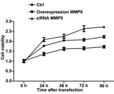
MTT analysis showed the cell viability in an experimental period of 96 h after transfection. As shown in Figure 1, the cell viability gradually increased with the increase of transfected time and showed significant differences of transfect-ed groups at 72 h after transfection compartransfect-ed with control group. Moreover, the cell viability of siRNA-MMP9 group was higher than the control group, while the cell viability of MMP9 overex-pression group was significantly lower than the control group.
Detection of cell apoptosis
Flow cytometry analysis showed the percent-age of apoptotic cells. The results showed that the apoptotic cells in MMP9 overexpression group significantly increased while that in siR-NA-MMP9 group obviously decreased when compared with that in control group (Figure 2).
The interaction between MMP9 and Ang2
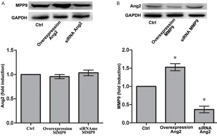
[image:3.612.91.283.71.230.2]To explore the interaction of MMP9 with Ang2, we performed Western blot to determine Ang2 expression in MMP9 overexpression group and siRNA-MMP9 group, as well as to determine
MMP9 expression in Ang2 overexpression gr- oup and siRNA-Ang2 group. The results show- ed that there were no significant differences of Ang2 expression in MMP9 overexpression group and siRNA-MMP9 group when compared with that in control group, respectively (Figure 3A). Notably, MMP9 expression in Ang2 overex-pression group significantly increased and obvious decreased in siRNA-Ang2 group when compared with that in control group (P < 0.05,
Figure 3B), respectively, indicting MMP9 was a downstream target of Ang2.
Analysis of MMP9 expression under different concentration of glucose
[image:4.612.93.525.73.224.2]The mRNA and protein expression level of MMP9 under different concentration of glucose were respectively determined by qRT-PCR and western blot analysis. Similar results were obtained that MMP9 expression in HG group significantly increased compared with that in control group (P < 0.05), while there were no significant differences between NG group and control group (Figure 4).
[image:4.612.92.517.267.534.2]Figure 2. The apoptosis of primary rat retinal Müller cells determined by flow cytometry.
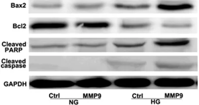
Expression of several apoptotic proteins under different concentration of glucose
Western blot analysis showed the expression of apoptotic proteins, such as Bax2, Bcl2, cleaved PARP and cleaved caspase3, in HG group and NG groups, respectively. The results showed the expression of Bax2, Bcl2, cleaved PARP and cleaved caspase3 in HG group increased compared with that in control group, while there were no significant differences between NG group and control group (Figure 5). HG could enhance the expression of MMP9 (Figure 4), implying that MMP9 might induce cell apopto-sis via enhancing the activity of these apoptotic proteins.
Discussion
The present study demonstrated the role of MMP9 in the development of DR and elucidat-ed potential regulatory mechanism by which MMP9 contributed to DR progression. The results showed that increased expression of MMP9 decreased cell viability and induced apoptosis in RMCs. Moreover, MMP9 was a downstream target of Ang2 and increased Ang2 expression promoted the expression of MMP9. Besides, the expression of MMP9 sig-nificantly increased under HG condition and consequently promoted the expression of sev-eral apoptotic proteins, such as Bax2, Bcl2, cleaved PARP and cleaved caspase3, thus leading to induce cell apoptosis.
MMP-9 as the largest member of the MMP fam-ily MMPs is shown to be elevated in the retina and always associated with many diabetes complications, including DR [13]. Kowluru et al.
also demonstrated that MMP9 can trigger apoptosis of retinal capillary cells via modulat-ed by a small molecular weight G protein, H-Ras [14]. Moreover, mitochondrial dysfunction is postulated to play a crucial role in the apopto-sis of retinal capillary cells in the development of DR [7]. Active MMP in cardiac myocytes is considered to degrade mitochondrial mem-brane potential and impair mitochondrial
[image:5.612.93.519.73.305.2]func-Figure 4. The expression level of MMP9 at mRNA and protein levels under different concentrations of glucose. Error bars indicate means ± SD and *indicates significant difference compared with control group (P < 0.05).
[image:5.612.91.287.360.464.2]tion [15]. Besides, Madsen-Bouterse et al.
demonstrated that inhibition of MMP-9 could prevent the continuation of the vicious cycle of mitochondrial damage and ultimately inhibited the of DR [16]. In our study, the results of MTT assay showed MMP9 could decrease cell viabil-ity. Moreover, flow cytometry analysis revealed that MMP9 could induce cell apoptosis. These results are consistent with previous findings and imply the apoptotic effect of MMP9 in the development of DR.
To further verify the apoptotic effect of MMP9 in the development of DR, we further explored the interaction of MMP9 with Ang2. Ang2, a member of the angiopoietins, is thought to play an important role in retinal neovascularization and angiogenesis in retinal development [17]. Increased Ang-2 can lead to persistent disrup-tion of the cellular cross-talk between pericytes and endothelial cells in early DR [18]. Moreover, Ang2 is reported to promote cell apoptosis [19, 20]. Ang2 is also proved to have proapoptotic activity and its increased expression may be associated with endothelial apoptosis [21]. Besides, high concentrations of Ang2 are con-sidered as an apoptosis survival factor for endothelial cells via activation of the phospha-tidylinositol 3’-kinase/Akt (PI3K/Akt) signal transduction pathway [22]. PI3K/Akt pathway is widely involved in high glucose-induced apoptosis in human vascular endothelial cells [23]. Inhibition of the PI3K/Akt pathway may have the potential therapeutic roles in patho-physiology of DR [24]. In combination, we spec-ulate that Ang2 be a key apoptosis factor to induce cell apoptosis in the development of DR. In our study, MMP9 expression was significant-ly regulated by Ang2 whereas no significant changes occurred in the expression of Ang2 when MMP9 expression changed, indicting MMP9 was a downstream target of Ang2. It can therefore be hypothesized that MMP9 may induce cell apoptosis in the development of DR via regulating by Ang2.
In addition, the expression of apoptotic pro-teins, such as Bax2, Bcl2, cleaved PARP and cleaved caspase3, in HG group was determined compared with that in NG group. Bax/Bcl-2 is regarded as a key mediator known to play a central role in cell apoptosis. Risso et al. revealed that Bax/Bcl-2 was involved in apop-tosis enhanced by intermittent HG in human umbilical vein endothelial cells [25]. Moreover,
PARP activation is considered a downstream effector of oxidative-nitrosative stress in the pathogenesis of diabetes complications [26]. PARP activation contributes to the inhibition of PI3K/Akt pathway and consequently inhibits cell apoptosis [27], thus increased expression of cleaved PARR may promote cell apoptosis. PARP activation is also thought to be a funda-mental step in the pathogenesis of DR via regu-lation of nuclear factor-κB (NF-κB) in the retinal cells [28]. Soufi et al. confirmed that reduced NF-kB activity by resveratrol could finally decrease apoptosis rates in the retinas of dia-betic rats in the development of DR [29]. Besides, caspase-3 is the executioner caspase involved in the proteolytic cascade during apop-tosis [30]. El-Asrar et al. demonstrated that the executioner caspase-3 was observed in gangli-on cells in diabetic retinas [31]. Kowluru et al. also confirmed that diabetes could induce activation of caspase-3 in retina, thus inhibiting apoptosis in the development of DR [32], sug-gesting increased expression of cleaved cas-pase-3 may promote the development of DR via inducing cell apoptosis. In our study, the expres-sion of these apoptotic proteins in HG group increased compared with that in control group. Considering that HG could enhance the expres-sion of MMP9, we speculate that MMP9 could induce cell apoptosis in the development of DR via regulating the activity of these apoptotic proteins.
In conclusion, our findings indicate that MMP9 may play an important role in the development of DR via inducing cell apoptosis. MMP may induce cell apoptosis via regulating by Ang2 or targeting apoptotic proteins, such as Bax2, Bcl2, cleaved PARP and cleaved caspase3. Understanding the apoptotic mechanism of MMP9 in the development of DR may help iden-tify novel molecular targets for future pharma-cological interventions of this disease.
Disclosure of conflict of interest
None.
References
[1] Joussen AM, Poulaki V, Le ML, Koizumi K, Esser C, Janicki H, Schraermeyer U, Kociok N, Fauser S and Kirchhof B. A central role for in-flammation in the pathogenesis of diabetic retinopathy. FASEB J 2004; 18: 1450-1452. [2] Barber AJ. A new view of diabetic retinopathy: a
neurodegenerative disease of the eye. Prog Neuro-Psychopharmacol Biol Psychiatry 2003; 27: 283-290.
[3] Ciulla TA, Amador AG and Zinman B. Diabetic retinopathy and diabetic macular edema pathophysiology, screening, and novel thera-pies. Diabetes Care 2003; 26: 2653-2664. [4] Petrovič D. Candidate genes for proliferative
diabetic retinopathy. BioMed Res Int 2013; 2013: 540416.
[5] Kowluru RA, Zhong Q and Santos JM. Matrix metalloproteinases in diabetic retinopathy: po-tential role of MMP-9. Expert Opin Investig Drugs 2012; 21: 797-805.
[6] Zhong Q and Kowluru RA. Regulation of matrix metalloproteinase-9 by epigenetic modifica -tions and the development of diabetic retinop-athy. Diabetes 2013; 62: 2559-2568.
[7] Kowluru RA, Mohammad G, dos Santos JM and Zhong Q. Abrogation of MMP-9 gene pro-tects against the development of retinopathy in diabetic mice by preventing mitochondrial damage. Diabetes 2011; 60: 3023-3033. [8] Mohammad G, Vandooren J, Siddiquei MM,
Martens E, El-Asrar AMA and Opdenakker G. Functional links between gelatinase B/matrix metalloproteinase-9 and prominin-1/CD133 in diabetic retinal vasculopathy and neuropathy. Prog Retin Eye Res 2014; 43: 76-91.
[9] Navaratna D, McGuire PG, Menicucci G and Das A. Proteolytic degradation of VE-cadherin alters the blood-retinal barrier in diabetes. Diabetes 2007; 56: 2380-2387.
[10] Kowluru RA and Kanwar M. Oxidative stress and the development of diabetic retinopathy: contributory role of matrix metalloprotein-ase-2. Free Radic Biol Med 2009; 46: 1677-1685.
[11] Giebel SJ, Menicucci G, McGuire PG and Das A. Matrix metalloproteinases in early diabetic retinopathy and their role in alteration of the blood–retinal barrier. Lab Invest 2005; 85: 597-607.
[12] Mohammad G and Kowluru RA. Diabetic reti-nopathy and signaling mechanism for activa-tion of matrix metalloproteinase-9. J Cell Physiol 2012; 227: 1052-1061.
[13] Shiau MY, Tsai ST, Tsai KJ, Haung ML, Hsu YT and Chang YH. Increased circulatory MMP-2 and MMP-9 levels and activities in patients with type 1 diabetes mellitus. Mt Sinai J Med 2006; 73: 1024-1028.
[14] Kowluru RA. Role of matrix metalloprotein-ase-9 in the development of diabetic retinopa-thy and its regulation by H-Ras. Invest Ophthalmol Vis Sci 2010; 51: 4320-4326. [15] Zhou HZ, Ma X, Gray MO, Zhu BQ, Nguyen AP,
Baker AJ, Simonis U, Cecchini G, Lovett DH and Karliner JS. Transgenic MMP-2 expression in-duces latent cardiac mitochondrial dysfunc-tion. Biochem Biophys Res Commun 2007; 358: 189-195.
[16] Madsen-Bouterse SA, Mohammad G, Kanwar M and Kowluru RA. Role of mitochondrial DNA damage in the development of diabetic reti-nopathy, and the metabolic memory phenom-enon associated with its progression. Antioxid Redox Signal 2010; 13: 797-805.
[17] Hackett SF, Ozaki H, Strauss RW, Wahlin K, Suri C, Maisonpierre P, Yancopoulos G and Campochiaro PA. Angiopoietin 2 expression in the retina: upregulation during physiologic and pathologic neovascularization. J Cell Physiol 2000; 184: 275-284.
[18] Cai J, Kehoe O, Smith GM, Hykin P and Boulton ME. The angiopoietin/Tie-2 system regulates pericyte survival and recruitment in diabetic retinopathy. Invest Ophthalmol Vis Sci 2008; 49: 2163-2171.
[19] Cohen B, Barkan D, Levy Y, Goldberg I, Fridman E, Kopolovic J and Rubinstein M. Leptin induc-es angiopoietin-2 exprinduc-ession in adipose tis-sues. J Biol Chem 2001; 276: 7697-7700. [20] Zagzag D, Amirnovin R, Greco MA, Yee H,
Holash J, Wiegand SJ, Zabski S, Yancopoulos GD and Grumet M. Vascular apoptosis and in -volution in gliomas precede neovasculariza-tion: a novel concept for glioma growth and angiogenesis. Lab Invest 2000; 80: 837-849. [21] Nag S, Papneja T, Venugopalan R and Stewart
DJ. Increased angiopoietin2 expression is as-sociated with endothelial apoptosis and blood-brain barrier breakdown. Lab Invest 2005; 85: 1189-1198.
[22] Kim I, Kim JH, Moon SO, Kwak HJ, Kim NG and Koh GY. Angiopoietin-2 at high concentration can enhance endothelial cell survival through the phosphatidylinositol 3’-kinase/Akt signal transduction pathway. Oncogene 2000; 19: 4549-4552.
[23] Ho FM, Lin WW, Chen BC, Chao CM, Yang CR, Lin LY, Lai CC, Liu SH and Liau CS. High glu-cose-induced apoptosis in human vascular en-dothelial cells is mediated through NF-κB and c-Jun NH 2-terminal kinase pathway and pre-vented by PI3K/Akt/eNOS pathway. Cell Signal 2006; 18: 391-399.
[25] Risso A, Mercuri F, Quagliaro L, Damante G and Ceriello A. Intermittent high glucose enhances apoptosis in human umbilical vein endothelial cells in culture. Am J Physiol Endocrinol Metab 2001; 281: E924-E930.
[26] Obrosova IG, Drel VR, Pacher P, Ilnytska O, Wang ZQ, Stevens MJ and Yorek MA. Oxidative-Nitrosative Stress and Poly (ADP-Ribose) Polymerase (PARP) Activation in Experimental Diabetic Neuropathy The Relation Is Revisited. Diabetes 2005; 54: 3435-3441.
[27] Veres B, Radnai B, Gallyas F, Varbiro G, Berente Z, Osz E and Sumegi B. Regulation of kinase cascades and transcription factors by a poly (ADP-ribose) polymerase-1 inhibitor, 4-hydroxy-quinazoline, in lipopolysaccharide-induced in-flammation in mice. J Pharmacol Exp Ther 2004; 310: 247-255.
[28] Zheng L, Szabó C and Kern TS. Poly (ADP-ribose) polymerase is involved in the develop-ment of diabetic retinopathy via regulation of nuclear factor-κB. Diabetes 2004; 53: 2960-2967.
[29] Soufi FG, Mohammad-nejad D and Ahmadieh H. Resveratrol improves diabetic retinopathy possibly through oxidative stress–nuclear fac-tor κB-apoptosis pathway. Pharmacol Rep 2012; 64: 1505-1514.
[30] Earnshaw WC, Martins LM and Kaufmann SH. Mammalian caspases: structure, activation, substrates, and functions during apoptosis. Annu Rev Biochem 1999; 68: 383-424. [31] El-Asrar AMA, Dralands L, Missotten L,
Al-Jadaan IA and Geboes K. Expression of apop-tosis markers in the retinas of human subjects with diabetes. Invest Ophthalmol Vis Sci 2004; 45: 2760-2766.