Original Article
HOTTIP is upregulated in acute myeloid leukemia
that indicates a poor prognosis
Shanfeng Hao, Rong Fu, Huaquan Wang, Yihao Wang, Mengying Zheng, Li You, Yang Zhang, Zonghong Shao
Department of Hematology, Tianjin Medical University General Hospital, Heping District, Tianjin, China
Received March 2, 2016; Accepted June 29, 2016; Epub August 1, 2016; Published August 15, 2016
Abstract: LncRNA HOTTIP (HOXA transcript at the distal tip) gene is located in physical contiguity (chr 7p15.2) with HOXA13 and directly controls the HOXA locus gene expression by interaction with the WDR5/MLL complex. HOX genes encode transcription factors regulating embryonic development and cell fate. Recent evidence highlighted HOTTIP plays a crucial role in some solid tumors. However, little is known about the role of HOTTIP in acute myeloid leukemia (AML). In this study, We evaluated HOTTIP expression in bone marrow of de novo AML patients, AML-CR (complete remission) patients and normal controls by real-time quantitative reverse transcription-PCR (qRT-PCR), and then we analyzed the relationship between the HOTTIP expression level and the clinicopathological parameters of AML. The results showed that HOTTIP is markedly upregulated in de novo AML patients compared with those of AML-CR patients and normal controls; the higher expression level of HOTTIP in AML patients was significantly cor-related with NCCN high risk group and higher blast cells. In conclusion, our study indicated that HOTTIP is highly expressed in AML patients, and the levels of HOTTIP are associated with AML patients’ clinical progression.
Keywords: Acute myeloid leukemia, long non-coding RNA, HOTTIP, prognosis
Introduction
Acute myeloid leukemia (AML) is the most com-mon acute leukemia in adults with an incidence of 3-4 per 100,000 persons per year. AML is a genetically heterogeneous disorder character-ized by the somatically acquired genetic chang-es in hematopoietic progenitor cells altering normal mechanisms of self-renewal, prolifera-tion, and differentiation. Greater insight into the genetic background of the disease fostered the extension of disease classification and pre-treatment risk-categorization by gene muta-tions. In order to improve outcome in AML, mul-tiple studies aimed at genetic characterization of AML have been performed inthe hopes of furthering our understanding of AML pathogen-esis and identifying new therapeutic approach-es [1, 2].
Long non-coding RNAs (lncRNAs) are a hetero-geneous class of RNAs that are generally defined as non-protein-coding transcripts lon-ger than 200 nucleotides. lncRNA which was considered as only transcriptional “noise” in
the homeobox pathways have been implicated in leukemogenesis [9].
Recently, HOTTIP has been determined to be a negative prognostic indicator in some solid-tumor patients [5, 10, 11]. Nevertheless, little is known about the impact of HOTTIP on AML. To understand the role of HOTTIP in AML, we investigated the expression level of HOTTIP in AML and analyzed its relationship to clinical pathological features.
Materials and methods
Patients and samples
A total of 34 AML cases were enrolled in our study including 21 de novo AML patients and 13 cases who had achieved complete remi- ssion (CR). 21 de novo AML patients were diag-nosed in the Hematology Department of Tianjin Medical University General Hospital between February 2014 and November 2014 according to the “2008 WHO adult acute myeloid leuke-mia (non acute pro-myelocytic leukeleuke-mia) diag-nosis guidelines” and “2008 WHO adult acute pro-myelocytic leukemia diagnosis guidelines”. 13 cases achieved complement remission (CR) were enrolled as AML-CR group. 16 iron-defi-ciency anemia (IDA) patients diagnosed accord-ing to the international criteria of IDA in our department were enrolled as control group. We collected 2 ml EDTA anticoagulant fresh bone marrow samples from each patient and BM sample of de novo AML patients were collected before intervention. We also collected the clini-cal and pathologiclini-cal characteristics for each patient, including age, sex, and blood routine
test. This study was approved by the Ethics Committee of Tianjin Medical University. In- formed written consent was obtained from all patients in accordance with the Declaration of Helsinki.
FCM analysis
Fresh heparinized BM samples (100 uL) were stained with CD34-FITC (BD Pharmingen, San Diego, CA, USA) and CD45-PerCP (BD Pharmin- gen, San Diego, CA, USA). After incubation in the dark for 30 min at 4°C, cells were incubat-ed with 1 ml erythrocyte lytic solution (BD PharMingen) for 10 min at room temperature and washed three times with PBS. Finally, at least 100000 cells were acquired and analyz- ed on a FACSC alibur flow cytometer (BD Bios- ciences). We gated on CD45/SSC to character-ize leukemic blasts. The blast population occu-pies the position with SSC in the low to moder-ate range, and CD45 of weak to modermoder-ate intensity.
RNA extraction and qRT-PCR analyses
[image:2.612.90.287.72.191.2]Total RNA from human bone marrow was ex- tracted using the TRIzol reagent (Life Techno- logies, Scotland, UK) according to the manufac-turer’s protocol. 1 μg total RNA was reverse transcribed in a final volume of 20 ul using ran-dom primers under standard conditions using the PrimeScript RT Master Mix (Takara, Dalian, China). After the RT reaction, qRT-PCR was per-formed using the BIO-RAD iQ5 Real-Time Sys- tem (BIO-RAD, Hercules, CA, USA), and SYBR Green (Tiangen, Beijing, China) was used as a double strand DNA-specific dye. The sequenc-es of primers specific for HOTTIP (forward, 5’-CCTAAAGCCACGCTTCTTTG-3’; reverse, 5’-TG- CAGGCTGGAGATCCTACT-3’) were synthesized by GENEWIZ (Suzhou, China). GAPDH was used as a housekeeping gene for standardizing the-expression of targeted mRNA. The sequences of primers specific for GAPDH (forward, 5’- CCGGGAAACTGTGGCGTGATGG-3’; reverse, 5’- AGGTGGAGGAGTGGGTGTCGCTGTT-3’) were al- so synthesized by GENEWIZ (Suzhou, China). qRT-PCR cycling program: 95°C for 15 min, followed by 40 cycles at 95°C for 10 s and 64.4°C for 30 s. After normalization of the data according to the expressionof GAPDH mRNA, the levels of HOTTIP were calculated using the 2-ΔΔCt method [(Ct, hot tip-Ct, GAPDH) sample- (Ct, hot tip-Ct, GAPDH) control].
Statistical analysis
Data were presented as mean ± standard devi-ation in the text and figures and were analyzed with GraphPad Prism 6 Software. Pearson’s correlation coefficient was used to determine the correlation between HOTTIP-mRNA level and the frequency of BM blast cells. Statistical significance was evaluated with the Student’s t-test for HOTTIP-mRNA level. A P value of <0.05 was considered statistically significant.
Results
Clinical data of patients
There were 21 cases of de novo AML patients, male 11 cases, female 10 cases, mean age 50.61±16.04 years old. Among them, 2 cases of acute myeloid leukemia differentiation type (M2), 7 cases of acute promyelocytic leukemia (M3), 7 cases of acute myelomonocytic leuke-mia (M4), 3 cases of acute monocytic leukeleuke-mia
the percentage of bone marrow blast cells and the percentage of CD34+ cells assayed with FCM were not shown.
HOTTIP was upregulated in de novo AML pa-tients
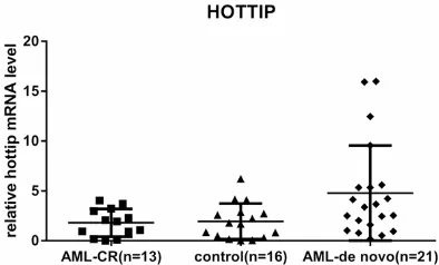
The level of HOTTIP was detected in 21 de novo AMLsamples, 13 AML-CR samples. And 16 con-trol samples by qRT-PCR, and normalized to GAPDH. HOTTIP expression was significantly up-regulated in de novo AML compared with AML-CR patients and controls. However, there were no significant differences between AML-CR patients and controls (Figure 1).
Correlations between elevated HOTTIP and clinicopathological characteristics
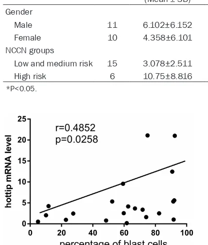
In order to examine the clinical importance of the HOTTIP overexpression, the correlation between clinicopathological parameters of de novo AML samples and level of HOTTIP expres-sion was investigated. Analyses showed that the elevated HOTTIP level had significantly cor-relation with NCCN high risk group. However, there were not any relationships between HOTTIP and patients’ gender or leukemia sub-type (Table 1). Moreover, in de novo AML group, there were positive correlations between the frequency of BM blast cells and the expression of HOTTIP Mrna (r=0.4852, P<0.05) (Figure 2); there were no correlations between the fre-quencies of BM CD34+ cells, blood HB level, PLT counts and WBC counts with the expres-sion of HOTTIP mRNA (data were not shown).
Discussion
[image:3.612.92.335.108.230.2]So far more than 10,000 lncRNAs have been identified in the human genome [3] and several
Table 1. Relationship between HOTTIP expression levels and clinicopathological parameters of de novo AML samples
Clinicopathologic
features Number
Relative expression of HOTTIP
(Mean ± SD) P-value
Gender 0.3589
Male 11 6.102±6.152 Female 10 4.358±6.101
NCCN groups 0.0466*
Low and medium risk 15 3.078±2.511 High risk 6 10.75±8.816
*P<0.05.
Figure 2. Relationship between the frequency of the BM blast cells and the hottip mRNA level in de novo AML group.
[image:3.612.87.301.139.390.2]lncRNAs have been associated with hemato-poietic cancers [12]. HOTAIRM1 (HOX antisense intergenic RNA myeloid 1) plays a role in the myelopoiesis through modulation of gene expression in the HOXA cluster, knockdown of HOTAIRM1 quantitatively blunted RA-induced expression of HOXA1 and HOXA4 during the myeloid differentiation of NB4 cells, and selec-tively attenuated induction of transcripts for the myeloid differentiation genes CD11b and CD18 [13]. In addition, the level of ANRIL was increased in many AML and ALL patients [14], and the abundance of MEG3 was decreased in myeloid leukemia [15] but BIC was increased in B cell lymphoma [16].
Up to date, 231 lncRNAs have been annotated within the 4 HOX loci [17].
Among these newly described lncRNAs, a lncRNA named HOTTIP located in physical con-tiguity (chr 7p15.2) with the HOXA13 gene has been recently functionally characterized [7]. HOTTIP is expressed from development to adulthood in lumbo-sacral anatomic regions and it can directly coordinate and control the activation of several 5’ HOXA genes by interact-ing with the WDR5/MLL complex [7]. Until now, many studies have showed that increased expression of HOTTIP is associated with malig-nant progression and poor survival in various solid cancer types. Hence, HOTTIP may be con-sidered as a potential target for diagnosis and treatment of various cancer types.
A study in hepatocellular carcinoma (HCC) indi-cates that HOTTIP was the most significantly up-regulated lncRNA in human HCCs, even in early stage of HCC formation. Functionally, knock-down of HOTTIP attenuated HCC cell pro-liferation in vitro and markedly abrogated tumourigenicity in vivo. In addition, knock-down of HOTTIP also inhibited migratory ability of HCC cells and significantly abrogated lung metastasis in orthotropic implantation model in nude mice. HOTTIP is an antisense lncRNA mapped to the distal end of the HOXA gene cluster. Knock-down of HOTTIP significantly suppressed the expression of a number of HOXA genes [10]. Quagliata et al also confirmed higher HOTTIP expression in non-neoplastic liver compared to
non-tumoral area tissue, the study also showed that high expression of HOTTIP is always coupled
with increased HOXA13 levels and conversely low HOTTIP levels correlate with low HOXA13 expression: the combination of clinico-patho-logical and expression data indicates that high levels of HOTTIP/HOXA13 expression are asso-ciated with metastasis formation and predict HCC patients’ clinical outcome [5]. Another study profiled 90 well-annotated mouse lnc- RNAs from cultured mouse keratinocytes after deleting the vitamin D receptor (VDR) and found that HOTTIP and several well-known oncogenes are significantly increased in VDR deleted kera-tinocytes, so this finding is a novel mechanism that predisposes the VDR deficient mice to skin cancer formation [11].
The results of our study indicated that the expression of HOTTIP was upregulated in de novo AML patients compared with normal con-trols and AML-CR patients. Moreover, HOTTIP expression was found to be significantly higher in NCCN high risk group. These findings indi-cate that HOTTIP may play a direct role in the modulation of AML progression and may be useful as a novel prognostic marker for AML. These results were also consistent with the findings about hot tip in other cancers before. In addition, we also found that BM blast cells and hot tip mRNA level were positively correlated.
However, it should be noted that the sample size of this study is not very big, thus each leu-kemia subtype has few cases, so we cannot thoroughly compare whether there is any differ-ence in HOTTIP level between them. Moreover, although several lncRNAs have been identified the association with hematopoietic cancers, the mechanism of the effect of these lncRNA on hematopoietic malignances is not very clear. Our study also failed to answer this question and we will precede thorough research on it in the future.
In summary the expression of HOTTIP was upregulated in de novo AML patients, and high-ly expressed HOTTIP is associated with a poor clinicopathological prognostic stratification, but its exact function and mechanism remains to be further studied.
Acknowledgements
81370607, 81400088 and 81400085), Tianjin Municipal Natural Science Foundation (12JC- ZDJC21500), Health Industry Research and Special Projects (201202017), Tianjin Science and Technology support key project plan (20140109), Health Industry Research and Special Projects (201202017), Tianjin Cancer Research of Major Projects (12ZCDZSY17- 900 and 12ZCDZSY18000) and Science Foun- dation of The Tianjin Education Commission (20140118).
Disclosure of conflict of interest
None.
Address correspondence to: Dr. Zonghong Shao, De- partment of Hematology, Tianjin Medical University General Hospital, 154 Anshan St, Heping District, Tianjin 300052, China. Tel: (86)2260362085; Fax: (86)2260362086; E-mail: shaozonghong@sina.com References
[1] Cancer Genome Atlas Research Network. Genomic and epigenomic landscapes of adult de novo acute myeloid leukemia. N Engl J Med 2013; 368: 2059-2074.
[2] Schlenk RF. Post-remission therapy for acute myeloid leukemia. Haematologica 2014; 99: 1663-1670.
[3] Guttman M, Amit I, Garber M, French C, Lin MF, Feldser D, Huarte M, Zuk O, Carey BW, Cassady JP, Cabili MN, Jaenisch R, Mikkelsen TS, Jacks T, Hacohen N, Bernstein BE, Kellis M, Regev A, Rinn JL and Lander ES. Chromatin signature reveals over a thousand highly conserved large non-coding RNAs in mammals. Nature 2009; 458: 223-227.
[4] Gao W, Chan JY and Wong TS. Long non-coding RNA deregulation in tongue squamous cell car-cinoma. Biomed Res Int 2014; 2014: 405860. [5] Quagliata L, Matter MS, Piscuoglio S, Arabi L,
Ruiz C, Procino A, Kovac M, Moretti F, Makowska Z, Boldanova T, Andersen JB, Hammerle M, Tornillo L, Heim MH, Diederichs S, Cillo C and Terracciano LM. Long noncoding RNA HOTTIP/HOXA13 expression is associated with disease progression and predicts out-come in hepatocellular carcinoma patients. Hepatology 2014; 59: 911-923.
[6] Bertani S, Sauer S, Bolotin E and Sauer F. The noncoding RNA Mistral activates Hoxa6 and Hoxa7 expression and stem cell differentiation by recruiting MLL1 to chromatin. Mol Cell 2011; 43: 1040-1046.
[7] Wang KC, Yang YW, Liu B, Sanyal A, Corces-Zimmerman R, Chen Y, Lajoie BR, Protacio A, Flynn RA, Gupta RA, Wysocka J, Lei M, Dekker J, Helms JA and Chang HY. A long noncoding RNA maintains active chromatin to coordinate homeotic gene expression. Nature 2011; 472: 120-124.
[8] Sasaki YT, Sano M, Kin T, Asai K and Hirose T. Coordinated expression of ncRNAs and HOX mRNAs in the human HOXA locus. Biochem Biophys Res Commun 2007; 357: 724-730. [9] Alharbi RA, Pettengell R, Pandha HS and
Morgan R. The role of HOX genes in normal he-matopoiesis and acute leukemia. Leukemia 2013; 27: 1000-1008.
[10] Tsang FH, Au SL, Wei L, Fan DN, Lee JM, Wong CC, Ng IO and Wong CM. Long non-coding RNA HOTTIP is frequently up-regulated in hepato-cellular carcinoma and is targeted by tumour suppressive miR-125b. Liver Int 2015; 35: 1597-606.
[11] Jiang YJ and Bikle DD. LncRNA profiling reveals new mechanism for VDR protection against skin cancer formation. J Steroid Biochem Mol Biol 2014; 144 Pt A: 87-90.
[12] Han BW and Chen YQ. Potential pathological and functional links between long noncoding RNAs and hematopoiesis. Sci Signal 2013; 6: re5.
[13] Zhang X, Lian Z, Padden C, Gerstein MB, Rozowsky J, Snyder M, Gingeras TR, Kapranov P, Weissman SM and Newburger PE. A myelo-poiesis-associated regulatory intergenic non-coding RNA transcript within the human HOXA cluster. Blood 2009; 113: 2526-2534. [14] Yu W, Gius D, Onyango P, Muldoon-Jacobs K,
Karp J, Feinberg AP and Cui H. Epigenetic si-lencing of tumour suppressor gene p15 by its antisense RNA. Nature 2008; 451: 202-206. [15] Eis PS, Tam W, Sun L, Chadburn A, Li Z, Gomez
MF, Lund E and Dahlberg JE. Accumulation of miR-155 and BIC RNA in human B cell lympho-mas. Proc Natl Acad Sci U S A 2005; 102: 3627-3632.
[16] Benetatos L, Hatzimichael E, Dasoula A, Dranitsaris G, Tsiara S, Syrrou M, Georgiou I and Bourantas KL. CpG methylation analysis of the MEG3 and SNRPN imprinted genes in acute myeloid leukemia and myelodysplastic syndromes. Leuk Res 2010; 34: 148-153. [17] Rinn JL, Kertesz M, Wang JK, Squazzo SL, Xu X,