Original Article
Effect of the conditional knockout of bone
marrow specific RIPK3 gene on bone
marrow hematopoiesis in mice
Yongfeng Chen, Zhongmin Wu, Xingjing Luo, Shi Bai, Lidong Zhao
Department of Basic Medical Sciences, Medical College of Taizhou University, Taizhou, China
Received October 6, 2017; Accepted October 28, 2017; Epub February 1, 2018; Published February 15, 2018
Abstract: Receptor-interacting serine-threonine kinase 3 (RIPk3) is a key signaling molecule in the regulation of cell apoptosis and necroptosis, it plays an important role in the pathophysiological changes of many hematologic dis-eases. However, the regulatory role of RIPk3 in programmed cell death (PCD) is not fully known. In this study, bone marrow-specific RIPk3 gene knockout homozygotes (RIPk3-/- mice) were established by homologous recombina -tion. The physiological index of peripheral blood, the morphology and structure of the bone marrow, the bone mar-row nucleated cells (BMNCs), the hemopoietic stem cells (HSCs), interleukin-6 (IL-6) level and the colony formation capacity of bone marrow hematopoietic progenitor cells were compared between RIPk3-/- mice and wild-type mice. The results showed that, the cell death rate of BMNCs in RIPk3-/- mice was significantly higher than that in control mice, indicated that RIPk3 gene knockout may cause damage to bone marrow cells to some extent. However, the bone marrow had normal structure and morphology in the bone marrow-specific RIPk3-knockout mice, and there were not significantly different between the two mice in most of the blood physiological indicators, and colony yields of hemopoietic stem/progenitor cells. Further study found that the bone marrow IL-6 level of the RIPk3-/- mice in-creased significantly, besides, the number of BMNCs and HSCs in the bone marrow of the RIPk3-/- mice inin-creased considerably as compared with the control mice. The findings implies that bone marrow RIPk3 gene knockout may lead to the increase of BMNCs cell death, however, increased secretion of hematopoietic cytokines such as IL-6 may promote the proliferation of hematopoietic stem/progenitor cells and thus maintain the stability of bone marrow hematopoiesis. This hypothesis and the detailed mechanisms remain to be further investigated.
Keywords: Bone marrow, RIPK3, gene knockout, hematopoiesis, mouse
Introduction
Programmed cell death (PCD) is a physiological
cell death process involved in the selective
elimination of unwanted cells. This process is
closely connected with the activation,
expres-sion and regulation of multiple genes and plays
an important role in maintaining homeostasis
and normal cell functions. Bone marrow is the
major site of hematopoiesis. The balance bet-
ween the proliferation and death of bone
mar-row cells is the basis of normal hematopoiesis.
When the regulatory mechanism of cell death is
in disorder, bone marrow hematopoiesis may
be affected by excessive proliferation or death
of bone marrow cells caused by PCD
abnormali-ties. This may further lead to various
hemato-logic diseases such as aplastic anemia,
leuke-mia and myeloproliferative diseases [1-5]. Un-
marrow PCD can facilitate hunting for
patho-genesis of hematologic diseases and provide
20]. However, the regulatory role of RIPk3 in
PCD is not fully known. Establishing the bone
marrow RIPk3 knockout mice may facilitate the
understanding on the molecular mechanism of
RIPk3-mediated PCD of bone marrows and the
pathogenesis of related hematologic diseases.
In this study, bone marrow-specific RIP3 knock
-out in the mouse model was induced by
homol-ogous recombination and the influence of bone
marrow-specific RIPk3 gene knockout on the
bone marrow hemopoietic function was obser-
ved. The purpose was to provide experimental
data for understanding about the pathological
role of RIPk3 in hematologic diseases.
Materials and methods
The study protocol was approved by the ethics
committee of the Institute of Tumor, Medical
College of Taizhou University and conformed to
the Guide for the Care and Use of Laboratory
Animals published by the Chinese National
Institutes of Health.
Reagents
Bacterial Artificial Chromosome (BAC) contain
-ing RIPk3 was purchased from BAC/PAC Re-
sources Center (Children’s Hospital Oakland
Research Institute, Oakland, CA, USA). pL-451
Plasmid and EL-350 Strain were provided by
Pengtao Liu (Welcome Trust Sanger Institute,
Cambridge, UK). pSC101-BAD-γβα-A-tet was
provided by Youming Zhang (Gene Bridges
GmbH, Germany). pBR322-2S and pDTA were
provided by Shanghai Research Center For Mo-
del Organisms. Restriction enzymes, T4 DNA
ligase, T4 DNA polymerase, Taq enzyme and
PCR kit were purchased from TaKaRa Bio-
technology (Dalian, China) and NEB company
(Carlsbad, CA, USA). Alexa Fluor 488 Annexin V
and a PI kit were purchased from KeyGEN
BioTECH (JiangXu, China). IL-6 ELISA kits, IMDM
substratum, erythropoietin (EPO),
thrombopoi-etin (TPO), granulocyte-macrophage colony sti-
mulating factor (GM-CSF), fluorescent antibody
PEc-kit and FITC-CD45 were all purchased from
the Boster Company (Wuhan, China).
Construction and identification of the targeting
plasmid
The loxp sequence was inserted to the intron 3
and 9 in the RIPk3 gene, respectively. Using
pGK-Neo gene as the positive selection marker
(flanked by frt sequence, with the deletion of
pGK-Neo gene using the flpe enzyme) and TK
gene in the targeting vector as the negative
selection marker, the targeting vector for exon
4-9 knockout was constructed (by reacting with
the Cre recombinase).
ES cell targeting, blastocyst microinjection and
transplantation
The linearized targeting vector was transferred
to the ES cells by electroporation. After that,
the ES cells were screened with 200 μg/mL
G418. Resistant ES cell clones were subjected
to further culture. Genomic DNA was extracted
from the resistance ES cells, and the positive
clones were screened by long fragment PCR.
The 5’ homology arm was identified using the
forward primer 5’-GGCAGGCTGGTTTCTGAGTT-
TG-3’, and the reverse primer 5’-GGCCTACCC-
GCTTCCATTGCTC-3; the 3’ homology arm was
identified using the forward primer
5’-CCGT-GCCTTCCTTGACCCTGG-3’, and the reverse pri-
mer 5’-CATGGGCAGGCAACAGTCACA-3’. Blasto-
cytes were harvested from female C57BL/6J
mice aged 3-4 weeks, the positive ES cells
were used for blastocyst microinjection after
check and transplanted to the mice during
spu-rious pregnancy.
Mice of F1 generation and genotyping
Coat color chimerism was determined in
new-born mice from the female C57BL/6J mice
transplanted with the blastocysts. Those with
high degree of chimerism were chosen for
breeding with the FLP mice to obtain the F1
generation. At 10-12 d after birth, the tail tip
was harvested, and 200 μl Tail buffer and 10 μl
protease K (20 μg/ml) were added. After diges
-tion at 55°C overnight, protease K was
deacti-vated at 95°C for 15 min and centrifuged. The
supernatant was collected, amplified by PCR
and sequenced. Identification of 5’ homology
arm was performed using the forward primer
5’-GGCAGGCTGGTTTCTGAGTTTG-3’, and the re-
verse primer 5’-ATTCATCTCCTGAGCCCATTCCA-
3’; identification of 3’ homology arm was per
-formed using 5’-CCCTCCACAGACTAAGACATCC-
CTAA-3’, and the reverse primer 5’-CATGGGC-
AGGCAACAGTCACA-3’. PCR reactions were
per-formed: 94°C for 2 min, then 98°C for 20 sec,
66°C for 20 sec and 68°C for 2.5 min, a total of
34 cycles; 68°C for 5 min.
Breeding
In theory, the breeding between RIPk3 loxp/+
contain loxp sites and express Cre
recombi-nase (i.e., loxp/+_cre/+). Similarly, the breeding
between RIPk3 loxp/loxp and loxp/+_cre/+
mice can lead to loxp/loxp_cre/+ individuals.
Study has shown that the binding of the loxp
site to Cre recombinase will result in gene
knockout [21]. Therefore, the loxp/+_cre/+
indi-viduals will be heterozygotes with bone mar
-row-specific knockout of RIPk3 gene (lyz-/+),
while loxp/loxp_cre/+ individuals will be homo-
zygotes.
Acquisition and identification of homozygotes
Tails were cut off from the mice reaching 2
weeks. DNA was extracted conventionally and
subjected to PCR and electrophoresis. The loxp
site was detected using the forward primer
5’-CTCCTTACCAGACGCCCTTCT-3’ and the
wild-type genomic site was detected using the
for-ward primer 5’-CAGCGACACCTTGTGATCTCC-3’.
The homozygous loxp site corresponded to a
629 bp band, and the heterozygous loxp site
was split into two bands, which were 629 bp
and 476 bp, respectively. The wild-type gene
containing no loxp site corresponded to one
476 bp band. That is, loxp/loxp = 629 bp; wt/
wt = 476 bp; wt/loxp = 476 bp + 629 bp. Cre
recombinase gene was detected using the
for-ward prime 5’-GACACGGCACTCCTTGGTAT-3’. If
Cre recombinase gene was inserted into the
genome, a 335 bp band would be produced;
otherwise, there would be no band.
Peripheral blood and bone marrow
examina-tion
Bone marrow-specific RIPk3 gene knockout
ho-mozygotes (RIPk3-/- mice) were obtained and
wild-type mice were taken as control. Routine
blood examination was conducted, then three
mice of each group were sacrificed, and the
femurs of each mouse were taken. One femur
was surgically dissected, the BMNCs
suspen-sion was prepared and number of BMNCs was
counted by flow cytometer.
60 μl of the BMNCs suspension was centri
-fuged, then washed with PBS and stained twice
with PE-c-kit and FITC-CD45, bone marrow
HSCs were counted by flow cytometer. 100 μl
BMNCs suspension was used for cell death
rate evaluation by annexin V and PI staining,
[image:3.612.90.525.70.326.2]and 100 μl suspension was used in ELISA for
Figure 1. Analysis of the vector and ES cell clones positive for homologous recombination. A. Map of restrictionenzyme analysis for the homologous recombination vector (For the digestion with SalI, the theoretical band was 3.7
kb and 14.2 kb in length, respectively; M: 1 kb DNA ladder). B, C. Electrophoretogram of PCR products for ES cell
the measurement of levels of IL-6. All
opera-tions step by kits instrucopera-tions.
Morphological evaluation of the bone marrow
Another femur were dissected and fixed in 10%
formalin solution for histopathological
evalua-tion. The femur was further decalcified in 5%
nitric acid solution for 7~12 h. Then,
paraffin-embedded sections were prepared routinely for
hematoxylin and eosin (HE) staining and
histo-pathological evaluation.
Hemopoietic progenitor cell culture in vitro
BMNCs were adjusted to a concentration of 5×
10
5cells/ml, cells were cultured at 37°C with
5% CO
2and saturated humidity. Three days
later, CFU-E was counted under the
micro-scope, and 7 days later, BFU-E, CFU-Meg and
CFU-GM were counted.
Statistical analysis
All data are expressed as the mean ± standard
deviation (SD). The t-test was used to evaluate
the difference between groups. A P value less
than 0.05 was considered statistically
signifi-cant.
Results
Identification of the homologous
recombina-tion vector using restricrecombina-tion enzyme cutting
The map of restriction enzyme analysis for the
homologous recombination vector is shown in
Figure 1A. The fragments obtained by
restric-Figure 2. Electrophoretogram of PCR products for the 5’ and 3’ homology arm of mice of F1 generation with Neo deletion. Number, serial number of mice of F1 generation with Neo deletion; wt, wild-type control; M, DNA marker, 1
[image:4.612.93.522.82.290.2]kb DNA ladder. A. Identification result of 5’ homology arm; B. Identification result of 3’ homology arm.
Figure 3. Electrophoretogram of PCR products of Rip3 floxed mice. (Number, serial number of mouse) Mice num
-bered 57, 59, 64, 65, 66, 67 and 68 were homozygous for loxp; those num-bered 56, 60 and 62 were homozygotes with bone marrow-specific knockout of the RIP 3 gene (RIPk3-/-). A total of 6 homozygous were obtained after
[image:4.612.89.519.352.445.2]tion enzyme digestion were of the same size as
expected, DNA sequencing also verified that
the vector was successfully constructed (see
Supplementary File 1).
PCR identification of ES cells positive for
ho-mologous recombination
After targeting the ES cells, screening with
G418 and further culture were performed. Thus
a total of 96 G418-resistant ES cell clones
were obtained. Genomic DNA extraction was
performed for these positive clones, which
were further identified using long fragment PCR
and sequenced (see Supplementary File 2).
One ES cell clone positive for homologous re-
combination was obtained (Figure 1B and
1
C
).
Identification of mice of F1 generation with
Neo deletion
Breeding between the male chimeras and
FLP mice yielded F1 generation. After PCR
identification (
Figure 2) and sequencing (see
Supplementary File 3
), four positive heterozy
-gous mice with Neo deletion (RIPk3 loxp/+)
were obtained. They were numbered as 7, 8, 10
and 11, respectively.
Acquisition and identification of homozygotes
Breeding between RIPk3 loxp/loxp and loxp/+_
cre/+ mice yielded two different individuals
homozygous for loxp, namely, individuals con
-taining only homozygous loxp sites (RIPk3 loxp/
loxp) and individuals containing homozygous
loxp sites and expressing Cre recombinase
(loxp/loxp_cre/+), and the latters are bone
mar-row-specific RIPk3 gene knockout homozygotes
(RIPk3-/-) (Figure 3).
Peripheral blood and bone marrow
examina-tion
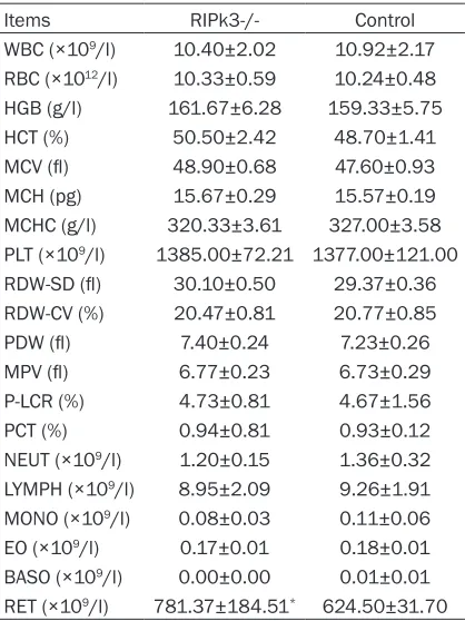
Detection of peripheral blood indicated that
RET increased considerably in the RIPk3
knock-out mice, while other peripheral blood
indica-tors were not significantly different from those
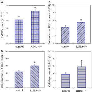
of the control mice (Table 1). Compared with
the control mice, the counts of BMNCs, HSCs
and bone marrow IL-6 level in the knockout
mice increased considerably. In addition, flow
cytometer showed that the cell death rate of
BMNCs in RIPk3-/- mice was significantly high
-er than that in control mice (Figure 4).
Morphological evaluation of the bone marrow
The bone marrow had normal structure and
morphology in the bone marrow-specific
RIPk3-knockout mice. Bone marrow cells proliferated
actively in mice of two different phenotypes.
A large number of hematopoietic cells were
observed, megakaryocytes were easy to be
seen (Figure 5). There was no sinusoidal
dilata-tion and the sinus wall was intact.
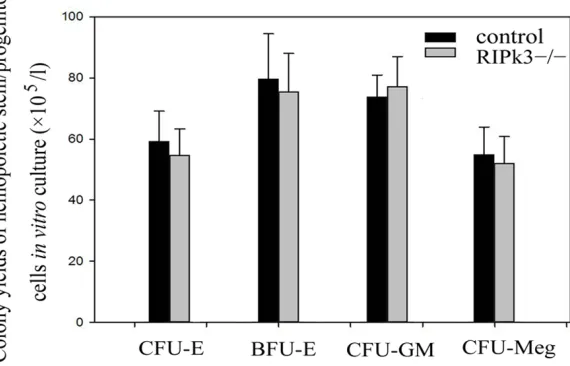
Colony yields of CFU-E, BFU-E, CFU-GM and
CFU-Meg
In vitro
cell culture indicated no significant dif
-ference between RIPk3-/- mice and control
mice in the colony yields of E, BFU-E,
CFU-GM and CFU-Meg (Figure 6).
Discussion
RIPk3 is a member of the receptor-interacting
[image:5.612.91.300.95.374.2]protein family and exhibits specific serine/thre
-onine kinase activity [22]. RIPk3 has been
found to be an important molecular switch for
PCD signaling. Excessive RIPk3 expression can
induce Caspase-mediated apoptosis of cells.
When Caspase activity is inhibited,
overexpres-sion of RIPk3 can promote the converoverexpres-sion from
Table 1. Peripheral blood and bone marrow
examination (
_
x
± s)
Items RIPk3-/- Control
WBC (×109/l) 10.40±2.02 10.92±2.17 RBC (×1012/l) 10.33±0.59 10.24±0.48
HGB (g/l) 161.67±6.28 159.33±5.75
HCT (%) 50.50±2.42 48.70±1.41 MCV (fl) 48.90±0.68 47.60±0.93
MCH (pg) 15.67±0.29 15.57±0.19
cell apoptosis to non-Caspase-medicated PCD
[23]. RIPk3 is involved in the pathogenesis of
many hematologic diseases [24, 25]. Through
preliminary experiments, it was found that
RIPk3-medicated PCD was involved in the cy-
clophosphamide- and busulfan-induced
aplas-tic anemia in mice [4]. Xiao et al. reported that
RIPk3-medicated PCD was related to the death
regulation of hematopoietic stem/progenitor
cells in Tak1-knockout mice. Moreover, it played
an important role in the progression of bone
marrow failure syndrome of Tak1(-/-) mice [26].
According to a recent study, defect in the
RIPk3-mediated PCD is related to the pathogenesis
of CLL and AML, and it is an important reason
for resistance to chemotherapy [14, 15]. Thus
RIPk3 is a valuable therapeutic target for
hema-tologic diseases.
Flox modification of the RIPk3 gene was per
-formed by targeting ES cells using homology
recombination. The Cre/Loxp system was used
for specific knockout of RIPk3 gene in the
mouse bone marrow. Cre recombinase is deriv-
ed from bacteriophage P1. Loxp is a 34 bp
pal-Thus the target gene remains normal in the
offspring. This method can prevent embryonic
death or severe developmental disorder at an
early developmental stage due to systemic
knockout in all cells and tissues [27-30].
Many reports have been published on the
spe-cific knockout by the breeding between mice
with specific Cre recombinase expression in the
bone marrow and Loxp transgenic mice. The
target gene is specifically knocked out in the
bone marrow of the offspring [21, 31]. The bone
marrow-specific RIPk3 knock-out mouse model
was successfully established using this
meth-od. However, this does not mean that the target
gene is removed from all bone marrow cells.
For example, the knockout rate is only
83%-98% in the mature macrophages, and
knock-out may be not limited to bone marrow cells.
Clausen et al. reported a 16% knockout rate in
a type of dendritic cells in the spleen [21]. More
researches are needed for the genotyping of
bone marrow-specific homozygous and hetero
-zygous RIPk3 knockout mice obtained in this
[image:6.612.91.387.70.366.2]study.
Figure 4. Bone marrow BMNCs and HSCs count, IL-6 level and cell death rate analysis. A. BMNCs count; B.HSCs count; C.IL-6 level; D.Cell death rate.*P < 0.05, compared with control group.
indromic sequence. Cre re-
combinase can specifically
recognize the Loxp
sequ-ence, causing
recombina-tion between 2 Loxp sequ-
ences and deletion of the
sequence between them.
DNA sequences in the im-
portant functional domain
of the target gene are
usu-ally flanked by 1 Loxp on
each side. The DNA se-
quence in the important
functional domain is mark-
ed by Loxp in the target
gene of the knockout mice.
Breeding between such
knockout mice and the
transgenic mice expressing
Cre recombinase in specific
Bone marrow is an important organ involved
in hematopoiesis and immune regulation, the
healthy bone marrow tissue is the structural
basis for maintaining normal hematopoiesis.
Our results showed that bone marrow had
normal structure and morphology for bone
marrow-specific RIPk3-knockout homozygotes.
However, flow cytometer showed that the cell
death rate of BMNCs in RIPk3-/- mice was
sig-nificantly higher than that in control mice, indi
-cated that bone marrow RIPk3 gene knockout
may lead to the increase of BMNCs cell death.
Physiological value of blood can reflect the
hematopoietic function of bone marrow to a
certain extent and also serves as an indicator
of the health status and genetic stability of
ani-mals. It is also an important basis for clinical
cant difference in the colony yields of
hemopoi-etic stem/progenitor cells of three lineages
between the two mice. This findings indicated
that the stability of the bone marrow
hemopoi-esis can be maintained under RIPk3 gene
knockout.
IL-6 is a kind of cytokine with a wide range of
biological activities, it is involved not only in
regulation of proliferation and differentiation of
early hemopoietic stem/progenitor, but also in
the progress of the stress reaction,
autoim-mune and neoplastic diseases of the body [32,
33]. In the present study, we found that the
bone marrow IL-6 level in RIPk3-/- mice was
increased significantly than that in control
mice. In addition, flow cytometry revealed that
[image:7.612.93.524.71.233.2]the number of BMNCs and HSCs in the bone
Figure 5. Histological analysis of bone marrow tissue in mice (100×), stained by HE. A. RIPk3-/- mice; B. Control.
Figure 6. Colony yields of hemopoietic stem/progenitor cells in vitro culture.
diagnosis. Our results sho-
wed that, compared with
the control mice, RET in
peripheral blood increased
significantly in the homozy
-gotes, while other
physio-logical indicators of
periph-eral blood did not change
significantly. All primitive
blood cells are derived from
bone marrow hemopoietic
stem/progenitor cells. Co-
lony yields of hemopoietic
stem/progenitor cells in vi-
tro
culture can reflect the
proliferative capacity of he-
mopoietic cells. Our results
of
in vitro culture showed
[image:7.612.99.384.284.467.2]-marrow of the RIPk3-/- mice increased
consid-erably, indicated that the positive
hematopoi-etic factors such as IL-6, may involved in the
promotion of proliferation, differentiation, and
maturation of HSCs in RIPk3-/- mice.
Taken together, the findings of our study implies
that bone marrow RIPk3 gene knockout may
lead to the increase of BMNCs cell death,
how-ever, increased secretion of hematopoietic
fac-tors such as IL-6 may promote the prolifera-
tion of hematopoietic stem/progenitor cells
and thus maintain the stability of bone marrow
hematopoiesis. This hypothesis and the detail-
ed mechanisms remain to be further investi-
gated.
Acknowledgements
This research was funded by the Public Wel-
fare Technology Application Research Project
of Zhejiang Province under Grant No. 2015-
C37122, Zhejiang, China. National natural
sci-ence foundation of China under Grant no.
81373139.
Disclosure of conflict of interest
None.
Address correspondence to: Yongfeng Chen, De- partment of Basic Medical Sciences, Medical Col- lege of Taizhou University, Taizhou, China. E-mail: [email protected]
References
[1] Caraballo JM, Acosta JC, Cortés MA, Albajar M, Gómez-Casares MT, Batlle-López A, Cuadrado MA, Onaindia A, Bretones G, Llorca J, Piris MA, Colomer D and León J. High p27 protein levels in chronic lymphocytic leukemia are associat-ed to low Myc and Skp2 expression, confer resistance to apoptosis and antagonize Myc effects on cell cycle. Oncotarget 2014; 5: 4694-4708.
[2] Chen Y, Zou Z, Wu Z, Zhao Z, Luo X, Xie C and Liang Y. TNF-α-induced programmed cell death in the pathogenesis of acquired aplastic ane-mia. Expert Rev Hematol 2015; 8: 515-526. [3] Zhang Y, Lu J, van den Berghe J and Lee SH.
Increased incidence of spontaneous apoptosis in the bone marrow of hyperdiploid childhood acute lymphoblastic leukemia. Exp Hematol 2002; 30: 333-339.
[4] Chen YF, Liu H, Luo XJ, Zhao Z, Zou ZY, Li J, Lin XJ and Liang Y. The roles of reactive oxygen
species (ROS) and autophagy in the survival and death of leukemia cells. Crit Rev Oncol He-matol 2017; 112: 21-30.
[5] Xia G, Chen B, Ding J, Gao C, Lu H, Shao Z, Gao F and Wang X. Effect of magnetic Fe3O4 nanoparticles with 2-methoxyestradiol on the cell-cycle progression and apoptosis of myelo-dysplastic syndrome cells. Int J Nanomedicine 2011; 6: 1921-1927.
[6] Moriwaki K, Balaji S, McQuade T, Malhotra N, Kang J and Chan FK. The necroptosis adaptor RIPK3 promotes injury-induced cytokine ex-pression and tissue repair. Immunity 2014; 41: 567-578.
[7] Newton K, Dugger DL, Wickliffe KE, Kapoor N, de Almagro MC, Vucic D, Komuves L, Ferrando RE, French DM, Webster J, Roose-Girma M, Warming S and Dixit VM. Activity of protein ki-nase RIPK3 determines whether cells die by necroptosis or apoptosis. Science 2014; 343: 1357-1360.
[8] Roychowdhury S, McMullen MR, Pisano SG, Liu X and Nagy LE. Absence of receptor inter-acting protein kinase 3 prevents ethanol-in-duced liver injury. Hepatology 2013; 57: 1773-1783.
[9] Wang Q, Liu Z, Ren J, Morgan S, Assa C and Liu B. Receptor-interacting protein kinase 3 con-tributes to abdominal aortic aneurysms via smooth muscle cell necrosis and inflamma -tion. Circ Res 2015; 116: 600-611.
[10] Meng L, Jin W, Wang Y, Huang H, Li J and Zhang C. RIP3-dependent necrosis induced inflam -mation exacerbates atherosclerosis. Biochem Biophys Res Commun 2016; 473: 497-502. [11] Afonso MB, Rodrigues PM, Carvalho T,
Cari-dade M, Borralho P, Cortez-Pinto H, Castro RE and Rodrigues CM. Necroptosis is a key patho-genic event in human and experimental mu-rine models of non-alcoholic steatohepatitis. Clin Sci (Lond) 2015; 129: 721-739.
[12] Mizumura K, Cloonan SM, Nakahira K, Bhashy -am AR, Cervo M, Kitada T, Glass K, Owen CA, Mahmood A, Washko GR, Hashimoto S, Ryter SW and Choi AM. Mitophagy-dependent ne- croptosis contributes to the pathogenesis of COPD. J Clin Invest 2014; 124: 3987-4003. [13] Chen YF, Zhao ZQ, Wu ZM, Zou ZY, Luo XJ, Li J,
Xie C and Liang Y. The role of RIP1 and RIP3 in the development of aplastic anemia induced by cyclophosphamide and busulphan in mice. Int J Clin Exp Pathol 2014; 7: 8411-8420. [14] Liu P, Xu B, Shen W, Zhu H, Wu W, Fu Y, Chen H,
[15] Nugues AL, El Bouazzati H, Hétuin D, Berthon C, Loyens A, Bertrand E, Jouy N, Idziorek T, Quesnel B. RIP3 is downregulated in human myeloid leukemia cells and modulates apopto-sis and caspase-mediated p65/RelA cleavage. Cell Death Dis 2014; 5: e1384.
[16] Bozec D, Iuga AC, Roda G, Dahan S and Yerets -sian G. Critical function of the necroptosis adaptor RIPK3 in protecting from intestinal tumorigenesis. Oncotarget 2016; 7: 46384-46400.
[17] Höckendorf U, Yabal M, Herold T, Munkhbaatar E, Rott S, Jilg S, Kauschinger J, Magnani G, Re-isinger F, Heuser M, Kreipe H, Sotlar K, Englei-tner T, Rad R, Weichert W, Peschel C, Ruland J, Heikenwalder M, Spiekermann K, Slotta-Hus-penina J, Groß O and Jost PJ. RIPK3 restricts myeloid leukemogenesis by promoting cell death and differentiation of leukemia initiating cells. Cancer Cell 2016; 30: 75-91.
[18] Xu, Y, Lin Z, Zhao N, Zhou L, Liu F, Cichacz Z, Zhang L, Zhan Q and Zhao X. Receptor interac-tive protein kinase 3 promotes cisplatin-trig-gered necrosis in apoptosis-resistant esopha-geal squamous cell carcinoma cells. PLoS One 2014; 9: e100127.
[19] Feng X, Song Q, Yu A, Tang H, Peng Z and Wang X. Receptor-interacting protein kinase 3 is a predictor of survival and plays a tumor sup-pressive role in colorectal cancer. Neoplasma 2015; 62: 592-601.
[20] Xin J, You D, Breslin P, Li J, Zhang J, Wei W, Can-nova J, Volk A, Gutierrez R, Xiao Y, Ni A, Ng G, Schmidt R, Xia Z, Pan J, Chen H, Patel MM, Kuo PC, Nand S, Kini AR, Zhang J, Chen J, Zhu J, Zhang J. Sensitizing acute myeloid leukemia cells to induced differentiation by inhibiting the RIP1/RIP3 pathway. Leukemia 2017; 31: 1154-1165.
[21] Clausen BE, Burkhardt C, Reith W, Renkawitz R and Förster I. Conditional gene targeting in macrophages and granulocytes using LysMcre mice. Transgenic Res 1999; 8: 265-277. [22] Park SM, Yoon JB and Lee TH. Receptor
inter-acting protein is ubiquitinated by cellular in-hibitor of apoptosis proteins (IAP1 and c-IAP2) in vitro. FEBS Lett 2004; 566: 151-6. [23] Zhang DW, Shao J, Lin J, Zhang N, Lu BJ, Lin
SC, Dong MQ and Han J. RIP3, an energy me-tabolism regulator that switches TNF-induced cell death from apoptosis to necrosis. Science 2009; 325: 332-336.
[24] Laubach JP, Fu P, Jiang X, Salter KH, Potti A and Arcasoy MO. Polycythemia vera erythroid pre-cursors exhibit increased proliferation and apoptosis resistance associated with abnor-mal RAS and PI3K pathway activation. Exp He-matol 2009; 37: 1411-1422.
[25] Li JP, Zheng CL and Han ZC. Abnormal immu-nity and stem/progenitor cells in acquired aplastic anemia. Crit Rev Oncol Hematol 2010; 75: 79-93.
[26] Xiao Y, Li H, Zhang J, Volk A, Zhang S, Wei W, Zhang S, Breslin P and Zhang J. TNF-α/Fas-RIP-1-induced cell death signaling separates murine hematopoietic stem cells/progenitors into 2 distinct populations. Blood 2011; 118: 6057-6067.
[27] Gui YS, Wang L, Tian X, Feng R, Ma A, Cai B, Zhang H, Xu KF. SPC-Cre-ERT2 transgenic mouse for temporal gene deletion in alveolar epithelial cells. PLoS One 2012; 7: e46076. [28] Katsuno T, Umeda K, Matsui T, Hata M,
Tamu-ra A, Itoh M, Takeuchi K, Fujimori T, Nabeshima Y, Noda T, Tsukita S and Tsukita S. Deficiency of zonula occludens-1 causes embryonic le -thal phenotype associated with defected yolk sac angiogenesis and apoptosis of embryonic cells. Mol Biol Cell 2008; 19: 2465-2475. [29] Yang SM, Hou ZH, Yang G, Zhang JS, Hu YY,
Sun JH, Guo WW, He DZ, Han DY, Young WY and Yang X. Chondrocyte-specific Smad4 gene conditional knockout results in hearing loss and inner ear malformation in mice. Dev Dyn 2009; 238: 1897-1908.
[30] Li C, Li YP, Fu XY and Deng CX. Anterior visceral endoderm SMAD4 signaling specifies anterior embryonic patterning and head induction in mice. Int J Biol Sci 2010; 6: 569-583.
[31] Sinclair GB, Jevon G, Colobong KE, Randall DR, Choy FY. and Clarke LA. Generation of a condi-tional knockout of murine glucocerebrosidase: utility for the study of Gaucher disease. Mol Genet Metab 2007; 90: 148-156.
[32] Möbius-Winkler S, Hilberg T, Menzel K, Golla E, Burman A, Schuler G, Adams V. Time-depen-dent mobilization of circulating progenitor cells during strenuous exercise in healthy individu-als. J Appl Physiol 2009; 107: 1943-1950. [33] Nishimoto N, Kishimoto T. Interleukin 6: from
Supplementary File 1. Plasmid sequencing results
1. M0827.0479-RIP3-RIP3_C1R_F09
TAAAGGAAAGGAAAGGGAAGGTATTAGATGAAGGGGGGGGTCAGAGATTTTTCCCTCTCTTGCTCCCCAGAAGATGCAGC AGCCTCAGCTTTTTGGAAGGTGTTATGGAGGACCAGAGGGAAGGTAAAGTCATTGAGAACTTAGCAGGAGATCTGAGTTG CTGATGGGCAGGCCTGAGATAAGTCAGCCGGATCCCCTCGAGGGACCTAATAACTTCGTATAGCATACATTATACGAAGT TATATTAAGGGTTATTGAATATGATCGGAATTGGGCTGCAGGAATTCGTCGACTGGGTCCGTGAGGGTGCATTAGGCCTG TGGAACTGAGCCAGGGGGTTAAGAAGGGCGTCTGGTAAGGAGGGTCACCTGGTATGACTAGAAATAAAACTCCTAGTTGG GTCTTGTCATGTGTGTAGTCTTGTATTCAGAGGGACAGGTACTCAACATGGTCCCAAGTCTACACTAGGTTGTAGATGGA CTAACCTTGGCGTGGAGCTCTGGATCCAGCAGAATGTTAGAGGGCTTGAGGTCCCGGTGCAGGAGCGGAGGGTTCAAGCT GTGTAGGTAGCACATCCCCAGCACCACTTCCTGCAGCAGGCGACAGAGGAGTGGCCAGGGCCGAGGGCACTCGGGTTGCA GCAGCCCTGCGAGGGAGCCATTCTCCATGAATCTTGTCACCAGAGCCTGCCCGGACACGAAGTCCCACTGGAGGTCCTCA GTGACCCCCAGCAGGAGCAGAACGTTCTCATTACGAAGATTAACCATAGCCTTCACCTCCCAGGATATCTTCTTCCTACA CGCCGAAAGGGGGCCAAAATTGGATTGCGCTCTGGAAGACCCCACCCTTGCCCCACTCAGCTCGTGACCACACTCTGACT ACAGCTGGGGTCACTCACGAGTTCACGATCTTGACTGCTACATCATGGTTCCATG
2. M0827.0480-RIP3-NEO_R_G09
CGCTCACCTGGCACGACGCGAGCTGCGGGGCGGGGGGGACTTCCTGACTAGGGGAGGAGTAGAAGGTGGCGCGAAGGGGC CACCAAAGAACGGAGCCGGTTGGCGCCTACCGGTGGATGTGGAATGTGTGCGAGGCCAGAGGCCACTTGTGTAGCGCCAA GTGCCCAGCGGGGCTGCTAAAGCGCATGCTCCAGACTGCCTTGGGAAAAGCGCCTCCCCTACCCGGTAGAATTTCGACGA CCTGCAGCCAAGCTAGCTTGGCTGGACGTAAACTCCTCTTCAGACCTGAAGTTCCTATACTTTCTAGAGAATAGGAACTT CGGAATTCTGCTGTTTGGGTGTCTTTGATGGGAGACCTTCTGGACTTGCTTATCAATAAAGGAACTGTGGGGGCTCACAG ACTCTGTGCTGGGAAAAGTCAGCCAATCCCGACTTTCTTTCGTTGTGTGACCTCAGTGGGATCCTCTCCTAGTGGATGTC ATGTTCCTCGTCTTAAACGAGGCAGATCGGGTTAGATGATCTCTAGTCATTCCTTCCCACCTCCAGAGTAACTCATGTCT ATAGATAAGTGCTGAGGAGGAAGGTGATGGGAGAGCTGAGCGGGGAGAGCAAGAGAGAATGAAGAATGGGTACCTGTCAT TGGATTCGGTGGGGTCCAGGGATACCAAGGAGTGCCGTGTCTTCCATCTCCCTGCAAACAACACAGAGCTCTCAGGCTTT CTATTCAAGAGCCTAATCTCATCACCGTGCAGTTCTCCTGGGTAAGAGGGTGTGCTCTAGTCTTGCCAGTGTCTCACCTG ATTCCTTTGGGGGTGAGGACCGGGAGTCTCAGTAAAGACTGGCCCAGGTGTTGTGCCTCTGAAGGGTAAAGTATGTGGAA TCTGAGGAGTGCCAGCCACGGGGTCAGAAGATGTCCTTGCTGGTGTGGCAGGCCCAACTGATGTGTCCTGTGCTTGCCTC TCAGGACATTTTCCAGGAACTGGTCCGGAGGGTTCCTCCAAATGCA
3. M0827.0481-RIP3-NEO_L_H09
Supplementary File 2. ES sequencing results
1. M0928.3699-2_5R_C11-NEO_R_A03
TGAATACTCTCAAGACGCGAGCTGCGGGGCGGGGGGGACTTCCTGACTAGGGGAGGAGTAGAAGGTGGCGCGAAGGGGCC ACCAAAGAACGGAGCCGGTTGGCGCCTACCGGTGGATGTGGAATGTGTGCGAGGCCAGAGGCCACTTGTGTAGCGCCAAG TGCCCAGCGGGGCTGCTAAAGCGCATGCTCCAGACTGCCTTGGGAAAAGCGCCTCCCCTACCCGGTAGAATTTCGACGAC CTGCAGCCAAGCTAGCTTGGCTGGACGTAAACTCCTCTTCAGACCTGAAGTTCCTATACTTTCTAGAGAATAGGAACTTC GGAATTCTGCTGTTTGGGTGTCTTTGATGGGAGACCTTCTGGACTTGCTTATCAATAAAGGAACTGTGGGGGCTCACAGA CTCTGTGCTGGGAAAAGTCAGCCAATCCCGACTTTCTTTCGTTGTGTGACCTCAGTGGGATCCTCTCCTAGTGGATGTCA TGTTCCTCGTCTTAAACGAGGCAGATCGGGTTAGATGATCTCTAGTCATTCCTTCCCACCTCCAGAGTAACTCATGTCTA TAGATAAGTGCTGAGGAGGAAGGTGATGGGAGAGCTGAGCGGGGAGAGCAAGAGAGAATGAAGAATGGGTACCTGTCATT GGATTCGGTGGGGTCCAGGGATACCAAGGAGTGCCGTGTCTTCCATCTCCCTGCAAACAACACAGAGCTCTCAGGCTTTC TATTCAAGAGCCTAATCTCATCACCGTGCAGTTCTCCTGGGTAAGAGGGTGTGCTCTAGTCTTGCCAGTGTCTCACCTGA TTCCTTTGGGGGTGAGGACCGGGAGTCTCAGTAAAGACTGGCCCAGGTGTTGTGCCTCTGAGGGGTAAAGTATGTGGAAT CTGAGGAGTGCCAGCCACCGGCGTCAAAGATGTCCTTGCTGGTGTGGCAGGCCCAACTGATGTGTCCTGTGCTTGCTCTC ACGACATTTTCAAGGAACTGGTCCGGAGGGTTCTCCAAATGCAGCGGTCCAGCATTTTATAACCATTGGTTTCCTTCGGG CAAT
2. M0928.3700-2_5R_C11-RIP3_C1R_B03
CGAAAATCGGATCTGATCTGATCCTGACCCTGACTGGGACCCTCCCTGAAACGTGGACAGGCCAAAATCTGCTAGCTGTA AAGGAAAGGAAAGGGAAGGTATTAGATGAAGGGGGGGGTCAGAGATTTTTCCCTCTCTTGCTCCCCAGAAGATGCAGCAG CCTCAGCTTTTTGGAAGGTGTTATGGAGGACCAGAGGGAAGGTAAAGTCATTGAGAACTTAGCAGGAGATCTGAGTTGCT GATGGGCAGGCCTGAGATAAGTCAGCCGGATCCCCTCGAGGGACCTAATAACTTCGTATAGCATACATTATACGAAGTTA TATTAAGGGTTATTGAATATGATCGGAATTGGGCTGCAGGAATTCGTCGACTGGGTCCGTGAGGGTGCATTAGGCCTGTG GAACTGAGCCAGGGGGTTAAGAAGGGCGTCTGGTAAGGAGGGTCACCTGGTATGACTAGAAATAAAACTCCTAGTTGGGT CTTGTCATGTGTGTAGTCTTGTATTCAGAGGGACAGGTACTCAACATGGTCCCAAGTCTACACTAGGTTGTAGATGGACT AACCTTGGCGTGGAGCTCTGGATCCAGCAGAATGTTAGAGGGCTTGAGGTCCCGGTGCAGGAGCGGAGGGTTCAAGCTGT GTAGGTAGCACATCCCCAGCACCACTTCCTGCAGCAGGCGACAGAGGAGTGGCCAGGGCCGAGGGCACTCGGGTTGCAGC AGCCCTGCGAGGGAGCCATTCTCCATGAATCTTGTCACCAGAGCCTGCCCGGACACGAAGTCCCACTGGAGGTCCTCAGT GACCCCCAGCAGGAGCAGAACGTTCTCATTACGAAGATTAACCATAGCCTTCACCTCCCAGGATATCTTCTTCCTACACG CCGAAAGGGGGCCAAAATTGGATTGCGCTCTGGAAGACCCCACCCTTGCCCCACTCAGCTCGTGACCACACTCTGACTAC AGCTGGGGTCACTCACGAGTTCACGATCTTGACTGCTACATCATGGTTCCATGTTCTGTGGTGTGCCCGGAACACGACTC CGAACCCTCCTTTACCCACAAACTCCAGCTTCTTCAGTTCTTCACGGCTCACCAGAGGAACCGCCTGACGCCCCAGTAGC CTGAAGAAAGGTTCTTGTCCGTAGGGGCCCATCCCCCAGCCTACGCATTCTCTGTCTCCAGCTCCGGATTCCTTGACTGC CGCGTGGCCAGCGTAGGCAGCGAG
3. M0928.3701-2_5R_H11-5RIP_111_C03
4. M0928.3714-2_3R_C11-3RIP_212_H04
CGCCCACATCATCTTGAGGAGGCAATGATTACCCTGAGCCTCTTATAAAGAAGGTGCTAAGAAGCTGCTCTTTCTGAGCT GCTGTCCCTTTCTGCCTCTTAGGTCCTACTGACCTGGTGCTTTTTCTCAGTGTCCAGCCTCCGGCGTGAGTGCATAGAGC TAAGAGCCTCCCGGAACGTGGCCCGCAATGCTCGCTCCATCAGGGCCTTGTGGTACCCTCCCACCACGTTCCCACACAGG AACAATACCGCGTTTGCTGCCAACTGTGGGCAAAGGCCAGACTTAAAAAGTGTACTACTCCCCTATCTCTACTCTGTGGG GGAGTCTATCTGTGTAGCTGGAGTTGGTTGCAAACACAGGTCGAGGAGAGCTGGCTCTATGGTTAAGGACCTAGGTATCT CAGAACAATAGGAGCCACTTTGTGACATCCATCCGGAAGAGTTGGTTGCTTTCCTTCTCTGCTCTCTGCTATAGACTCTT TCTTGGCTGTGGAACCAACCTGACCGCTTAGGCCTAGCTATCTTGCATGTATATTTCCTCTGTCTACAGTGCTATTCTTC CATATTCATCTCCTGAGCCCATTCCAGCTGCAAGAAACTACTCCAGTTCTGATTCACGTTTTCCACTAATGCTCCTCGGT TTACTTCTGTACCCGCGAGAGAATCTTGACTGATTTCCCAATGGCTATAGTGCCCTCTTCTGACAAAGGTAGGCATGATG CACTGATGTACCAACGACACTAGAGGTCGCTATAGGGGTGTTATCAATGCTTGGCTCCTGCGAGGGCCGAGCCATGTCTT GGATAATGCTCACAGCATTACTAAACAGCTGACCCAGCTCTGGGCACACTATCGACACCACTTGCATTTTTACCTGTGGT AGCAGAGCTCTCTGTGACTCAAGCTGCCACCCAAGATACAGTCCAAGGACCAGCACGTGCGAGAGGGATGAGATGACTCC CGCAGCAGCAGCATCCCGCATGCCCAAGGCAGCATGGCGTACACGGTGAAGATGATGAAGAGGAAAAAAAGACACCTGGA GAAGGGTGTGGGAAGCTGAATGCCCGTGATGTTGCGCCCAGGGTCTAAATTTGAAGGTTCAACTTCCCTCAAGCCTTGAT TTTCCTCACCTGGATCCAAGGCGCTCACCACACCCCCCAGTGCACAAGAAAATTCCATAGGCCAAAGGCGGTAATGCCAC CCGATATTGC
5. M0928.3715-2_3R_C11-NEO_L_A05
Supplementary File 3. Sequencing results of F1 generation
Results of no. 1:
Results of no. 2:
Results of no. 3:
Results of no. 4:
Results of no. 5: