Original Article
High FEV1 and BMI as protective factors in reducing the
infection incidence of lung cancer in patients
who underwent thoracoscopic surgery
Ming Wang1, Pengfei Zou2
1Department of Thoracic Surgery, Shulan (Hangzhou) Hospital, Hangzhou, Zhejiang Province, China; 2Department of Infectious Diseases, Shulan (Hangzhou) Hospital, Hangzhou, Zhejiang Province, China
Received May 30, 2018; Accepted July 20, 2018; Epub December 15, 2018; Published December 30, 2018 Abstract: Objective: Thoracoscopic surgery has become an important treatment approach for lung cancer in China. However, postoperative complications seriously affect surgical outcomes and prognoses. This study explored the risk factors involved in perioperative concurrent infections. Methods: A total of 253 patients with lung cancer
un-dergoing video-assisted thoracoscopic surgeries were classified into infected (47) and non-infected (206) groups.
Univariate analysis and logistic regression were used to analyze factors associated with concurrent infection. Results: Single factor analysis on preoperative factors showed that age, smoking index, diabetes mellitus, forced
expiratory volume in the first second (FEV1), ejection fraction (EF) value, body mass index (BMI), and albumin levels influenced the incidence of infections after the thoracoscopic surgery in patients with lung cancer. Single factor analysis on intraoperative factors showed that operation time and intraoperative blood loss may influence the
incidence of infection following thoracoscopic surgery. Single factor analysis on postoperative factors showed that
postoperative drainage time also influenced the incidence of postoperative infections in patients with lung cancer.
On multivariate analysis, age, operative time, postoperative drainage time, and diabetes mellitus were risk factors
for higher incidence of postoperative infections in patients with lung cancer (P<0.05), whereas high FEV1 and BMI
were protective factors in reducing postoperative infection (P<0.05). Conclusion: Age, operative time, postoperative drainage time, and diabetes mellitus are risk factors for infections following a thoracic surgery in patients with lung
cancer. High FEV1 and BMI are protective factors in reducing the incidence of postoperative infections.
Keywords: Thoracoscopic surgery, perioperative infections, lung cancer, preoperative interventions
Introduction
Lung cancer is the first leading cause of cancer
deaths worldwide. Approximately 1.1 million people have been reported to die from lung cancer, and approximately 1.2 million new cases were diagnosed each year worldwide. In China, lung cancer is also the most common malignant tumor. Currently, surgical treatment remains one of the primary treatment methods. In recent years, with the development of mini-mally invasive techniques with the aim to improve patients’ quality of life, thoracoscopic surgery has become an important treatment alternative, especially in patients with early-stage lung cancer. However, due to factors such as underlying diseases, surgical factors, comor-bidities, and compromised immunity, patients
may experience postoperative complications [1, 2], such as infections, atelectasis, arrhyth-mias, respiratory failure, heart failure, and pul-monary embolism, among others. The occur-rence of postoperative complications can seri-ously affect the surgical outcomes and patient prognosis [3, 4] and may also seriously endan-ger patients’ lives. Among these complications, postoperative infection is one of the most com-mon, with an incidence of about 20% [5]. Once a patient acquires an infection, it often leads to exacerbations and is not conducive to a favor-able prognosis.
analysis on risk factors for postoperative infec-tion after a thoracoscopic surgery can provide the theoretical basis for the prevention and treatment of concurrent infections in patients undergoing thoracoscopic procedures. Previous screening on risk factors has focused on post-operative-related factors. However, the under-lying health status of preoperative patients also carries the potential risk of coinfection. Therefore, this study evaluated the risk factors of perioperative concurrent infections by ana-lyzing the patient’s basic preoperative status and related factors postoperatively, with the aim of providing a basis for early intervention and treatment of postoperative infections. Materials and methods
Patient eligibility
We retrospectively analyzed the data of 253 patients with lung cancer who underwent tho-racoscopic surgery at Shulan (Hangzhou) Hospital. All patients were diagnosed with lung cancer by pathological examination and had no prior thoracoscopic surgery. Exclusion criteria included the following: (1) patients with acute and chronic infection preoperatively; (2) aller-gies to antibiotics; (3) secondary surgery for lung cancer recurrence; (4) previous history of other malignant tumors; and (5) incomplete data.
Treatment method
All patients underwent lung surgery after com-pleting preoperative examinations, adequate smoking cessation, lung function training, and airway preparation. All surgeries were per-formed under thoracoscopic non-contrast vision. The decision to perform lung lobe, lung segment, wedge resection, or pneumonectomy depends on the patient’s condition. Lobectomy was performed using Wang’s method. The sub-lobectomy margin was required to be not sm-
aller than the tumor diameter or ≥2 cm.
Mediastinal lymph node sampling was per-formed on some wedge resections, while sys-temic lymph node dissection was performed for the remainder.
Based on whether infection occurred postop -eratively, patients were divided into infected
(n=47) and non-infected (n=206) groups. In- fections included lung infections and incision infections.
Data collection
Preoperative factors: For all patients, basic
data were routinely collected upon admission and after preoperative examinations, which included pulmonary function,
echocardiogra-phy, natriuretic peptide (BNP) levels, liver and
renal function tests, and hematuria examina-tion. The following indicators were included in the statistical analysis: (1) general information:
age, sex, and smoking index (>400, ≤400); (2)
concomitant diseases: chronic diseases such as diabetes mellitus, coronary heart disease, hypertension, and stroke; (3) pulmonary
func-tion: FEV1% (the actual ratio of the forced expi
-ratory volume in the 1st s ratio >60%, ≤60%); (4) heart function: EF (>55%, ≤55%) measured by cardiac ultrasound and blood BNP levels (>500 pg/mL, ≤500 ng/mL); and (5) nutritional indicators: body mass index [BMI] (>24 kg/m2,
≤24 kg/m2) and serum albumin level (>35 g/L,
≤35 g/L).There were 12 factors in total. The
smoking index was calculated as follows: num-ber of cigarettes smoked per day × years of
smoking. BMI was defined as weight/height2.
Intraoperative factors: A total of three intraop-erative factors were included: the type of sur-gery (pulmonary resection, sub-lobectomy,
lobectomy), operative time (>3 h, ≤3 h), and intraoperative blood loss (>300 mL, ≤300 mL).
Postoperative factors: A total of four postopera-tive factors were included: pathological type (squamous cell carcinoma, adenocarcinoma, small cell carcinoma, large cell carcinoma,
oth-ers), pathological stage (I, II, III, IV), pathological
differentiation (low differentiation, moderately well-differentiated), and drainage time (>60 h,
≤60 h).
Statistical analysis
test, and rank data were computed with the sum test. In multivariate logistic regression analysis, a P<0.05 indicated statistical signi-
ficance.
Results
Analysis of surgical management
A total of 253 patients underwent a surgery; among them 182 (71.94%) underwent thoraco-scopic lobectomy and 71 underwent sub-lobar resection. Out of the 71 patients who had sub-lobar resection, 25 (9.88%) underwent seg-mental resection and 46 (18.18%) wedge resection. The postoperative infection rate was 18.58% (47/253). None of the patients died during the perioperative period. Postoperative histological types included 116 cases (45.85%) of adenocarcinoma, 80 cases of squamous cell carcinoma (31.62%), 19 cases of small cell car-cinoma (7.51%), 21 cases of large cell carcino-ma (8.30%), and 17 cases of undifferentiated carcinoma. Pathological differentiation was determined as poor in 86 cases (33.99%) and moderate in 167 cases (66.01%). The postop-erative pathological stages included 129 cases (50.99%) in stage I, 95 cases (37.55%) in stage II, 24 cases (9.49%) in stage III, and 5 cases
(1.98%) in stage IV. Among the 47 infected
patients, one case had incision infection and one had incision infection combined with lung infection (Figure 1).
Single factor analysis
Single factor analysis of the preoperative fac-tors in the two groups showed that was the dif-ference in the incidence of infections with regard to sex; presence of hypertension,
coro-nary heart disease, stroke; or BNP levels was not significant between the infected and
non-infected groups (P>0.05). However, a
statisti-cally significant difference was observed with
regard to age, smoking index, diabetes
melli-tus, FEV1%, EF, BMI, and albumin levels
(P<0.05), which suggested that these factors
may influence the incidence of infections in
patients with lung cancer after a thoracoscopic surgery (Table 1).
The proportion of patients with operative time of >3 h and blood loss of >300 mL in the
infect-ed group was significantly higher than that in
the non-infected group (Table 2). The drainage
time of the infected group was significantly
higher than that of the non-infected group (Table 3), which suggested that operative time, intraoperative blood loss, and postoperative
drainage time may be the influencing factors for
infections in patients with lung cancer after a thoracoscopic surgery.
Multivariate analysis
[image:3.612.89.520.71.276.2]Therefore, preoperative assessment of a
patient’s nutritional status by measuring BMI
can have a certain predictive value for the occurrence of postoperative infection (Table 4). Discussion
Lung infection in patients undergoing thoraco-scopic surgery is a common postoperative com-plication and affects postoperative recovery [7, 8]. A large number of studies [9, 10] have shown that pulmonary complications will affect long-term outcomes in patients undergoing lung surgery. Due to various factors, concurrent infection in patients undergoing thoracoscopic surgery for lung cancer remains a complication with high postoperative incidence. Therefore,
clinical prevention is of utmost significance to identify factors that may influence the inci -dence of concurrent infections in patients with lung cancer who underwent thoracoscopic sur-gery. This study found that age, operative time, postoperative drainage time, and diabetes mel-litus were risk factors for postoperative
infec-tion after a thoracic surgery in patients with
lung cancer, while high levels of FEV1% and BMI
may be protective factors in preventing postop-erative infection.
Preoperative evaluation is closely related to the risk of surgical treatment. A comprehensive preoperative evaluation [11] includes tumor staging, cardiopulmonary function assess-ment, and nutritional status assessassess-ment, among others. The statistical analysis of preop-erative indicators in this study included
assess-ment of FEV1%, EF values, BNP levels, BMI, and
albumin levels. The cardiopulmonary function of patients was comprehensively evaluated to investigate the correlation between heart and lung functions and postoperative infection. Univariate analysis showed that age, smoking
index, diabetes, FEV1%, EF values, BMI, and albumin levels may be influencing factors of
[image:4.612.93.521.99.387.2]infections in patients with lung cancer after a thoracoscopic surgery. Single factor analysis on intraoperative and postoperative indexes showed that differences in operation time, Table 1. Analysis of preoperative factors in patients with lung cancer with and without infection after undergoing thoracoscopic surgery
Terms Infection group n=47 Non-infection group n=206 Statistics P Gender
Male 26 (55.32%) 117 (56.80%)
Female 21 (44.68%) 89 (43.20%) 0.034▲ 0.854
Age (years, n)
<60 11 (23.40%) 83 (40.29%)
≥60 36 (76.60%) 123 (59.71%) 2.158Δ 0.031
Smoking index (n)
<400 13 (27.66%) 116 (56.31%)
≥400 34 (72.34%) 90 (43.69%) 3.538Δ 0.000
Combined diseases (n)
Diabetes (with/without) 31/16 65/141 19.237▲ 0.000
Hypertension (with/without) 28/19 126/80 0.041▲ 0.840
Coronary heart disease (with/without) 26/21 87/119 2.652▲ 0.103
Stroke (with/without) 6/41 16/190 1.205▲ 0.272
FEV1% (n)
>60% 17 (36.17%) 152 (73.39%)
≤60% 30 (63.83%) 54 (26.21%) 4.931Δ 0.000
EF value (%) 54.23±6.05 56.14±5.39 2.142# 0.033
BNP level (pg/mL) 357.28±112.53 316.57±153.91 1.711# 0.088
BMI (kg/m2) 17.93±5.28 20.39±4.32 3.373# 0.001
Albumin (g/L) 30.56±6.19 37.83±5.31 8.204# 0.000
intraoperative blood loss, and postoperative drainage time between the two groups were
statistically significant (P<0.05).
Further multivariate logistic regression analysis
showed that age, operative time, postoperative drainage time, and diabetes mellitus were risk factors for infection after a thoracic surgery in patients with lung cancer (P<0.05), while
ele-vated levels of FEV1% and BMI were protective
factors in reducing the incidence of postopera-tive infection (P<0.05). Smoking is closely relat-ed to the occurrence of lung cancer [12] and
can lead to chronic respiratory inflammatory
reactions, including bronchial glandular hyper-plasia, goblet cell proliferation, and destruction
of the normal physiological structure of the lung tissues. Likewise, smoking can damage the endothelial cells of the lung, resulting in re- duced alveolar compliance, postoperative lung
insufflation, poor drainage, and postoperative
complications such as infection. Studies [13] have shown that the incidence of postoperative complications in patients with a smoking index
of ≥400 is 2.4 times higher than those with a
smoking index of <400. Therefore, in patients
with a smoking index of ≥400, preoperative
respiratory tract preparation should be rein-forced in order to reduce respiratory mucosal
inflammatory responses and improve the repair of the mucosal epithelium. Furthermore, respi -Table 2. Analysis of intraoperative conditions in lung cancer patients with and without infection after undergoing thoracoscopic surgery
Terms Infected group n=47 Non-infected group n=206 Statistics P Surgery type (n)
Sub-lobar resection 17 (36.17%) 54 (26.21%)
Lobectomy 30 (63.83%) 152 (73.79%) 1.879▲ 0.170
Operation time (n)
≤3 h 21 (44.68%) 154 (74.76%)
>3 h 26 (55.32%) 52 (25.24%) 4.021Δ 0.000
Intraoperative blood loss (n)
≤300 mL 28 (59.57%) 185 (89.81%)
>300 mL 19 (40.43%) 21 (10.19%) 5.116Δ 0.000
[image:5.612.90.521.96.229.2]Note: ▲The statistical value is χ2 value; ΔThe statistical value is Z value.
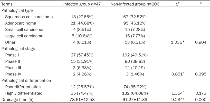
Table 3. Analysis of postoperative factors in lung cancer patients with and without infection after undergoing thoracoscopic surgery
Terms Infected group n=47 Non-infected group n=206 χ2 P
Pathological type
Squamous cell carcinoma 13 (27.66%) 67 (32.52%)
Adenocarcinoma 21 (44.68%) 95 (46.12%)
Small cell carcinoma 4 (8.51%) 15 (7.28%)
Large cell carcinoma 5 (10.64%) 16 (7.77%)
Other 4 (8.51%) 13 (6.31%) 1.036▲ 0.904
Pathological stage
Phase I 27 (57.45%) 102 (49.51%)
Phase II 15 (31.91%) 80 (38.83)
Phase III 3 (6.38%) 21 (10.19)
Phase IV 2 (4.26%) 3 (1.46%) 0.851Δ 0.395
Pathological differentiation
Poor differentiation 12 (25.53%) 74 (35.92%)
Highly differentiated 35 (74.47%) 132 (64.08%) 1.354Δ 0.176
Drainage time (h) 78.61±12.58 61.27±11.39 9.234# 0.000
[image:5.612.90.521.287.500.2]ratory function training should be incre- ased to facilitate postoperative lung lobe expansion and sputum discharge. Pulmonary function is an important indicator for the risk assessment during thoracic surgery [14].
Because elderly patients with lung cancer
have decreased chest wall compliance and decreased respiratory muscle strength, their combined lung function reserve capacity is often reduced, and postoperative chest pain can further limit the compensation of lung func-tion; therefore, airway resistance, lung tissue elasticity, and thoracic and respiratory muscle function should be fully assessed using pre-
operative lung function measurements. By
strengthening preoperative exercises and post-operative care, patients’ lung function can be improved to a certain extent, and the risk of postoperative infections can be reduced. Diabetes mellitus is a risk factor for postopera-tive infections, which induced microcirculatory disturbances. The incidence of postoperative pulmonary infection in patients with diabetes
and lung cancer has been reported to be signifi -cantly higher than that in patients without dia-betes [14-17], and diadia-betes is associated with postoperative wound infections. Therefore, in patients with lung cancer with diabetes, preop-erative blood pressure reduction is recom-mended, diet should be adjusted, postopera-tive glucose levels closely monitored, surgical incisions observed, dressings changed regular-ly, and strict aseptic conditions maintained [18, 19].
Surgical time is also a risk factor for posto-
perative infection. Because the entire proce -dure requires mechanical ventilation, the lon-ger the operation time is, the higher the risk of lung infection. Therefore, the anatomical
struc-ents postoperatively reduces the body’s immune response and increases the risk of infection [21]. The present study showed that
BMI and albumin levels in the infected group were significantly lower than those in the
non-infected group, indicating that nutritional
sta-tus is an influencing factor for postoperative
infection. Multivariate analysis showed that
BMI is a protective factor to reduce postopera -tive infection. However, albumin levels in the multivariate analysis did not show any
statisti-cal significance. The reason may be related to higher BMI. However, [22] the decline in albu -min levels has been reported to be associat- ed with postoperative infection. Therefore, a patient’s albumin levels should be monitored postoperatively, and if necessary, hypoprotein-emia should be promptly corrected. In addition, patients should be encouraged to consume a high-protein diet and increase nutritional
intake. In the univariate analysis, the EF value
observed in this study was higher in the non-infected group than that in the non-infected group.
However, no statistical significance was dem -onstrated in the multivariate analysis, which is likely due to the small sample size.
In conclusion, age, operative time, postopera-tive drainage time, and diabetes mellitus are risk factors for postoperative thoracic surgery in patients with lung cancer. Elevated levels of
[image:6.612.89.369.96.202.2]FEV1% and BMI are protective factors in reduc -ing the incidence of postoperative infection. Patients with lung cancer with these associat-ed risk factors should be managassociat-ed with appro-priate preoperative intervention and posto- perative care, reduced operation time, strong- er emphasis on the recognition of relevant risk factors, and effective measures to avoid and reduce the occurrence of concurrent infections.
Table 4. Multivariate analysis of risk factors for concurrent infec-tion in lung cancer patients undergoing thoracoscopic surgery
Factor β S.E. Wals χ2 OR 95% CI P
Age 1.122 0.651 4.236 1.127 1.012~9.204 0.013 Operation time 1.585 0.835 6.594 1.216 2.933~35.267 0.002 Smoking index 0.759 0.124 7.525 2.763 1.125~11.938 0.000
BMI -0.521 0.328 4.921 0.875 0.219~2.623 0.021
FEV1% -0.093 0.167 3.617 0.729 0.348~1.269 0.023 Diabetes 1.036 0.598 3.872 1.172 1.018~3.216 0.025 Drainage time 1.235 0.716 4.383 3.528 1.293~13.117 0.000
ture of the patient’s lesion should be fully assessed pre- operatively to predict risks and potential emergencies and min-imize the operation time. Mal- nutrition is an important factor affecting postoperative recov-ery in elderly patients [20].
Because of blood loss during
surgery, limited postoperative
fluid consumption, nutritional
nutri-Disclosure of conflict of interest
None.
Address correspondences to: Pengfei Zou, Depart- ment of Infectious Diseases, Shulan (Hangzhou) Hospital, 848 East New Road, Xiacheng District, Hangzhou 310000, Zhejiang Province, China. Tel: +86-13858115506; E-mail: zpfzou@163.com References
[1] Sganga G, Tascini C, Sozio E, Carlini M, Chirletti
P, Cortese F, Gattuso R, Granone P, Pempinello C and Sartelli M. Focus on the prophylaxis, epi -demiology and therapy of methicillin-resistant Staphylococcus aureus surgical site infections and a position paper on associated risk fac-tors: the perspective of an Italian group of sur-geons. World J Emerg Surg 2016; 11: 26. [2] Yamanashi K, Marumo S, Fukui M and Huang
CL. Nontuberculous Mycobacteria Infection and Prognosis after Surgery of Lung Cancer: a Retrospective Study. Thorac Cardiovasc Surg 2017; 65: 581-585.
[3] Tomono T, Kajita M, Yano K and Ogihara T. Adenovirus vector infection of non-small-cell lung cancer cells is a trigger for multi-drug
re-sistance mediated by P-glycoprotein. Biochem Biophys Res Commun 2016; 476: 183-187.
[4] Monti M, Diano D, Allegrini F, Delmonte A, Fausti V, Cravero P, Marcantognini G and Frassineti GL. Bordetella bronchiseptica pneu -monia in a patient with lung cancer; a case
re-port of a rare infection. BMC Infect Dis 2017;
17: 644.
[5] Pei G, Zhou S, Han Y, Liu Z and Xu S. Risk fac-tors for postoperative complications after lung resection for non-small cell lung cancer in el-derly patients at a single institution in China. J Thorac Dis 2014; 6: 1230-1238.
[6] Ito H, Nakane S, Nakamura S, Koyama J, Machida K and Matsuo M. The treatment of coexisting lung cancer and <i>mycobacterium
avium</i> complex infection with gefitinib, ri -fabutin, clarithromycin, and ethambutol, con-comitant with the measurement of the blood
gefitinib concentration: a case report. Haigan
2016; 56: 355-360.
[7] Lee HY, Kim SJ, Kim D, Jang J, Sung H, Kim MN and Choi CM. Catheter-related bloodstream Infection due to lodderomyces elongisporus in a patient with lung cancer. Ann Lab Med 2018; 38: 182-184.
[8] Sawada S, Suehisa H, Ueno T and Yamashita M. Changes in post-operative complication and mortality rates after lung cancer resection
in the 20-year period 1995-2014. Acta Med Okayama 2016; 70: 183-188.
[9] Lugg ST, Agostini PJ, Tikka T, Kerr A, Adams K,
Bishay E, Kalkat MS, Steyn RS, Rajesh PB and
Thickett DR. Long-term impact of developing a postoperative pulmonary complication after lung surgery. Thorax 2016; 71: 171-176. [10] Nojiri T, Hamasaki T, Inoue M, Shintani Y,
Takeuchi Y, Maeda H and Okumura M. Long-term impact of postoperative complications on cancer recurrence following lung cancer sur-gery. Ann Surg Oncol 2017; 24: 1135-1142. [11] Guan Y, Huang J, Xia T, You X, He J and He J.
Preoperative evaluation of stage T3, central-type non-small cell lung cancer with double sleeve lobectomy under complete video-assist-ed thoracoscopic surgery using spiral comput-ed tomography post-processing techniques. J Thorac Dis 2016; 8: 1738.
[12] Jia M, Li J, Lin H, Zou X and Zhao P. Effect of smoking on lung cancer histology and its
epi-demiology in Chinese Male. Zhongguo Fei Ai Za
Zhi 2017; 20: 516-521.
[13] Zhang ZM, Lu HY and Wang JY. Nursing of 53 lung cancer cases having cryoablation. Journal of Practical Nursing 2002; 11: 17.
[14] Zhang Y, Li S, Cao K, Feng Y, Zhang X, Xiao Y
and Li J. Mechanism research on combination of decoction for reinforcing lung Qi and argon helium lancet in treatment of non-small cell lung cancer. J Tradit Chin Med 2013; 33: 307-311.
[15] Sato KK, Hayashi T, Harita N, Koh H, Maeda I, Endo G, Nakamura Y, Kambe H and Kiyotaki C. Relationship between drinking patterns and the risk of type 2 diabetes: the kansai fealth-care study. J Epidemiol Community Health 2012; 66: 507-511.
[16] Bamba R, Gupta V, Shack RB, Grotting JC and
Higdon KK. Evaluation of diabetes mellitus as a risk factor for major complications in pa-tients undergoing aesthetic surgery. Aesthet Surg J 2016; 36: 598-608.
[17] Zhang Y, Zheng QJ, Wang S, Zeng SX, Zhang YP,
Bai XJ and Hou TY. Diabetes mellitus is associ -ated with increased risk of surgical site infec-tions: a meta-analysis of prospective cohort studies. Am J Infect Control 2015; 43: 810-815.
[18] Zwiebel S and Becker D. Risk of postoperative
infection following carpal tunnel release in pa-tients with diabetes mellitus: a review of 658 surgeries. Plastic and Reconstructive Surgery 2014; 134: 37.
[19] Ogawa S, Okawa Y, Sawada K, Goto Y, Ya-
mamoto M, Koyama Y, Baba H and Suzuki T.
by-pass grafting using bilateral internal mammary artery grafts: a propensity-matched analysis. Eur J Cardiothorac Surg 2015; 49: 420-426. [20] Kudo D, Miyakoshi N, Hongo M, Kasukawa Y,
Ishikawa Y, Mizutani T and Shimada Y. Relationship between preoperative serum rap-id turnover proteins and early-stage surgical wound infection after spine surgery. Eur Spine J 2017; 26: 3156-3161.
[21] Bennett KM, Levinson H, Scarborough JE and Shortell CK. Validated prediction model for se -vere groin wound infection after lower
extremi-ty revascularization procedures. J Vasc Surg
2016; 63: 414-419.
[22] Horie H, Okada M, Kojima M and Nagai H.