Original Article
Correlation between cellular
immune function and prognosis of gastric cancer
Xinhui Yang, Lin Liu, Fa Fang, Darebai Redati, Haijiang Wang
Department of Gastrointestinal Surgery, Cancer Hospital, Affiliated to Xinjiang Medical University, 789 East Suzhou Street, Urumqi 830011, Xinjiang Uyghur Autonomous Region, P. R. China
Received September 14, 2016; Accepted October 10, 2016; Epub January 15, 2017; Published January 30, 2017
Abstract: Objective: To investigate the functions of Treg cells and immune cells of the cancer tissues, para-cancer-ous tissues and normal tissues. Method: Tumor tissues, para-carcinoma tissue and normal tissues from 265 gastric patients were collected, and the expression of Treg cells, CD3, CD4, CD8, NK cell and DC cells (dendritic cells) were evaluated. Through statistical methods of t test, chi-square test, Cox univariate survival analysis and Multivariate survival analysis, we studied the correlation between varieties of immune cells with clinical prognosis. Result: (1) The average number Foxp3+ Treg cell and CD3, CD4, CD8, NK cell, DCs in tumor tissues and para-carcinoma tissues was higher than that of normal tissues (P<0.05), but there was no difference in the number of immune cells be-tween tumor tissues and para-carcinoma tissues (P>0.05). (2) The regional lymph node metastasis was significantly different between high and low infiltration groups (P<0.05). (3) The recurrence, metastasis of tumor and survival condition between high and low infiltration groups were not statistically significant (P>0.05). (4) Cox univariate survival analysis and multivariate survival analysis indicated the content and infiltrating of Foxp3+ Treg cells is an independent factor for the prognosis of patients with gastric cancer. Conclusion: The number of immune cells in
tumor tissue and para-carcinoma tissues were significantly more than those in normal tissues. Foxp3+ Treg cell
infiltration in tumor tissue may indicate the survival prognosis of gastric cancer patients and will benefit for cancer
immunotherapy against tumors.
Keywords: Immune microenvironment, T-Lymphocytes, regulatory lymphocytes, tumor-infiltrating, gastric cancer,
radical operation
Introduction
Gastric cancer is one of the most common can-cers that derived from epithelial malignant tumors, namely gastric adenocarcinoma, which accounts for 95% of the malignant gastric tumors [1]. Gastric cancer is more commonly seen in middle-aged men. In recent years, with the development of modern life style, as well as the bad living habits and environmental pol-lution, there is increasing number of young stomach cancer patients [2]. Long-term sumption of the roast and salt food, which con-tains carcinogens such as nitrite, mycotoxins, polycyclic aromatic hydrocarbons, would result in high incidence of distal gastric cancer; smok-ers have 50% higher risk of stomach cancer than the non-smokers [3]. Gastric cancer has high incidence in East Asia with an obvious regional difference. Especially in China, the
huge population base, the higher morbidity and mortality has made gastric cancer a serious threat to people’s life and health.
symp-toms like hematemesis, black stool and gastro-intestinal bleeding. Continuous pain in abdo-men often suggests tumor extending beyond the lining of the stomach, with symptoms like supraclavicular lymph node enlargement, asci-tes, jaundice, abdominal mass, rectal before concave ammonites and lump, etc [6]. Advan- ced gastric cancer patients often show symp-toms like anemia, emaciation, malnutrition, and even cachexia, etc. Proliferation and meta- stasis of gastric cancer has following appro- aches: direct infiltration, hematogenous metas-tasis, peritoneal planting metastasis and lym-phatic metastasis.
In recent years, studies have shown that local immune microenvironment and prognosis of malignant tumors have a close relationship [7]. As reported in literature: Treg cells can inhibit immune cells from killing the tumor cells by inhibiting the CD4+ CD25+ activation and
inhib-iting CD8+ cell proliferation, resulting in poor
prognosis [8]. Study also showed that the expression of Treg cells can be used as an independent prognostic factor for some can-cers, and it plays an important role in the sec- retion of cytokines in spleen tumor [9]. This study observed Treg cell infiltration in gastric cancer and detected the expression of various cytokines in order to explore the relationship between Treg cells and prognosis of gastric cancer.
Material and methods
Patients
Two hundreds and sixty-five patients with gas-tric cancer treated in Xinjiang Medical Univer- sity Affiliated Tumor Hospital between January 2013 and January 2016 were included in this study. The patients were consisted of 174 male and 91 female, with an average age of 65 years old (range from 48 to 82 years old). Exclusion criteria include: (1) patients with malfunctions in heart, lung, liver, kidney and other organs; (2) patients with other endocrine, metabolic, and autoimmune diseases; (3) patients with severe postoperative complications. Inclusion criteria: (1) pathologically confirmed gastric cancer patients; (2) no previous history of radiothera-py, chemotherapy or biological immunotherapy; (3) patents with KARNOFSKY score (KPS) over 60 points; (4) patients without history of auto-immune disease [10, 11]. The surgeries were
performed by the same surgical team to guar-antee the consistency of surgical quality. The study was approved by Ethical Committee of Xinjiang Medical University Affiliated Tumor Hospital, and the written informed consent was obtained from each participant.
Methods
Tissues from tumor lesions, tissues adjacent to tumor lesions (1 cm), and normal gastric wall tissues were obtained during the surgery to perform immunohistochemical staining for the observation of TNM stage, differentiation degree, and infiltration as well as metastasis.
Regents
Primary antibodies include: mouse anti human S100 monoclonal antibody (labeled by DC cell marker); mouse anti human CD3 monoclonal antibody (labeled by CD3 cell); mouse anti human cd45rot monoclonal antibody (labeled by memory T-lymphocyte); mouse anti human cd8t monoclonal antibody (labeled by cytotoxic T lymphocyte); and mouse anti human CD57 monoclonal antibody (labeled by NK cell) (titers were 1:100, all monoclonal antibodies were purchased from Beijing Zhongshan Biotechno- logy Co. company); foxp3-fitc (PCH 101), fox P3 staining buffer ( purchased from bioscience company) (Foxp3 is a specific human Treg cell antibody); Secondary antibodies were derived from Goat anti Mouse and anti-rabbit; DAB solution (purchased from Beijing Zhongshan Biotechnology Co., Ltd.).
Immunohistochemical staining: Elivision two-step process: the tissues were fixed by 10% neutral formalin, embedded by paraffin, and then sliced with thickness of 4 μm; de-waxed then soaked in 3% H2O2; add primary antibody and incubated in 4°C refrigerator overnight before adding secondary antibody; dye with DAB first, and then washed with l% HCl alcohol before secondary dye with hematoxylin.
Observe Foxp3+ Treg cells and CD3, CD4, CD8,
NK cells, DCs under Optical microscope.
The concentration of Foxp3+ Treg cells was
(21.77 ± 3.38) kg/m2. Ninety-two cases were
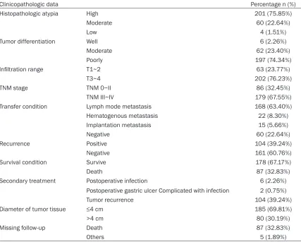
lost during follow-up with a ratio of 34.72%, in which 87 patients died within a year after sur-gery with a mortality rate of 32.83%. The main causes of death were postoperative tumor recurrence and metastasis. One hundred and twelve patients were readmitted for surgical factors, including 6 cases of postoperative in- fection, 2 cases of gastric ulcer complicated with infection, and 104 cases of recurrence after surgery. For details, please see Tables 1 and 2.
Immunohistochemical staining was performed to observe the positive expression of Foxp3+
Treg cells, CD3, CD4, CD8, NK cells, and DCs in tumor tissues, para-carcinoma tissues and nor-mal gastric tissues. The average numbers of above cells in tumor tissues and para-carcino-ma tissues were higher than that of norpara-carcino-mal gas-tric tissues (all P<0.05), see Table 3.
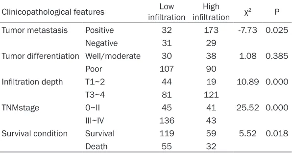
Regarding on tumor metastasis, patients with high infiltration of positive Foxp3+ Treg cells,
CD3, CD4, CD8, NK cells, and DCs were signifi-cantly more than those with low infiltration (P<0.05), however, there was no difference between high and low infiltration group regard-ing on tumor differentiation, infiltrative depth, TNM stage (P>0.05). For details, see Table 4. In the immune microenvironment of gastric cancer, the comparison between different degree of Foxp3+ Treg cells infiltration in tumor
metastasis, infiltration depth, TNM stage and survival condition was statistically significant (P<0.05), the high infiltration of Foxp3+ Treg
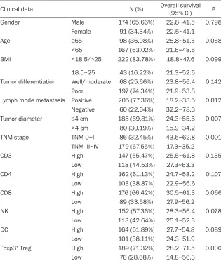
cells suggests the presence of metastasis and deep infiltration, poor TNM staging, may lead to poor survival status. For details, see Table 5. Cox univariate analysis showed that factors including lymph node metastasis, tumor diam-eter, TNM staging, and infiltration of Foxp3+
Treg cells were closely related with the progno-sis of gastric cancer patients; however, age, gender, BMI and histological differentiation degree, CD3/CD4/CD8 cell content and NK cell and DC cell content had no correlation with the prognosis. For details, see Table 6.
According to the Cox univariate analysis, lymph node metastasis, tumor diameter, TNM staging and Treg infiltration had a significant relation-ship with prognosis of the disease. Therefore, the above four factors were taken into Cox mul-incubated at 4°C in the dark environment. Add
Foxp3-FITC after washing with PBS, resusp- ended and then performed flow cytometry. The mean fluorescence intensity (M FI) represents the Foxp3+ Treg concentration.
Flow Cytometry: The cell membrane and cyto-plasmic staining appears brown ( DC and NK cells were larger, round or irregular in shape as positive cells, while the memory T cell/cytotoxic T cells/CD3 cell was smaller. Selected lympho-cyte-intensive cells as positive cells in low mag-nification randomly, counts the total number of positive cells in 400× magnification to repre-sents the number of immune cells in the gastric tissue microenvironment. Calculate the num-ber of positive cells by two pathologists under double-blind according to the computer system of image acquisition and image analysis soft-ware (NIH ImageJ 1.46 bundled with 64-bit Java).
Statistical analysis
Statistical analysis was performed with SPSS 21.0. Measurement data were presented by mean ± standard deviation (x ± s), and exam-ined by T test. Enumeration data were present-ed by percentage and testpresent-ed by chi-squaretest. COX univariate regression analysis [13] was performed to analyze the relationship between immune parameters and the survival. Cox mul-tivariate regression analysis was performed to analyze the correlation between activity of immunefunction and the prognosis. P<0.05 was regarded as statistically significant.
Results
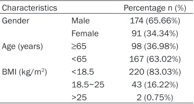
[image:3.612.91.289.84.191.2]Among the 265 patients, there were 174 male and 91 female, the average age of the patients was (65.2 ± 17.2) years old. Pre-hospital BMI of patients was (18.85 ± 2.83) kg/m2, and the BMI one year after surgery was
Table 1. Clinical data of patients
Characteristics Percentage n (%)
Gender Male 174 (65.66%)
Female 91 (34.34%)
Age (years) ≥65 98 (36.98%)
<65 167 (63.02%) BMI (kg/m2) <18.5 220 (83.03%) 18.5~25 43 (16.22%)
tivariate analysis, and the results showed that the four factors could be used as independent factors for the prognosis in tumor tissues. For details, see Table 7.
Discussion
Tumor development and immune function are closely related with each other, especially in
tissues was higher than that in normal gastric tissues (P<0.05), however, the comparison between infiltration status in tumor tissues and para-carcinoma tissues was not statistically significant (P>0.05). It’s commonly thought to be related to the following 3 mechanisms: (1) Stimulation of tumor associated antigens (TAAs) induced activation and proliferation of various kinds of immune cells; (2) The chemo-Table 2. Clinicopathologic data
Clinicopathologic data Percentage n (%)
Histopathologic atypia High 201 (75.85%)
Moderate 60 (22.64%)
Low 4 (1.51%)
Tumor differentiation Well 6 (2.26%)
Moderate 62 (23.40%)
Poorly 197 (74.34%)
Infiltration range T1~2 63 (23.77%)
T3~4 202 (76.23%)
TNM stage TNM 0~II 86 (32.45%)
TNM III~IV 179 (67.55%)
Transfer condition Lymph mode metastasis 168 (63.40%)
Hematogenous metastasis 22 (8.30%)
Implantation metastasis 15 (5.66%)
Negative 60 (22.64%)
Recurrence Positive 104 (39.24%)
Negative 161 (60.76%)
Survival condition Survive 178 (67.17%)
Death 87 (32.83%)
Secondary treatment Postoperative infection 6 (2.26%)
Postoperative gastric ulcer Complicated with infection 2 (0.75%)
Tumor recurrence 104 (39.24%)
Diameter of tumor tissue ≤4 cm 185 (69.81%)
>4 cm 80 (30.19%)
Missing follow-up Death 87 (32.83%)
[image:4.612.94.521.86.429.2]Others 5 (1.89%)
Table 3. Immune cell infiltration in different site (μ ± S)
Immune cell types Tumor tissues Para-carcinoma tissues gastric tissuesNormal DCs 41.35 ± 6.25* 37.38 ± 5.03# 27.65 ± 2.02 NK cells 88.38 ± 5.68* 84.46 ± 4.88# 63.89 ± 3.58 CD4T-lymphocyte 95.08 ± 5.38* 93.35 ± 5.64# 75.89 ± 6.12 CD8T-lymphocyte 87.28 ± 5.52* 87.92 ± 6.21# 63.31 ± 4.05 CD3 T cells 97.21 ± 3.85* 93.82 ± 4.09# 38.36 ± 4.21 Foxp3+ Treg cells 11.36 ± 0.97* 8.95 ± 4.12# 4.35 ± 0.82
*tumor tissues group, shows obvious different from normal gastric tissues group; #para-carcinoma tissues, shows obvious different from normal gastric tissues group.
immune microenvironment of a tumor. Although the immune mic- roenvironment of a tumor cannot directly reflect the immune function of the body, it is useful for analyzing the progression of tumor and prog-nosis [14].
The mechanism of the aggregation of various types of immune cells
Infiltration of Foxp3+ Treg cells, CD3,
[image:4.612.92.349.464.568.2]kines secreted by tumor cells induced the aggregation of immune cells in tumor lesion; (3) Secretion and induction of some cytokines [15]. Maybe the Treg cells gathering in tumor tissues is the basis of its immunosuppressive effect, and the suppression function further promotes the accumulation of Treg cells [16].
Correlations between various types of immune cells and clinical pathological features
The immune cell infiltration in tumor tissues was positively correlated to regional lymph node metastasis, and higher infiltration indi-cated the possibility of lymph node metastasis (P<0.05); however, the immune cell infiltration was not obviously correlated to the degree of tumor differentiation, infiltrative depth, TNM stage as well as survival (P<0.05). The possible mechanism might be that the abnormal
expres-Correlations between Treg cells and clinical pathological features
Treg cells have two functions: immune anergy and immune suppression [19]. Treg cells can inhibit the tumor specific T cell responsesin the tumor microenvironment, thus impeding the anti-tumor reaction. The study showed that infil-tration of Foxp3+ Treg cells in tumor tissue was
increased significantly compared with those in normal gastric tissue (P<0.05), confirming the significance of Foxp3+ Treg cells in promoting
[image:5.612.91.524.96.256.2]the proliferation of tumorcells. Effective anti-tumor immunity can inhibit potential invasive ability of tumor cells [20]. In contrast, Treg cells with immunosupression ability may allow inva-sion, proliferation and degeneration of tumor cells [21], that was confirmed by the significant correlation of high infiltration of Foxp3+ Treg
Table 4. Relationship between the immune cell infiltration and clinicopathological status in each group
Clinicopathological features T cells (CD3/CD4/CD8) NK cells DCs
High Low P High Low P High Low P
Tumor metastasis Yes 155 50 0.000 151 54 0.000 132 73 0.022
No 32 28 29 31 25 35
Tumor differentiation Well/moderate 31 37 0.363 36 32 0.382 35 33 0.452
Poor 141 56 128 69 109 88
Infiltration depth T1~2 41 22 0.142 37 26 0.445 33 30 0.893
T3~4 133 69 137 65 142 60
TNM stage 0~II 48 38 0.163 50 36 0.121 53 33 0.092
III~IV 134 45 133 46 136 43
Survival condition Death 64 23 0.332 63 24 0.188 60 27 0.373
Survival 125 53 115 63 106 72
Table 5. Relationship between infiltration level of Foxp3+ Treg cell and
clinicalpatholocical features
Clinicopathological features infiltrationLow infiltrationHigh χ2 P
Tumor metastasis Positive 32 173 -7.73 0.025
Negative 31 29
Tumor differentiation Well/moderate 30 38 1.08 0.385
Poor 107 90
Infiltration depth T1~2 44 19 10.89 0.000
T3~4 81 121
TNMstage 0~II 45 41 25.52 0.000
III~IV 136 43
Survival condition Survival 119 59 5.52 0.018
Death 55 32
[image:5.612.91.389.304.460.2]Table 6. Cox univariate analysis of factors affecting the prognosis of pa-tients with gastric cancer
Clinical data N (%) Overall survival (95% CI) P
Gender Male 174 (65.66%) 22.8~41.5 0.798
Female 91 (34.34%) 22.5~41.1
Age ≥65 98 (36.98%) 25.8~51.5 0.058
<65 167 (63.02%) 21.6~48.6
BMI <18.5/>25 222 (83.78%) 18.8~47.6 0.099
18.5~25 43 (16.22%) 21.3~52.6
Tumor differentiation Well/moderate 68 (25.66%) 23.8~56.4 0.142
Poor 197 (74.34%) 21.9~53.8
Lymph mode metastasis Positive 205 (77.36%) 18.2~33.5 0.012 Negative 60 (22.64%) 32.2~78.3
Tumor diameter ≤4 cm 185 (69.81%) 24.3~55.6 0.007
>4 cm 80 (30.19%) 15.9~34.2
TNM stage TNM 0~II 86 (32.45%) 43.5~62.8 0.001
TNM III~IV 179 (67.55%) 17.3~35.2
CD3 High 147 (55.47%) 25.5~61.8 0.135
Low 118 (44.53%) 27.3~63.3
CD4 High 162 (61.13%) 24.7~58.2 0.107
Low 103 (38.87%) 22.9~56.6
CD8 High 176 (66.42%) 30.5~61.3 0.066
Low 89 (33.58%) 27.9~56.2
NK High 152 (57.36%) 28.3~56.4 0.078
Low 113 (42.64%) 25.1~52.3
DC High 164 (61.89%) 27.7~54.8 0.089
Low 101 (38.11%) 24.3~51.9
Foxp3+ Treg High 189 (71.32%) 28.2~71.5 0.000
Low 76 (28.68%) 14.8~56.3
Table 7. Cox Multivariate survival analysis of gastric cancer patients
Affective factors HR (95% CI)Survival P
Center of tumor tissue Lymph mode metastasis (P/N) 3.42 0.15~0.43 0.012
Tumor diameter (≤4/>4 cm) 2.51 2.85~8.83 0.000 TNM stage (T 0~II/T III~IV) 0.48 3.02~9.97 0.000 Foxp3+ Treg infiltration (high/low) 2.89 2.27~7.98 0.001 Peritumoral 1 cm Lymph mode metastasis (P/N) 0.35 0.83~1.35 0.089
Tumor diameter (≤4/>4 cm) 0.012 0.78~1.42 0.093 TNM stage (T 0~II/T III~IV) 0.003 1.12~1.43 0.133 Foxp3+ Treg infiltration (high/low) 0.025 1.08~1.56 0.215 Normal gastric tissues Lymph mode metastasis (P/N) 0.000 1.03~1.11 0.562
Tumor diameter (≤4/>4 cm) 0.000 0.96~1.07 0.628 TNM stage (T 0~II/T III~IV) 0.000 0.98~1.05 0.891 Foxp3+ Treg infiltration (high/low) 0.006 1.12~1.25 0.245
cells with metastasis, deep infiltration, poor TNM stage and poor survival.
COX regressionanaly-sis on the factors af-fecting the prognosis of gastric cancer
The prognosis of gas-tric cancer is influen- ced by many factors; Cox univariate analy-sis showed that lym- ph node metastasis, tumor diameter, TNM stage and Foxp3+ Treg
invasion were the fac-tors related to the prognosis of the dis-ease. It is reported that Foxp3+ Treg cells
in tumor tissue can be used to predict the prognosis of tumor such as colorectal ca- ncer and ovarian can-cer [22]. Shen et al.
also found that Fox- p3+ Treg infiltration in
hepatocellular carci-noma was significant-ly correlated with the prognosis of cancer. The possible mecha-nisms might be: 1. direct or indirect inhi- biting T cell prolifer- ation and activation through a variety of ways: 2. inhibiting the killing effect of CD8+
T cells and NK cells on tumor cells; 3. co- mpetitively inhibiting IL-2 to influence the immune response of T cells to tumor cells [23].
[image:6.612.89.409.505.690.2]addition, target inhibiting tumor inflammatory factors and chemokines to reduce the infiltra-tion of Foxp3+ Treg cells may be effect to
achieve tumor growth inhibition.
Disclosure of conflict of interest
None.
Address correspondence to: Haijiang Wang, Dep- artment of Gastrointestinal Surgery, Cancer Hos-
pital, Affiliated to Xinjiang Medical University, 789
East Suzhou Street, Urumqi 830011, Xinjiang Uyghur Autonomous Region, P. R. China. Tel: +86-0991-7819093; E-mail: [email protected]
References
[1] ICH Harmonised Tripartite Guideline. Statisti-cal principles for cliniStatisti-cal trials. International Conference on Harmonisation E9 Expert Work-ing Group. Stat Med 1999; 18: 1905-1942. [2] Jan M, Chao MP, Cha AC, Alizadeh AA, Gentles
AJ, Weissman IL and Majeti R. Prospective sep-aration of normal and leukemic stem cells based on differential expression of TIM3, a hu-man acute myeloid leukemia stem cell marker. Proc Natl Acad Sci U S A 2011; 108: 5009-5014.
[3] Huang X, Bai X, Cao Y, Wu J, Huang M, Tang D, Tao S, Zhu T, Liu Y, Yang Y, Zhou X, Zhao Y, Wu M, Wei J, Wang D, Xu G, Wang S, Ma D and Zhou J. Lymphoma endothelium preferentially expresses Tim-3 and facilitates the progres-sion of lymphoma by mediating immune eva-sion. J Exp Med 2010; 207: 505-520.
[4] Sato S, Ouellet M, St-Pierre C and Tremblay MJ. Glycans, galectins, and HIV-1 infection. Ann N Y Acad Sci 2012; 1253: 133-148.
[5] Wu FH, Yuan Y, Li D, Lei Z, Song CW, Liu YY, Li B, Huang B, Feng ZH and Zhang GM. Endothe-lial cell-expressed Tim-3 facilitates metastasis of melanoma cells by activating the NF-kappaB pathway. Oncol Rep 2010; 24: 693-699. [6] DeKruyff RH, Bu X, Ballesteros A, Santiago C,
Chim YL, Lee HH, Karisola P, Pichavant M, Ka-plan GG, Umetsu DT, Freeman GJ and Casas-novas JM. T cell/transmembrane, Ig, and mu-cin-3 allelic variants differentially recognize phosphatidylserine and mediate phagocytosis of apoptotic cells. J Immunol 2010; 184: 1918-1930.
[7] Chiba S, Baghdadi M, Akiba H, Yoshiyama H, Kinoshita I, Dosaka-Akita H, Fujioka Y, Ohba Y, Gorman JV, Colgan JD, Hirashima M, Uede T,
Takaoka A, Yagita H and Jinushi M. Tumor-infil -trating DCs suppress nucleic acid-mediated in-nate immune responses through interactions
between the receptor TIM-3 and the alarmin HMGB1. Nat Immunol 2012; 13: 832-842. [8] Fourcade J, Sun Z, Benallaoua M, Guillaume P,
Luescher IF, Sander C, Kirkwood JM, Kuchroo V and Zarour HM. Upregulation of Tim-3 and PD-1 expression is associated with tumor
anti-gen-specific CD8+ T cell dysfunction in mela -noma patients. J Exp Med 2010; 207: 2175-2186.
[9] Sakuishi K, Apetoh L, Sullivan JM, Blazar BR, Kuchroo VK and Anderson AC. Targeting Tim-3 and PD-1 pathways to reverse T cell exhaus-tion and restore anti-tumor immunity. J Exp Med 2010; 207: 2187-2194.
[10] Wherry EJ. T cell exhaustion. Nat Immunol 2011; 12: 492-499.
[11] Yang ZZ, Grote DM, Ziesmer SC, Niki T, Hirashi-ma M, Novak AJ, Witzig TE and Ansell SM. IL-12 upregulates TIM-3 expression and induces T cell exhaustion in patients with follicular B cell non-Hodgkin lymphoma. J Clin Invest 2012; 122: 1271-1282.
[12] Zhou Q, Munger ME, Veenstra RG, Weigel BJ, Hirashima M, Munn DH, Murphy WJ, Azuma M, Anderson AC, Kuchroo VK and Blazar BR.
Coexpression of Tim-3 and PD-1 identifies a
CD8+ T-cell exhaustion phenotype in mice with disseminated acute myelogenous leukemia. Blood 2011; 117: 4501-4510.
[13] Gao X, Zhu Y, Li G, Huang H, Zhang G, Wang F, Sun J, Yang Q, Zhang X and Lu B. TIM-3 expres-sion characterizes regulatory T cells in tumor tissues and is associated with lung cancer pro-gression. PLoS One 2012; 7: e30676.
[14] Zhao JZ, Zhang RP, Yu JP, Li H, Wang G, Wang XJ, Xue Q, Li FX, Ren XB. Relationship between Immunocytes in the Tumor Microenvironment and Prognosis of Gastric Carcinoma. Chinese Journal of Clinical Oncology 2010; 37: 1290-1292.
[15] Zhang Y, Ma CJ, Wang JM, Ji XJ, Wu XY, Moor-man JP and Yao ZQ. Tim-3 regulates pro- and
anti-inflammatory cytokine expression in hu -man CD14+ monocytes. J Leukoc Biol 2012; 91: 189-196.
[16] Brown KE, Freeman GJ, Wherry EJ and Sharpe AH. Role of PD-1 in regulating acute infections. Curr Opin Immunol 2010; 22: 397-401. [17] Gleason MK, Lenvik TR, McCullar V, Felices M,
O’Brien MS, Cooley SA, Verneris MR, Cichocki F, Holman CJ, Panoskaltsis-Mortari A, Niki T, Hirashima M, Blazar BR and Miller JS. Tim-3 is an inducible human natural killer cell receptor that enhances interferon gamma production in response to galectin-9. Blood 2012; 119: 3064-3072.
[18] Ndhlovu LC, Lopez-Verges S, Barbour JD, Jones
RB, Jha AR, Long BR, Schoeffler EC, Fujita T,
natural killer cell maturation and suppresses cell-mediated cytotoxicity. Blood 2012; 119: 3734-3743.
[19] da Silva IP, Jimenez-Baranda S, Gallois A, Kuchroo V, Osman I and Bhardwaj N. Tim-3 ex-pression and function in natural killer cells from advanced melanoma patients. Journal of Clinical Oncology 2012: 8571.
[20] Zhuang X, Zhang X, Xia X, Zhang C, Liang X, Gao L and Ma C. Ectopic expression of TIM-3 in lung cancers: a potential independent prognostic factor for patients with NSCLC. Am J Clin Pathol 2012; 137: 978-985.
[21] Li C, Yu X, Zhu Y, Wu X, Ma X, Liu H, Ye L, Ma C, Xia R, Sun A, Ruan C, Chen S, Depei W. TIM-3 Is Highly Expressed On Blast Cells In Patients With Acute Myeloid Leukemia. Blood 2013; 21: 4931-4931.
[22] Ichihara F, Kono K, Takahashi A, Kawaida H, Sugai H and Fujii H. Increased populations of regulatory T cells in peripheral blood and
tu-mor-infiltrating lymphocytes in patients with
gastric and esophageal cancers. Clin Cancer Res 2003; 9: 4404-4408.