Thesis by
Eric W. Burkholder
In Partial Fulfillment of the Requirements for the degree of
Doctor of Philosophy
CALIFORNIA INSTITUTE OF TECHNOLOGY
Pasadena, California
2019
© 2019
Eric W. Burkholder ORCID: 0000-0001-7420-4290
ACKNOWLEDGEMENTS
Just as active matter can only move by pushing against a substrate or embedding
medium, a thesis cannot be written without a strong base of support from which
to move oneself forward. First and foremost I must thank my advisor, John Brady.
John is a researcher of unparalleled physical insight and mathematical abilities, and I am privileged to have had the opportunity to learn from and work with him. I am
grateful for his integrity, professionalism, and the opportunities I have had—from
attending a summer conference in Corsica to presenting at the Society of Rheology
Meeting—with his support. I am most grateful for his respect for and encouragement
of my independence as a researcher.
I would like to thank the members of my thesis committee: Zhen-Gang Wang,
Richard Flagan, and Julia Kornfield. Professor Wang has been not only an ardent supporter during my time at Caltech, but perhaps the best teacher I’ve ever known.
All of his lectures in Thermodynamics, Statistical Mechanics, and Polymer Physics
are perfectly crafted and clear. At the end of class, I often felt as if I had meditated on
the subject matter for the past 90 minutes. I thank Professor Kornfield for her passion
and enthusiasm in her committee service, and for challenging me and teaching me
to defend my work. Professor Flagan—thank you for your service, and for keeping
the lofty ideas of a theorist grounded in reality.
A special note of thanks to a Brady group alumna and my undergraduate research advisor, Roseanna Zia. From her I learned the fundamentals of
microhydrodynam-ics, microrheology, and fluid mechanics that allowed me to be successful in graduate
school. Even as an undergraduate, she held me to high expectations and supported
me and encouraged me to strive for great things. I thank Ron Phillips, Joel
Chris-tenson, Stephanie Dungan, Robert Guy, and Becca Thomases for their hospitality
during my visits at the University of California Davis.
I am grateful to all members and visitors of the Brady group: Austin Dulaney,
Camilla Kjeldbjerg, Hyeongjoo Row, Zhiwei Peng, Sho Takatori, Kevin Marshall, Ahmad Omar, Kevin Shen, Charlie Slominski, Wen Yan, Mu Wang, Mikey Phan,
Marco Heinen, Stewart Mallory, Karol Makuch, Markus Gruber, Mario Sandoval,
Charles Schroeder, Sara Abdelsalam, and Tyler Ross. I owe a particular debt of
gratitude to Mikey for helping me learn how to use the cluster, and to Sho and
advice over the years. I would have been lost had I not been able to bombard them
with questions. I would like to thank Suresh Guptha for helping me navigate the foreign world of computer clusters, and Kathy Bubash and Allison Oulette for their
assistance throughout my PhD.
Thank you to my friends and family for all their love and support. Thank you Mom
and Dad for listening to me rant about problems at work, validating my grievances,
and being supportive of all the decisions I’ve made throughout my academic career.
Thank you Patrick and Emily for always taking my phone calls when I was upset
about something, and thank you to Ben and Devon for always being willing to go on an adventure when I needed a break from work. Thank you to all the members
of my incoming class at Caltech for providing much-needed commiseration and
celebration as appropriate.
To Jason LeGrand—my partner and most vehement supporter—I owe all of my
successes at Caltech and in my future endeavors. Thank you for encouraging me to
celebrate the small victories, keeping me grounded when faced with small problems,
and giving me strength and love in times of adversity. Words can scarcely express
ABSTRACT
“Active matter” refers to a broad class of materials in which the constituent
parti-cles or organisms are able to self-propel (swim) by some internal physicochemical
mechanism. Though the origin of this self-propulsive motion is a rich area of study,
we are primarily interested in the collective effects of this motion on the physical properties—and in particular, the rheology—of the active material as a whole. As
such we model self-propulsive motion using the minimal active Brownian particle
(ABP) model: a particle of sizea, swims in a directionqwith a speedU0, and the
direction of its motion changes randomly over some time scaleτR.
On a macroscopic scale, active motion leads to unique hydrodynamic and mechanical
stresses exerted by the particles on their embedding medium. These stresses arise
from the microscopic force associated with particle locomotion—the swim force Fsw i m. Though the idea of the swim force is widely recognized in the abstract, little attention has been given to the characterization and mechanical consequences of this
force. In this work we are particularly interested the role of the swim force in the
effective motion of passive constituents in active environments, and how the swim
force affects long-ranged hydrodynamic interactions (HI) in active suspensions. We
examine these issues through the lens of microrheology: tracking the motion of a
colloidal probe particle through an active medium, and using its motions to infer the
effective viscoelastic properties of the suspension.
Using generalized Taylor dispersion theory, we find an activity-driven enhancement
to the diffusion of the probe in an active medium. This first-principles theory
unites many experimental observations of tracer diffusion, and provides simple
physical descriptions of the problem that do not rely on the specific self-propulsion
mechanism of the swimmer. This same framework is then used to compute the
suspension microviscosity (as measured by the drag on the probe particle), and the
fluctuation-dissipation relation in an active system. We find that activity reduces the
drag on the probe, but the drag is still larger than it would be in a Newtonian fluid;
this stands in contrast to experimental measurements of reduced shear viscosities. We show that the microviscosity of a suspension is reduced—and may even become
negative!—due to HI, and that this effect isnotdue to the fluid velocity disturbance
PUBLISHED CONTENT AND CONTRIBUTIONS
1E. W. Burkholder, and J. F. Brady, “Tracer diffusion in active suspensions”, Phys.
Rev. E - Stat. Nonlinear, Soft Matter Phys. (2017) 10 . 1103 / PhysRevE . 95 . 052605,
E.W.B. participated in the conception of the project, performed the calculations, analyzed the data, and participated in the writing of the manuscript.
2E. W. Burkholder, and J. F. Brady, “Do hydrodynamic interactions affect the swim
pressure?”, Soft Matter14, 3581–3589 (2018),
TABLE OF CONTENTS
Acknowledgements . . . iii
Abstract . . . v
Published Content and Contributions . . . vi
Table of Contents . . . vii
List of Illustrations . . . ix
Chapter I: Introduction . . . 1
1.1 Locomotion, stress, and pressure . . . 2
1.2 Suspension mechanics & microrheology . . . 6
1.3 Contributions and outlooks . . . 8
Chapter II: Tracer diffusion . . . 16
2.1 Mechanical model . . . 17
2.2 Active Diffusivity . . . 19
Chapter III: Fluctuation-dissipation in active matter . . . 35
3.1 Model system . . . 39
3.2 Probe speed and self-drag . . . 43
3.3 Microstructure . . . 52
3.4 Conclusions . . . 54
Chapter IV: Closure of the field equations . . . 68
4.1 Full smoluchowski equation: no closure . . . 70
4.2 Finite-element results, d= 2 . . . 73
4.3 Conclusions . . . 78
Chapter V: Active and nonlinear microrheology in active suspensions . . . . 82
5.1 Theoretical framework . . . 86
5.2 Nonlinear microrheology . . . 88
5.3 Suspension microstructure . . . 92
5.4 Conclusions . . . 96
Chapter VI: Hydrodynamic interactions in active suspensions . . . 104
6.1 Suspension mechanics and the swim force . . . 104
6.2 Single-particle, equilibrium problems . . . 107
6.3 External perturbations . . . 113
6.4 Anisotropic particles . . . 117
Chapter VII: Force on a boundary: the osmotic pressure . . . 124
7.1 Momentum balance . . . 125
7.2 Model system . . . 127
7.3 Flat plate . . . 128
7.4 Pressure on a fixed spherical body . . . 131
7.5 Conclusions . . . 133
8.2 Linear response . . . 142
8.3 Finite size effects & fixed-force vs. fixed-velocity . . . 153
8.4 Nonlinear response . . . 155
8.5 Conclusions . . . 158
Chapter IX: Fixed-probe microrheology: an orienting external field . . . 169
9.1 Model system . . . 170
9.2 Microviscosity: force on the probe . . . 173
9.3 Linear response: no hydrodynamic interactions . . . 174
LIST OF ILLUSTRATIONS
Number Page
1.1 Sketch of the half-surface of a squirming organism as described by
Blake’s model of ciliary propulsion [37]. The black (dotted)
hemi-spherical line is the equilibrium (stationary) surface of the organism
R0, and the stationary polar angle for the organism is θ. The blue
(solid) line is a surface modulation (modeN =22 in Eqn. 19 of [37]) at some initial time with amplitude R0/10, and the red (dot-dashed)
line is the same surface modulation at 0.8 of the beat period later. The surface wave travels to the left, which results in a net propulsion
of the organism to the right. . . 3
1.2 Sketch of the fluid flow fields created by a pusher (left), mover
(cen-ter), and puller (right). . . 3
1.3 Sketch of a generic N particle colloidal suspension. Each particle αhas a position xα and orientation qα in the laboratory coordinate frame. Each particle moves with a velocity Uα and rotates with an
angular velocityΩα. Note that the particle orientation may not be in
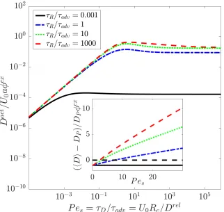
the direction of motion. . . 6 2.1 Active diffusivity of the probe as a function of the ratio of the
pair-diffusion time to the advection time Pes = τD/τadv = U0Rc/Dr el, where U0 is the swim speed, Rc is the center-to-center separation
distance of the probe and swimmer upon contact, and Dr el is the
rel-ative thermal diffusivity of the probe-swimmer pair. The ratioτD/τR
indicates the strength of Brownian motion relative to the
reorien-ations of the swimmers. The active diffusivity is non-dimensionalized
by the probe’s SES diffusivity DP times the active volume fraction φex = (4π/3)n∞
R2ca/2, where a is the swimmer size and n∞ is the number density of swimmers. . . 20
2.2 Active diffusivity of the probe non-dimensionalized by(ksTs/ζP)(R/Rc)φex
as a function of the ratio of the diffusion time to the swimmer
reorien-tation time τD/τR = R2c/τRDr el for various values of the mechanical
2.3 Active diffusivity of the probe non-dimensionalized byU0aas a
func-tion ofPes =τD/τadv =U0Rc/Dr el. The ratioτR/τadv =U0τR/Rc =
`/Rc reflects the speed of reorientation relative to advection. The inset shows the total O(φex) change in the probe’s diffusivity,
non-dimensionalized by DPφex, where DP is the bare diffusivity of the
probe. . . 23
2.4 Depiction of the model system. There is a Brownian probe of size R
immersed in a dispersion of ABPs with sizeaat number densityn∞.
The ABPs swim in a direction qwith speedU0, and reorient with a
characteristic timeτR. . . 26
3.1 Sketch of the fluctuation-dissipation theorem (FDT) in a colloidal suspension. LEFT: A tracer particle diffuses in a suspension of bath
particles due to random Brownian motion. The time derivative of
the mean squared-displacements due to Brownian motion hx0x0iis
proportional to the self-diffusivity of the particle hDi. RIGHT: The
same tracer particle moves through the same suspension under the
action of an external forceFext. The speed of this particle is linear
in the external force, with the constant of proportionality being the
average mobilityhMUFi. The FDT states that these two problems are
fundamentally related byhDi · hMU Fi−1= kBTI, wherekBTis the temperature of the system and is independent of all other suspension
properties (composition, interparticle interactions, etc.). . . 36
3.2 Schematic of the model system: a Brownian probe particle of size
Rimmersed in a suspension of ABPswith sizeaat number density
n∞—the center-to-center separation distance upon a collision is
de-noted by Rc = R+a. The ABPs swim in a directionqat speedU0;
q changes randomly on a time scale characterized byτR. The probe
3.3 Particle contribution to the probe drag (d =3) scaled by its value in a passive suspensionζPφex. The Stokes drag of the probe isζP, and the
excluded volume fraction of the suspension isφex =2πa(R+a)2n∞/3, wheren∞ is the number density,ais the swimmer size, andRis the
probe size. This scaled drag contribution is plotted as a function
of `/Rc ≡U0τR/(R+a), where U0 is the speed of the swimmers, andτRis their reorientation time. Different colors indicate different
strengths of swimming: 6Dswim/Dr el = U02τR/Dr el, where Dr el is
the relative thermal diffusivity. Squares are for the closure Q0 = 0
and crosses are for the closure B0 = 0. The dashed lines serve as
guides for the eye. . . 45 3.4 Particle contribution to the probe drag (d = 2) scaled by its value in
a passive suspension ζPφexA. The Stokes drag of the probe isζP, and
the excluded area fraction of the suspension isφexA = 2πa(R+a)n∞A, wheren∞A is the areal number density,ais the swimmer size, andRis
the probe size. This scaled drag contribution is plotted as a function
of `/Rc ≡U0τR/(R+a), where U0 is the speed of the swimmers, andτRis their reorientation time. Different colors indicate different
strengths of swimming: 2Dswim/Dr el = U02τR/Dr el, where Dr el is
the relative thermal diffusivity. Squares are for the closure Q0 = 0
and crosses are for the closure B0 = 0. The dashed lines serve as
guides for the eye. . . 46
3.5 Sketch of swimmer trajectories upon collision with the probe particle
for various regimes of`/Rc =U0τR/(R+a), whereU0is the speed of the swimmer,τRis its reorientation time,ais its size, andRis the
size of the probe. The background arrows indicate the direction of
fluid flow. . . 47
3.6 Comparison between our theoretical predictions (red crosses) and the
3.7 Contour plots of the 3-D (axisymmetric) perturbed microstructure for
weak external forcing; the direction of the external force is indicated with a solid black arrow. The background green colors indicates a
uniform microstructure fluctuation: n0,m0 ∼ 0. Red (warm) colors
indicate an accumulation of particles, and blues (cold colors) indicate
a depletion. For the polar orderm0k, concentration gradient∇kn0, and
fluxjTk, the color indicates how strongly the field is aligned with (reds)
or against (blues) the external force. . . 52
3.8 Contour plots of the 3-D (axisymmetric) perturbed microstructure
for weak external forcing. The background green colors indicates
a uniform microstructure fluctuation: n0,m0,Q0 ∼ 0. Red (warm) colors indicate an accumulation of particles, and blues (cold colors)
indicate a depletion. For the nematic order Q0, the color indicates
how strongly the swimmers’ alignments are correlated—small black
arrows are included to help with visualizing the three components of
Q0: Q0k=uˆ·Q0·uˆ, whereuˆ= ezis the unit vector in the direction of
the external force (as indicated in the concentration disturbance plot),
Q0⊥ =e⊥·Q0·e⊥, wheree⊥ =cosψex+sinψey, andQ0× =e⊥·Q0·uˆ. The azimuthal coordinate isψ. . . 54
4.1 Sketch of the probe-swimmer coordinate system. The probe has a position xP and orientation ex in the fixed coordinate frame. The
swimmer has a position xs and orientationqs (parametrized by the
angle ψ) in the fixed frame. The relative separation between the
particles is r, and the polar angle of the swimmer’s position with
respect to the probe is θ. The orientation of the swimmer relative to
the probe is parametrized by the angle β= ψ−θ. . . 71 4.2 Example of adapted finite-element mesh and solution for `/δ = 50
4.3 Equilibrium distribution contact as a function of the angle of the
swimmer relative to the probe unit normal β: ∫ P0(1, θ, β)dθ/2πn∞ for`/δ= 10, `/Rc =3. Solid lines are moments derived from the full solution for P, square symbols are mean-field predictions forQ= 0, crosses are the mean-field predictions forB = 0, and circles are for the closure Q ∼ ∇m. The red data truncate the series expansion
at i = 2, green data truncate ati = 3 and the blue line contains all moments. . . 75
4.4 Microviscosity (d = 2) as a function of`/Rc at`/δ =10 for various closures of the moments expansion of P. The squares and crosses
are from finite difference solutions of the 2-D governing equations in MATLAB, and the circles are from finite element simulations of the
full Smoluchowski equation in FreeFEM++ [11]. . . 76
4.5 Linear disturbance to P as a function of β for various closures at `/δ =10,`/Rc= 3. The legend is the same as that in Fig. 4.3. . . 77 5.1 Schematic of the model system: a Brownian probe particle of size
R immersed in a suspension of active Brownian particles (ABPs)
with size a at number density n∞—the center-to-center separation
distance upon a collision is denoted by Rc= R+a. The ABPs swim in a direction q at speed U0; q changes randomly on a time scale characterized by τR. The probe translates under the action of some
external forceFex t = hζiUpr o be, which may be constant or specified such that the resulting probe velocityUpr o be is constant. The force and velocity are related by the effective drag coefficient hζi. . . 85
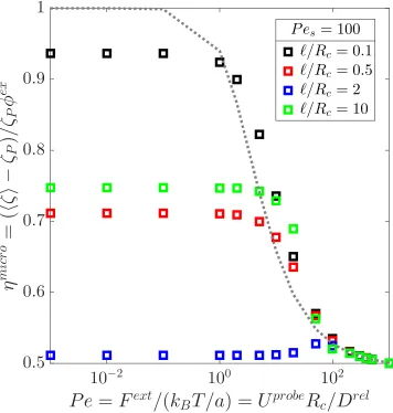
5.2 Nonlinear intrinsic microviscosity (d =3)ηmicr oplotted as a function ofPe=Upr obeRc/Dr elfor various`/Rc. The probe’s speed isUpr obe,
Dr el is the relative thermal, Rc = R+a, where Ris the probe size,
a is the swimmer size, U0 is the speed of the swimmers, and τR is
their reorientation time. Different colors indicate different strengths of swimming: `/δ=
q
U02τR/Dr el. . . 89
5.3 Nonlinear apparent microviscosity (d = 3) ηmicr o, scaled by its passive value plotted as a function of Pe = Upr obeRc/Dr el for
Pes = U0Rc/Dr el = 100. The swimmers’ speed is U0, Dr el is the relative thermal diffusivity of the probe-swimmer pair, and the
center-to-center separation of a probe-swimmer pair at contact isRc.
5.4 RIGHT: Microviscosity as a function of th external Péclet number
Pe = Fext/(kBT/a), where Fext is the external force moving the probe (indicated by the black arrow) and a is the size of the bath
particles, and the swim Péclet number Pes = U0Rc/Dr el whereU0 is the swim speed, Rc is the center-to-center separation distance of a
swimmer and the probe upon contact, and Dr el is the relative
trans-lational diffusivity of the pair. The activity level is fixed at`/δ =10 (ksTs/kBT ∼ 100). LEFT: Contour plots of the 3-D (axisymmetric)
microstructure for various degrees external forcing Pe and activity
levels Pes. The background light blue color indicates a uniform
microstructure: n = n∞,m = 0. Red (warm) colors indicate an accumulation of particles, and darker blues (cold colors) indicate a
depletion. . . 93
5.5 RIGHT: Microviscosity at Pe = 100 as a function of `/Rc and
U0/Upr obewhereU0is the swim speed of the bath particles andUpr obe
is the speed of the probe. LEFT: Contour plots of the 3-D
(axisym-metric) microstructure at Pe = 100 for various ratios ofU0/Upr obe. The rows vary the value of`/Rcas in Fig. 5.2. The background light
blue color indicates a uniform microstructure: n = n∞,m = 0. Red (warm) colors indicate an accumulation of particles, and darker blues (cold colors) indicate a depletion. . . 95
5.6 Nonlinear apparent microviscosity (d= 2)ηmicr oplotted as a function of Pe = Upr obeRc/Dr el for various `/Rc. Legend is the same as in Fig. 5.2 . . . 100
7.1 Schematic of the active-diffusive boundary-layer (BL) in a 1D
geom-etry. The black line is the concentration profile of swimmers,λ−1is
the BL thickness, anda∆is where the no-flux condition applies. The
dimensionless translational and rotational diffusivities ˆDTT and ˆDRR
7.2 Pressure at the wall scaled by the bulk pressure Π∞ = n∞kBT(1+
Dswim/DT) . The number density of swimmers is n∞ , DT is the thermal diffusivity far from the wall, andDswimis the swim diffusivity. This quantity is plotted against ∆, which characterizes the strength
of HI; HI are strong for ∆ 1, and weak for ∆ 1. The square
symbols indicate the pressure calculations neglecting the nematic
order (hqqi = I/3), and crosses include nematic order (hqqqi = hα·qi/5, where α is the fourth-order isotropic tensor. INSET:
Active contribution to the wall pressure scaled by the prediction from
the boundary-layer analyis: ζ is their translational drag, U0 is the
swimming speed, and`is the run length of their active random walk. 129 7.3 Active contribution to the pressure on a fixed spherical cavity of sizeR
scaled byn∞ζ(∆)U0`(∆)/6. The swimmer number density isn∞,kBT
is the thermal energy,ζis the translational drag,U0is the swim speed,
` is the run length, and∆characterizes the strength of hydrodynamic interactions (HI). We plot this against`/Rc = U0τR(∆)/(R+a)(1+
∆), where a is the size of the swimmer and τR(∆) is its (thermal)
reorientation time. The colors represent different strengths of HI;
square symbols are for a/R = 1/8, crosses are for a/R = 1, and circles are for a/R = 8. The dashed line is the analytical prediction from [25]. . . 132
7.4 Same legend as Fig. 7.3 withQ, 0. . . 133
8.1 Sketch of a sphere of size R moving with velocityUpr o be through fluid containing a dilute dispersion of point ABPs with swim velocity
U0. The probe-bath hydrodynamic interactions are modeled by an
excluded annulus potential, which manifests as a no-flux condition at
Rc. The fluid velocity stream-lines incdicated by the blue arrows are
plotted in a probe-fixed reference frame. . . 139
8.2 The hydrodynamic contribution to to the intrinsic microviscosity (in the absence of probe motion), scaled by the result for passive colloidal
suspensions, is plotted as a function of the swimmers’ activity`/R=
U0/D⊥RR, whereU0is the swim speed,DR⊥is the inverse reorientation time, andRis the size of the probe. Different colors represent different
8.3 Sketch of the distribution and orientation of swimmers around the
probe leading to the increase in the high-frequency shear viscosity ηH
i,0. Hydrodynamic interactions are strong for∆ 1 and moderate for∆∼O(1). . . 145
8.4 The interparticle contribution to the intrinsic microviscosity, scaled
by the result for passive colloidal suspensions, plotted as a function
of the (spherical) swimmers’ activity `/R = U0/D⊥RR, where U0 is the swim speed, D⊥R is the inverse reorientation time, and R is the
size of the probe. Different colors represent different strengths of
hydrodynamic interactions. Square symbols include fluid vorticity,
and crosses neglect vorticity. . . 146 8.5 Sketch of a swimmer’s trajectory around the probe when`/Rc ∼O(1).
The figure on the left illustrates flow in the absence of HI, and the
figure on the right illustrates the additional fluid velocity disturbance
of the probe when HI are included. . . 147
8.6 The interparticle contribution to the intrinsic microviscosity, scaled
by the result for passive colloidal suspensions, plotted as a function of
the swimmers’ activity`/R=U0/D⊥RR, whereU0is the swim speed, D⊥R is the inverse reorientation time, and Ris the size of the probe.
Different colors represent different swimmer aspect ratiosξ = L/d. Hydrodynamic interactions are strong: ∆ = 0.2. Square symbols include anisotropy in the thermal diffusivity and crosses neglect it. . . 148
8.7 Intrinsic interparticle microviscosity, scaled by its value in
passive-suspensions, as a function of `/R for various aspect ratios in the
absence of hydrodynamic interactions∆→ ∞. . . 149
8.8 Sketch of the flux of swimmers due to strain-alignment in the fluid
for prolate (left) and oblate (right) swimmers. . . 150
8.9 The effective viscosity of suspension of motile bacteria (B. subtilis),
scaled by the effective viscosity of a suspension of nonmotile bacteria at the same concentration, plotted as a function of activity `/R.
The run-length is the product of the bacteria’s swimming speedU0
and reorientation time τR ∼ 1s. The probe size R is 50 µm, the
aspect ratio of the bacteria is 7, and the number density of bacteria
isn∞ ∼ 1×10¹⁰/mL. We compare the experimental data (in black)
8.10 Intrinsic interparticle microviscosity (fixed-velocity mode) as a
func-tion of the swimmer activity`/Rcfor various swimmer-to-probe size ratios β. Hydrodynamic interactions are strong ∆ = 0.2, and the swimmers are spherical. . . 154
8.11 Ratio of the particle contribtuion to the fixed-velocity microviscosity
hηiV−ηsto the fixed-force particle contribution to the microviscosity
hηiF −ηs as a function of bath particle to probe size ratio β = a/R.
Different colors indicate different strength of HI. . . 155
8.12 Viscosity ratio as a function of the swimmer-to-probe particle size
ratio β. Different colors indicate different levels of activity, whereas
the different symbols denote different values of∆. Squares are∆ = 0.2, crosses are∆=1, and Circles are∆= 5. . . 156 8.13 Intrinsic interparticle microviscosity (in pseudo-2-D) as a function
of Pe, scaled by its Pe 1 value in the absence of activity. The
dashed grey line is a guide for the nonlinear response observed when `/R = 0, the symbols connected by dashed lines are at the activity level `/R = 1. Different colors represent different particle aspect ratiosξ = L/d. . . 157 8.14 The hydrodynamic contribution to to the intrinsic microviscosity (in
the absence of probe motion) scaled by the result for passive col-loidal suspensions, is plotted as a function of the strength of
hydro-dynamic interactions ∆. Square symbols are for swimmer activity `/R =U0/D⊥RR= 0.1 and crosses are for`/R = 1; U0 is the swim speed, D⊥R is the inverse reorientation time, and Ris the size of the
probe. Different colors represent different swimmer aspect ratios ξ = L/d. . . 160 8.15 The hydrodynamic contribution to the intrinsic microviscosity, scaled
by the result for passive colloidal suspensions, plotted as a function of
8.16 The hydrodynamic contribution to the intrinsic microviscosity, scaled
by the result for passive colloidal suspensions, plotted as a function of the swimmers’ activity`/R=U0/D⊥RR, whereU0is the swim speed,
D⊥R is the inverse reorientation time, and Ris the size of the probe. Different colors represent different swimmer aspect ratiosξ = L/d. Hydrodynamic interactions are weak: ∆= 5. . . 162 8.17 The interparticle contribution to to the intrinsic microviscosity (in
the absence of probe motion) scaled by the result for passive
col-loidal suspensions, is plotted as a function of the strength of
hydro-dynamic interactions ∆. Square symbols are for swimmer activity `/R =U0/D⊥RR= 0.1 and crosses are for`/R = 1; U0 is the swim speed, D⊥R is the inverse reorientation time, and Ris the size of the
probe. Different colors represent different swimmer aspect ratios ξ = L/d. . . 163 8.18 The interparticle contribution to the intrinsic microviscosity, scaled
by the result for passive colloidal suspensions, plotted as a function of
the swimmers’ activity`/R=U0/D⊥RR, whereU0is the swim speed, D⊥R is the inverse reorientation time, and Ris the size of the probe.
Different colors represent different swimmer aspect ratiosξ = L/d. Hydrodynamic interactions are moderate: ∆=1. . . 164 8.19 The interparticle contribution to the intrinsic microviscosity, scaled
by the result for passive colloidal suspensions, plotted as a function of
the swimmers’ activity`/R=U0/D⊥RR, whereU0is the swim speed, D⊥R is the inverse reorientation time, and Ris the size of the probe.
Different colors represent different swimmer aspect ratiosξ = L/d. Hydrodynamic interactions are weak: ∆= 5. . . 165 9.1 Schematic of the fixed-probe microrheology of an active suspension
under the influence of an external field H. The probe of size R is
fixed, while swimmers of size a swim with speed U0 and reorient their direction of motion q due to random Brownian fluctuations
and a torque Lext induced by the external field. The swimmer’s
displacement from the probe is r; n is the outward-pointing unit
9.2 Plot of the fixed probe microviscosity for weak external fields: χR = HτR/ζR 1, where H is the external field moving the probe,ζRis
the rotational Stokes drag of the swimmer, andτRis the reorientation
time. The dashed lines are values of the microviscosity as measured
by the translating probe. Different colors correspond to different
C h a p t e r 1
INTRODUCTION
“How can the events in space and time which take place within the spatial
bound-ary of a living organism be accounted for by physics and chemistry?” Since
Schrödinger’s philosophizing on this subject [1], countless physicists, chemists,
mathematicians—even engineers—have spent their careers in pursuit of simple
phys-ical models that describe emergent behaviors in biologphys-ical systems. Schrödinger
understood that the quantum and statistical interactions between the atoms and molecules which comprise all matter must be responsible for emergent phenomena
in living organisms. In this dissertation, we take the same philosophical approach to
the description of active suspensions—a subject of active research among soft-matter
physicists today.
“Active suspensions” denote a class of materials in which collections of organisms,
particles, or even molecules are capable of converting chemical energy in to
me-chanical work. A further distinction is made between wet active suspensions, in
which the particles are immersed in a (usually continuous) suspending medium that mediates long-ranged interactions between constituents [2, 3], and dry active
suspensions (e.g. vibrated granular media) in which the constituents generally do
not interact through the substrate off of which they self-propel.1 Common examples
of wet active matter are collections of bacteria, suspensions of chemically reactive
nanoparticles [5], and bundles of cytoskeletal filaments (e.g. actin) bound together
by motor proteins [6]. Such materials are known to exhibit fascinating dynamic
behavior such as spontaneous collective motion [7–13], phase-separation [4, 10,
14–23], and enhanced diffusion [24–30]. Of particular interest to us is the
rheologi-cal behavior of active suspensions. Viscosity reduction [31–35] is but one observed behavior not typically found in passive suspensions of colloids.
In this work, we develop coarse-grained descriptions of the mechanics of active
suspensions. To do so, we need only to think carefully about how phenomenological
1A common misconception regarding dry active systems is that they do not conserve momentum
models of biological and diffusiophoretic locotmotion [36–39] can be incorporated
into existing microscopic frameworks used to describe the behavior of passive colloidal suspensions [40–43]. In doing so, we need never concern ourselves with the
details of how the active constituents convert chemical into mechanical energy, only
the nature of the emergent motion. From these models, we make predictions about
the microrheological behavior of active materials—how the microscopic interactions
between active and passive constituents inform the osmotic pressure, diffusivity, and
viscosity of the medium.
1.1 Locomotion, stress, and pressure
At its core, biological motion is a mechanics problem, and thus beholden to
the the same fundamental laws introduced in any introductory physics course—
conservation of mass and momentum. Consider a body of size a and density ρP
moving at a speedU in a continuous medium of viscosityηand density ρ. For an
incompressible medium, conservation of mass requires that the velocity field is
di-vergence free∇·u=0. We consider motions at low Reynolds numberRe ≡ ρUa/η
and low Stokes numberSt ≡ (ρP/ρ)Re. In this regime, both the particles and fluid
have no inertia, and thus all motion is force-free: Newton’s second law requires that
the sum of the forces (and torques) on the body be equal to zero, and the Stokes
equations then require that the the suspension stress is divergence free∇ ·σ = 0.
Taylor [44], Lighthill [36], and later Blake [37] were the first to recognize the relevance of low Reynolds number fluid mechanics, in particular the consequences
of negligible inertia, to the problem of locomotion on microscopic length scales.
In low Reynolds number flows all motions are instantaneous and time-reversible;
thus microscopic organisms are unable to swim (with non-zero net displacement)
unless they make non-reciprocal motions, such as the chiral rotations of a flagellum
inE. coli[45, 46]. This is commonly known as Purcell’s scallop theorem. Because
local time-reversal symmetrymustbe broken to achieve net self-propulsion, active
systems are inherently non-equilibrium and cannot be described in terms of typical
thermodynamic relations.
Blake’s squirmer model provides a phenomenological description of the swimming
of ciliated organisms, which move through time-asymmetric modulations of their
surfaces (see Fig. 1.1) [37]. Blake expresses this motion as a series of small
harmonic modulations to the surface of a spherical organisms—e.g. parasites of
Figure 1.1: Sketch of the half-surface of a squirming organism as described by Blake’s model of ciliary propulsion [37]. The black (dotted) hemispherical line is the equilibrium (stationary) surface of the organism R0, and the stationary polar angle for the organism is
θ. The blue (solid) line is a surface modulation (mode N = 22 in Eqn. 19 of [37]) at some initial time with amplitude R0/10, and the red (dot-dashed) line is the same surface
modulation at 0.8 of the beat period later. The surface wave travels to the left, which results in a net propulsion of the organism to the right.
Figure 1.2: Sketch of the fluid flow fields created by a pusher (left), mover (center), and puller (right).
and the hydrodynamic stress σH exerted on the fluid due to the particles
self-propulsion. Because the particle motion is force-free, the swim force—the internal
body force which drives the self-propulsive motion of the particles [3, 21, 47]—is
precisely balanced by the fluid drag. The fluid velocity disturbance associated with
self-propulsion is thus dipolar and decays as 1/r2 (or faster) [48]. This has been
experimentally verified by particle-image velocimetry [49, 50]. This velocity field
experimental measurements of the particle’s self-propulsive swim force Fsw i m to predict the magnitude σ0 of σH: σ0 = ±Fswima, where a is the particle size. If
σ0 > 0, the organism is said to be a puller likeC. reinhardtii, ifσ0 <0 it is a pusher likeB. subtilisorE. coli, and for σ0 = 0, it is a mover, likeVolvox. Flow fields for the various types of swimmers are sketched in Fig. 1.2. Nearly all descriptions of
the rheology of active suspensions rely on the calculation ofσH—the work of [51]
and this thesis being notable exceptions. Viscosity reductions and instabilities are
thus attributed to a negative active hydrodynamic stresslet.
However, there is a key feature missing from the squirmer model. As observed by Berg [46], flagellated microorganisms do not simply swim, they also tumble;
tumbling is a sudden change in their swimming direction associated with unbundling
of the flagella. Similarly, chemically active colloids will change their direction due
to rotary Brownian motion. This observation informs the active Brownian particle
(ABP) and run-and-tumble particle (RTP) models, which neglect the hydrodynamic
details associated with the particles’ self-propulsion, and simply ascribe a swim
velocityU0and a characteristic time for reorientations (tumbles)τR[38]. For ABPs,
the reorientation process is continuous on scales long compared to the momentum
relaxation time of the fluid whereas the reorientations of RTPs are discrete [52].
RTPs are commonly modeled as having a Poisson-distributed orientation with a mean that is usually matched to empirical measurements (for example, the average
tumbling angle for E. coliis 68.5 degrees, with a similar variance [27]). At times
long compared to τR, these models are equivalent; as we are primarily concerned
with steady-state behaviors, we always assume a diffusive reorientation process in
this work—the tumbles are not correlated at long times.
This run-and-tumble motion is described by an effective mechanical “swim”
diffu-sivity Dswim ∼ `2/τR ∼ U02τR, where the ` = U0τR is the run-length (the distance
traveled between reorientations) [21, 46]. Diffusive motion implies an unbounded
(linear) growth of the mean-squared displacements of the particle at long times—a
particle diffusing in an unbounded medium will tend to “run away” in space. Thus,
if one wishes to consider an unbounded suspension of active particles at a constant
number densityn, there must be a stress in the fluidσsw i m = −nζDsw i m that pre-vents the particles from diffusing off to infinity (ζ is the drag on a particle). Though
this is a new mechanical stress unique to active systems, it should be noted that even
Brownian particles have a similar Brownian stress associated with their thermal
diffusive process has a corresponding stress of the same form. The swim stress
scales asFswim` ∼ (`/a)σ0. The run length is typically large compared to the body size, and thus this stress is much larger than the active stresslet that characterizes
the swimming gait; it should be emphasized that this stress exists for all
swim-mers. Indeed Takatori and Brady have used this idea extensively and shown that
phase-separation, and “superfluidity” can both be explained by this swim stress [51,
56].
Where there is stress, there is pressure: Π =−tr(σ)/d, wheredis the spatial dimen-sion; this implies the existence of a unique particle-phase pressureΠswim= nζDswim
given that these particles diffuse. Takatori et al. showed that one can use the swim
pressure to explain the phase separation of active systems [21], and even give an
analytic prediction for the mechanical instability of active suspensions [56]. This
concept of the swim pressure has been highly contentious2, despite its clear physical
origins [57]. Though one can always define a mechanical pressure from stress, it
has been argued that this pressure is not necessarily equal to the average force the
suspension exerts on confining boundaries as it dependents on the particle-boundary
interactions. This mechanical pressure is thus saidnotto be a function of state,
ren-dering attempts to use it as a description of phase behavior invalid [57]. We address
this in detail in Chapter 6, but there is a key point that counters this criticism. If mass and momentum are conserved, one can show that the force per unit area on the
confining boundariesmust be equal to the pressure in the bulk of the suspension.
It must simply be recognized that this conservation statement applies to the total
system pressure, and not necessarily each component individually. The same is
true for a box of air—we may not know how the pressure of each component gas is
distributed in the container, only that the total pressure is uniform for a given volume
and temperature. Even in dry active matter, the second ‘component’ of the active
suspension is the substrate from which the constituents are able to self-propel. Thus,
any container-dependent particle-wall interactions cannot cause spatial variations in the total pressure in a container, so the total pressure in active suspensions is a
function of state.
2Most hotly debated have been attempts at thermodynamic descriptions of active matter—
Figure 1.3: Sketch of a generic N particle colloidal suspension. Each particle α has a positionxα and orientationqα in the laboratory coordinate frame. Each particle moves with a velocityUαand rotates with an angular velocityΩα. Note that the particle orientation may not be in the direction of motion.
1.2 Suspension mechanics & microrheology
Though stresses provide a continuum-level description of active materials, we are
primarily interested in the effects of acitvity on the suspension microstructurePN— the N-particle probabability distribution for finding the suspension in a certain
configuration at a given time. From PN, one can compute the mean physical
properties of the suspension, e.g. the viscometric functions and the swim stress.
The microstructure for a fixed number of particles is a conserved quantity; thus the
dynamics are described by a Smoluchoski equation:
∂PN
∂t + N Õ
α=1
∇xα·jαT +
N Õ
α=1
∇Rα ·jαR =0, (1.1)
where the positionxαand orientationqαof each particleαare measured in a fixed
(or laboratory) reference frame, and∇Rα = qα× ∇qα is the appropriate rotational operator for a particleαthat can be characterized by a single orientation vectorqα. The translational flux of particleαisjαT =UαPand its rotational flux isjαR =ΩαP, whereUα andΩαare, respectively, the velocity and angular velocity of particleα.
Low Reynolds number flows are linear, and thus the velocity of each particle is
linearly proportional to the forces and torques acting on all other particles in the
suspension—this includes the swim force. In Chapter 6 we derive a systematic way to
incorporate the self-propulsive swim force into the typicalN−body hydrodynamics
due to higher-order moments of a particle squirming set [37], and for particle
reorientations due to a phenomenological, non-hydrodynamic mechanism. From this framework, one can (in principle) model all emergent behaviors in biological
and active matter systems.
In real systems, organisms and active colloids do not move in isolation. Consider
the interior of a cell, modeled as a colloidal suspension. The cytoplasm is a dense
polydisperse suspension (volume fractionφ ∼ 30−50% [58, 59]) of both passive
and active constituents—organelles, motor proteins, etc.—embedded in a gel-like
matrix comprised of cytosol and filaments. Furthermore, this suspension is confined by a selectively-permeable, flexible membrane. While we do not claim to be—nor
desire to be—modeling the interior of a cell, we do wish to address the fundamental
idea that active constituents are often embedded in materials with other passive
constituents. Bacteria may exchange signaling proteins, autophoretic nanoparticles
may be in the presence of tracers or finite-sized fuel molecules, and motor proteins
move in complex environments. The activity of certain constituents directly impacts
the average motion of the passive counterparts, thus altering effective properties of
the medium in which these particles are embedded. In biological systems, these
mean properties are linked to important physiological indicators of disease [60].
In synthetic active matter systems—e.g. chemically reactive nanoparticles used in environmental remediation [5]—these active-passive interactions may inform
crucial design parameters such as the surface patterning of the chemically active
material.
The study of the microscopic motion of a particle through some medium is known
as microrheology [60, 61]. Using the same framework as above to describe the
motions of all particles in a suspension, one (or multiple) “probe” particle(s) is
(are) selected [62], and its (their) passive (diffusive) and active (moving under the influence of a targeted external field or force) motions are tracked. Standard
correlations such as mean-squared displacements and average particle velocities
are then related to an effective tracer diffusivity [63] and viscosity of the medium
[64, 65]. In the linear response regime, colloidal suspensions are known to obey
a generalized Stokes-Einstein-Sutherland relation (GSESR) relating the drag and
diffusivity—namely the product of the drag and diffusivity is always equal to the
system temperaturekBT[66, 67]. These suspension properties characterize the mean
viscoelastic behavior of a material on length scales comparable to the tracer particles
measures the material properties on length scales much larger than the interparticle
spacing. For many biological realizations of active materials, the microrheological response is often more relevant to understanding how particles move through their
environment.
1.3 Contributions and outlooks
Few experimental [31, 68], computational [69], or theoretical studies have attempted
to address problems of passive particle motion in active matter—the problem of
tracer diffusion being a notable exception. The next two chapters aim to unify
the numerous predictions of tracer diffusion in active suspensions with the local
viscous response as measured by a tracer being dragged through the suspension by
an external force. We compute the long-time self-diffusivity of a passive tracer using
generalized Taylor dispersion theory [63, 70]. From the same theory, we compute the average velocity of the tracer particle as it actively moves through the suspension
under the action of an external force. When the external force is weak compared to
Brownian fluctuations, this should be related to the tracer diffusivity via a GSESR.
However, because active matter systems do not obey detailed balance, this relation
is not guaranteed a priori (and indeed has been shown to be violated in certain
instances [68]). We show that an equivalent GSESR is obeyed in active systems
when the run length of the swimmers is much smaller than the size of the tracer
particle` R: the product of the drag on the tracer and its self-diffusivity is equal to
the system temperature, plus the kinetic energy of the active bath particles. We then
investigate the nonlinear viscoelastic behavior via microrheology, and discover that highly active suspensions are nearly Newtonian, as measured in some experiments
[31].
The results in Chapters 2 and 3 rely on a mean-field description of active systems
that accounts for particle reorientations through average vector (polar) and tensor
(nematic) order; this method is favored by Saintillan in his work on rheology in active
suspensions [33, 71, 72]. In Chapter 4, we analyze the validity of this mean-field
rep-resentation through comparison to exact numerical results for the 2-D microrheology problem. We show that—for problems in steady-state microrheology—representing
the probability distribution in phase space as position-space concentration, polar
order, and nematic order fields is sufficient for correct quantitative predictions.
Chapters 2-5 describe dry active systems, in that we neglect the hydrodynamic
Chapters 6-8 focus on an outstanding problem in the field of soft, active matter: the
role of hydrodynamic interactions (HI). Most descriptions of HI in active systems rely on ad hoc approximations of the effects of activity on the fluid mechanics. The
most common assumption is that the particle-phase stress is completely described
by the active hydrodynamic stresslet [28, 33, 35, 73, 74], or a distribution of such
stresslets along the length of a body [27]. They largely neglect interparticle and
Brownian contributions to the rheological behavior. In Chapter 6 we systematically
incorporate self-propulsion into the framework familiar to those who study colloidal
suspensions. In Chapter 7, we apply this framework to a simple problem: the force
on a boundary in a suspension of movers. In this approximation, any disturbances
to the fluid velocity field are a result of the Brownian forces on the particles and the interactions with the wall; there is no contribution due to active stresses, just the
swim force. We show that HI produce a substantial quantitative effect in the swim
pressure measured at the wall, but that the pressure is qualitatively no different from
that in the absence of HI.
In Chapter 8, we investigate the role of HI in the microrheology of active
suspen-sions. Again, we neglect active stresses, as the necessary mobility functions for
these are limited 3. Here we find that HI are necessary to recover reports of
de-creased viscosities in active suspensions (though all previous reports refer to shear
or extensional viscosity). In fact, we find that the microviscosity of the medium
may become zero—or even negative!—as previously measured and predicted for the shear macroviscosity of active materials [34, 51]. In Chapter 9, we briefly
investi-gate a mode of “fixed probe” microrheology, wherein the probe is held fixed, and an
external field biases the motion of the active particles—similar (but not identical)
to holding the probe fixed in a uniformly flowing suspension.
These findings open up an exciting realm of exploration in the micromechanics of
biological systems. If self-propulsion makes a material effectively less viscous, how
does it change the elasticity, and then what are the implications for physiological
indicators of disease in cells? Perhaps increased compliance of infected cells is
simply due to a viscosity reduction by bacteria that have invaded a host cell. Motility is also essential to processes associated with healthy cellular function, so what
do these viscoelastic effects imply for cellular divison? These findings also give
key design cues to those interested in developing ‘smart’ active, or autonomous
materials. One can control the activity level with fuel concentration, but it is
3Ishikawa and co-workers have devised descriptions of the basic pair particle hydrodynamics for
important to know that this in turn affects drag and changes the material viscoelastic
response. Indeed, despite the glut of research activity in soft active materials, numerous phenomena have yet to be fully explored, and even more have yet to be
BIBLIOGRAPHY
1E. Schrödinger, “What Is Life?”, Bioscience41, 631–634 (1991).
2M. C. Marchetti, J. F. Joanny, S. Ramaswamy, T. B. Liverpool, J. Prost, M. Rao,
and R. A. Simha, “Hydrodynamics of soft active matter”, Rev. Mod. Phys. 85, 1143–1189 (2013).
3D. Saintillan, and M. J. Shelley, “Theory of Active Suspensions”, in Complex
fluids biol. syst. Edited by S. Spagnolie, (Springer, New York, 2015) Chap. 9, pp. 319–355.
4Y. Fily, Y. Kafri, A. P. Solon, J. Tailleur, and A. Turner, “Mechanical pressure and
momentum conservation in dry active matter”, ArXiv e-prints (2017).
5S. Ebbens, “Active colloids: Progress and challenges toward realising autonomous
applications”, Curr. Opin. Colloid Interface Sci.21, 14–23 (2016).
6F. G. Woodhouse, and R. E. Goldstein, “Spontaneous circulation of confined active
suspensions”, Phys. Rev. Lett.109(2012)10.1103/PhysRevLett.109.168105.
7G. Subramanian, and D. L. Koch, “Critical bacterial concentration for the onset
of collective swimming”, J. Fluid Mech.632, 359 (2009).
8D. L. Koch, and G. Subramanian, “Collective Hydrodynamics of Swimming
Mi-croorganisms: Living Fluids”, Annu. Rev. Fluid Mech.43, 637–659 (2011).
9A. Sokolov, and I. S. Aranson, “Physical properties of collective motion in
sus-pensions of bacteria”, Phys. Rev. Lett.109, 1–5 (2012).
10A. Zöttl, and H. Stark, “Hydrodynamics Determines Collective Motion and Phase
Behavior of Active Colloids in Quasi-Two-Dimensional Confinement”, Phys. Rev. Lett.112, 1–5 (2014).
11F. Alarcón, and I. Pagonabarraga, “Spontaneous aggregation and global polar
ordering in squirmer suspensions”, J. Mol. Liq.185, 56–61 (2013).
12D. Grossman, I. S. Aranson, and E. Ben-Jacob, “Emergence of agent swarm
migration and vortex formation through inelastic collisions”, New J. Phys. 10 (2008)10.1088/1367-2630/10/2/023036.
13J. M. Yeomans, “The hydrodynamics of active systems”, Riv. del Nuovo Cim.40,
1–31 (2017).
14J. Tailleur, and M. E. Cates, “Statistical mechanics of interacting run-and-tumble
bacteria”, Phys. Rev. Lett.100, 3–6 (2008).
15J. Stenhammar, A. Tiribocchi, R. J. Allen, D. Marenduzzo, and M. E. Cates,
16T. Speck, “Collective behavior of active Brownian particles: From microscopic
clustering to macroscopic phase separation”, Eur. Phys. J. Spec. Top.225, 2287– 2299 (2016).
17J. Bialké, H. Löwen, and T. Speck, “Microscopic theory for the phase separation
of self-propelled repulsive disks”, EPL103, 30008 (2013).
18M. E. Cates, D. Marenduzzo, I. Pagonabarraga, and J. Tailleur, “Arrested phase
separation in reproducing bacteria creates a generic route to pattern formation”, Proc. Natl. Acad. Sci. U. S. A.107, 11715–11720 (2010).
19G. S. Redner, A. Baskaran, and M. F. Hagan, “Reentrant phase behavior in active
colloids with attraction”, Phys. Rev. E - Stat. Nonlinear, Soft Matter Phys.88, 1–5 (2013).
20T. Vicsek, A. Czirk, E. Ben-Jacob, I. Cohen, and O. Shochet, “Novel type of phase
transition in a system of self-driven particles”, Phys. Rev. Lett. 75, 1226–1229 (1995).
21S. C. Takatori, W. Yan, and J. F. Brady, “Swim pressure: Stress generation in
active matter”, Phys. Rev. Lett.113, 1–5 (2014).
22T. Brotto, J. B. Caussin, E. Lauga, and D. Bartolo, “Hydrodynamics of confined
active fluids”, Phys. Rev. Lett.110, 1–5 (2013).
23A. Patch, D. Yllanes, and M. C. Marchetti, “Kinetics of motility-induced phase
separation and swim pressure”, Phys. Rev. E - Stat. Nonlinear, Soft Matter Phys. 95, 1–9 (2017).
24X.-l. Wu, and A. Libchaber, “Particle Diffusion in a Quasi-Two-Dimensional
Bacterial Bath”, Phys. Rev. Lett.84, 3017–3020 (2000).
25M. J. Kim, and K. S. Breuer, “Enhanced diffusion due to motile bacteria”, Phys.
Fluids16, 1–5 (2004).
26T. Ishikawa, J. T. Locsei, and T. J. Pedley, “Fluid particle diffusion in a semidilute
suspension of model micro-organisms”, Phys. Rev. E - Stat. Nonlinear, Soft Matter Phys.82, 1–15 (2010).
27T. V. Kasyap, D. L. Koch, and M. Wu, “Hydrodynamic tracer diffusion in
suspen-sions of swimming bacteria”, Phys. Fluids26, 081901 (2014).
28A. Morozov, and D. Marenduzzo, “Enhanced diffusion of tracer particles in dilute
bacterial suspensions.”, Soft Matter10, 2748–58 (2014).
29A. Jepson, V. a. Martinez, J. Schwarz-Linek, A. Morozov, and W. C. K. Poon,
“En-hanced diffusion of nonswimmers in a three-dimensional bath of motile bacteria”, Phys. Rev. E - Stat. Nonlinear, Soft Matter Phys.88, 3–7 (2013).
30A. E. Patteson, A. Gopinath, P. K. Purohit, and P. E. Arratia, “Particle diffusion in
A. Sokolov, and I. S. Aranson, “Reduction of viscosity in suspension of swimming bacteria”, Phys. Rev. Lett. (2009)10.1103/PhysRevLett.103.148101.
32Y. Hatwalne, S. Ramaswamy, M. Rao, and R. A. Simha, “Rheology of
Active-Particle Suspensions”, Phys. Rev. Lett.92, 1–4 (2004).
33D. Saintillan, “The Dilute Rheology of Swimming Suspensions: A Simple Kinetic
Model”, Exp. Mech.50, 1275–1281 (2010).
34H. M. López, J. Gachelin, C. Douarche, H. Auradou, and É. Clément, “Turning
Bacteria Suspensions into Superfluids”, Phys. Rev. Lett.115, 028301 (2015).
35M. Moradi, “Rheology of active suspensions with hydrodynamic interactions”,
1–5 (2017).
36M. J. Lighthill, “On the Squirming Motion of Nearly Spherical Deformable Bodies
through Liquids at Very Small Reynolds Numbers”, Commun. Pure Appl. Math. 5, 109–118 (1952).
37J. Bialké, “A spherical envelope approach to ciliary propulsion”, J. Fluid Mech.
46, 199 (1971).
38L. Schimansky-Geier, M. Mieth, H. Rosé, and H. Malchow, “Structure formation
by active Brownian particles”, Phys. Lett. A207, 140–146 (1995).
39J. F. Brady, “Particle motion driven by solute gradients with application to
au-tonomous motion: continuum and colloidal perspectives”, J. Fluid Mech. 667, 216–259 (2011).
40S. Kim, and S. J. Karilla, Microhydrodynamics : Principles and Selected
Appli-cations(1991).
41J. K. G. Dhont, “Fundamental Equations of Motion”, inAn introd. to dyn. colloids,
1st ed. (Elsevier, 1996) Chap. 4, pp. 172–195.
42D. McQuarrie, “Theory of Brownian Motion”, in Stat. mech. (2000) Chap. 20,
pp. 452–260.
43É. Guazzelli, and J. F. Morris, A physical introduction to suspension dynamics
(2011), pp. 1–229.
44G. Taylor, “Analysis of the Swimming of Microscopic Organisms”, Proc. R. Soc.
A Math. Phys. Eng. Sci.209, 447–461 (1951).
45E. Purcell,Life at low Reynolds number, 1977.
46H. C. Berg, and R. A. Anderson, “Bacteria swim by rotating their flagellar
fila-ments”, Nature245, 380–382 (1973).
47W. Yan, and J. F. Brady, “The swim force as a body force”, Soft Matter11, 6235–
6244 (2015).
48G. K. Batchelor, “The stress system in a suspension of force-free particles”, J.
49K. Drescher, R. E. Goldstein, N. Michel, M. Polin, and I. Tuval, “Direct
measure-ment of the flow field around swimming microorganisms”, Phys. Rev. Lett. 105, 1–4 (2010).
50K. Drescher, J. Dunkel, L. H. Cisneros, S. Ganguly, and R. E. Goldstein, “Fluid
dynamics and noise in bacterial cell – cell and cell – surface scattering”, Proc. Natl. Acad. Sci. U. S. A.108, 10940–10945 (2011).
51S. C. Takatori, and J. F. Brady, “Superfluid Behavior of Active Suspensions from
Diffusive Stretching”, Phys. Rev. Lett.118, 018003 (2017).
52A. P. Solon, M. E. Cates, and J. Tailleur, “Active brownian particles and
run-and-tumble particles: A comparative study”, Eur. Phys. J. Spec. Top.224, 1231–1262 (2015).
53A. Einstein, “Neue Bestimmung der Moleküldimensionen”, Ann. Phys., 1–5
(1906).
54M. von Smoluchowski, “Zur kinetischen Theorie der Brownschen
Molekularbe-wegung und der Suspensionen”, Ann. Phys.326, 756–780 (1906).
55W. Sutherland, “A dynamical theory of diffusion for non-electrolytes and the
molecular mass of albumin”, Philos. Mag. Ser. 69, 781–785 (1905).
56S. C. Takatori, and J. F. Brady, “A theory for the phase behavior of mixtures of
active particles”, Soft Matter11, 7920–7931 (2015).
57A. P. Solon, Y. Fily, A. Baskaran, M. E. Cates, Y. Kafri, M. Kardar, and J. Tailleur,
“Pressure is not a state function for generic active fluids”, Nat. Phys.11, 673–678 (2015).
58R. P. Sear, “The cytoplasm of living cells: A functional mixture of thousands of
components”, J. Phys. Condens. Matter17, 3587–3595 (2005).
59J. Spitzer, and B. Poolman, “How crowded is the prokaryotic cytoplasm?”, FEBS
Lett.587, 2094–2098 (2013).
60D. Wirtz, “Particle-tracking microrheology of living cells: principles and
appli-cations.”, Annu. Rev. Biophys.38, 301–326 (2009).
61T. M. Squires, and T. G. Mason, “Fluid Mechanics of Microrheology”, Annu. Rev.
Fluid Mech.42, 413–438 (2010).
62J. C. Crocker, and B. D. Hoffman, Multiple-Particle Tracking and Two-Point
Microrheology in Cells, 2007.
63R. N. Zia, and J. F. Brady, “Single-particle motion in colloids: force-induced
diffusion”, J. Fluid Mech.658, 188–210 (2010).
64T. M. Squires, and J. F. Brady, “A simple paradigm for active and nonlinear
A. S. Khair, and J. F. Brady, “Single particle motion in colloidal dispersions: a simple model for active and nonlinear microrheology”, J. Fluid Mech. 557, 73 (2006).
66T. G. Mason, and D. A. Weitz, “Optical measurements of frequency-dependent
linear viscoelastic moduli of complex fluids”, Phys. Rev. Lett. 74, 1250–1253 (1995).
67T. G. Mason, K. Ganesan, J. H. Van Zanten, D. Wirtz, and S. C. Kuo, “Particle
tracking microrheology of complex fluids”, Phys. Rev. Lett.79, 3282–3285 (1997).
68D. T. N. Chen, A. W. Lau, L. A. Hough, M. F. Islam, M. Goulian, T. C. Lubensky,
and A. G. Yodh, “Fluctuations and rheology in active bacterial suspensions”, Phys. Rev. Lett.99, 1–4 (2007).
69C. Reichhardt, and C. J. O. Reichhardt, “Active microrheology in active matter
systems: Mobility, intermittency, and avalanches”, Phys. Rev. E - Stat. Nonlinear, Soft Matter Phys.91, 1–7 (2015).
70I. Frankel, and H. Brenner, “Taylor diserpsion of orientable Brownian particles in
unbounded homogeneous shear flows”, J. Fluid Mech.355, 129–156 (1993).
71D. Saintillan, and M. J. Shelley, “Active suspensions and their nonlinear models”,
Comptes Rendus Phys.14, 497–517 (2013).
72R. Alonso-Matilla, B. Ezhilan, and D. Saintillan, “Microfluidic rheology of active
particle suspensions: Kinetic theory”, Biomicrofluidics10(2016).
73S. Nambiar, P. R. Nott, and G. Subramanian, “Stress relaxation in a dilute bacterial
suspension”, J. Fluid Mech.812, 41–64 (2017).
74T. M. Bechtel, and A. S. Khair, “Linear viscoelasticity of a dilute active
suspen-sion”, Rheol. Acta56, 149–160 (2017).
75T. Ishikawa, M. P. Simmonds, and T. J. Pedley,Hydrodynamic interaction of two
C h a p t e r 2
TRACER DIFFUSION
1E. W. Burkholder, and J. F. Brady, “Tracer diffusion in active suspensions”, Phys.
Rev. E - Stat. Nonlinear, Soft Matter Phys. (2017) 10 . 1103 / PhysRevE . 95 . 052605,
Diffusive and rheological properties of active suspensions are important for
un-derstanding many biological systems and processes, such as transport within cells.
Active Brownian particles (ABPs), which move with a self-propulsive velocityU0
and randomly reorient with a characteristic time scaleτR, provide a minimal model
for active suspensions; even the precise mechanism of their autonomous motion need
not be specified. The motion of these active particles, or “swimmers,” affects not only material properties (e.g. viscosity), but also the motion of passive constituents,
such as nutrients or signaling proteins that may be important for cell survival.
In a passive suspension where particles lack the ability to self-propel, it is well
known that “collisions” between a probe and the bath particles sterically hinder the long-time diffusive motion of a probe; the effective long-time diffusivity is less than
the isolated Stokes-Einstein-Sutherland (SES) value [1, 2]. By contrast, experiments
have confirmed that colloidal tracers (both Brownian and non-Brownian) in active
bacterial suspensions undergo enhanced diffusive motion at long times due to bath
activity. This is observed not only in liquid cultures, but also in porous media and on
agar surfaces [3–5]. As a result, recent theoretical and experimental investigations
have been motivated to understand the character of this enhanced diffusive motion
and to provide models that describe this behavior [6–15]. For example, Kasyap et al.
[10] developed a mean-field hydrodynamic theory to describe the effects of binary interactions between point tracers and ellipsoidal bacterial swimmers. This theory
predicts a net enhancement of tracer diffusivity arising from the fluid flow induced
by the swimming bacteria, which was shown to be a non-monotonic function of a
Péclet number relating the strength of bacterial advection to the Brownian motion
of the tracer. Experiments have also observed a non-monotonicity in Péclet number
when varying the size of the tracer particle [16]. Other theory and experiments
propose that the enhancement to the diffusivity is linear in the “active flux” due to
of tracers in the swimmers’ flow field is primarily responsible for this enhancement
[13, 14].
Here we show that these same qualitative features are recovered without considering
hydrodynamic interactions (HI)—the enhanced diffusivity of passive particles may
be understood as a result of the activity of the bath particles and excluded volume
interactions alone. This does not mean the HI are not important, only that their effect
is quantitative, not qualitative. We use a Smoluchowski-level analysis to model the
active suspension and compute the long-time diffusivity of a passive probe using
generalized Taylor dispersion theory and expansions in orientational tensor harmon-ics [2, 17, 18]. The derivation and complete expressions for the active diffusivity of
the probe are given in the appendix; here we focus on limiting behaviors.
Addition-ally, we show that these excluded volume interactions have important implications
for experimental measurements of activity-enhanced diffusion: steric hindrance to
passive diffusion is in competition with active enhancement and both effects must
be considered when designing and analyzing experiments.
2.1 Mechanical model
Consider a passive Brownian particle of size R moving through a bath composed
of a Newtonian solvent of viscosity η, and a dispersion of ABPs of size a, swim
speedU0, and reorientation time τR. This reorientation time may be the Brownian
reorientation time, or a characteristic tumbling time of a swimmer. The origin of fluctuations in swimmer orientation is unimportant; at long times (t τR), it may
be effectively modeled by a diffusive process. In the absence of the probe, the
swimmers undergo both a thermal and an active random-walk, where the thermal
walk is characterized by the SES diffusivity DT, and the random walk due to their
self-propulsion is characterized by a swim diffusivity Dswim = U02τR/6. We define
the mechanical activity of the bath as the Stokes drag times the swim diffusivity:
ksTs = ζsDswim, just as kBT = ζsDT [19]. The volume fraction of swimmers is
φ = 4πa3n∞/3, where n∞ is the uniform number density of swimmers far from the probe. The probe has a thermal diffusivity DP = kBT/ζP, and the probe-swimmer pair has a relative thermal diffusivity Dr el = DT + DP. The competition between swimming and Brownian motion is governed by the swim Péclet number:
Pes = U0Rc/Dr el = U0R/DT = U0a/DP, and Rc = R+ a is the center-to-center separation distance of the probe and swimmer upon contact.
reflect the distance from the particle centers where their no-flux surfaces lie. This
excluded annulus model is commonly used in colloidal suspensions to easily tune the strength of hydrodynamic interactions [2, 20–22]. We neglect hydrodynamic
interactions in this chapter, which means that the ‘contact’ distanceRcmay actually
be many times the true body sizes (where the no-slip condition in the fluid applies).
In this sense,Rcis truly the appropriate length scale, and reflective of the interparticle
spacing in the fluid. In later chapters, we discuss this model in greater detail.
In the absence of activity, the (passive) bath particles hinder the probe’s motion
due to steric interactions [1]. For dilute suspensions the active contribution to the diffusivity is hDac ti ≡ hDi − DPI(1 − φex), where hDi is the effective long-time self-diffusivity of the probe and φex ≡ φ(Rc/a)2/2 measures the number of
swimmers colliding with the probe (which can be much larger than the actual volume
fractionφ for large probes). The diffusivity of a probe in a suspension of inactive
swimmers isDPI(1−φex). When the probe and ABP are the same size,φex =2φ, and the steric reduction is 1−2φ, a well-known result in the absence of HI. (With
full HI, the reduction is 1−2.1φ[1].) Both the effective and active diffusivities are
isotropic.
We can predict Dact with simple scaling arguments. The kinematic definition of
the diffusivity isDact = N(U0)2τ, whereU0is the magnitude of the probe’s velocity fluctuations due to collisions with the swimmers,τis the time scale over which these
fluctuations become decorrelated, andN is the number of swimmers colliding with
the probe. Upon collision a swimmer pushes the probe with its propulsive swim
force Fswim = ζsU0, while the solvent resists this motion via the probe’s Stokes drag. Thus, the magnitude of velocity fluctuations is U0 ∼ ζsU0/ζP. (When the
probe is small compared to the swimmers, the velocity fluctuations scale with the
swim speed,U0∼U0.) On average the probe will experienceN ∼n∞Rc3collisions,
whereR3c is the volume occupied by a swimmer-probe pair. Therefore,
Dact ∼ n∞R3c ζ
s
ζP 2


![Figure 3.6: Comparison between our theoretical predictions (red crosses) and the simulationsof [29] (black squares).](https://thumb-us.123doks.com/thumbv2/123dok_us/951618.608334/68.612.117.472.84.344/figure-comparison-theoretical-predictions-crosses-simulationsof-black-squares.webp)