The Caenorhabditis elegans ALA neuron:
its transcriptome and role in inducing sleep
Thesis by
Elly Suk Hen Chow
In Partial Fulfillment of the Requirements
for the Degree of
Doctor of Philosophy
California Institute of Technology
Pasadena, California
2014
© 2014
To Nathan,
Acknowledgments
First and foremost I would like to express my sincere gratitude to my advisor, Paul Sternberg, for having accepted me as a student in his laboratory. It is a privilege to work with Paul. This thesis is a sum of Paul’s influence, insights, and inputs. I am
grateful for the latitude of freedom he afforded me to explore, to learn, and the luxury to learn from my mistakes, while providing me with encouragement and timely guidance.
His kindness to others, scientific insights, endless passion in science and life, and fearlessness in concurring challenges inspire me throughout the journey and beyond. Thank you, Paul, for trusting in me, and for being such an awesome mentor.
I would also like to thank the members of my thesis committee, David Prober, Angela Stathopoulos, and John Allman for kindly reviewing this work. I appreciate their
guidance, advice, energy, and time over the years. David is also a collaborator on the zebrafish project; I thank him for the opportunity, inspirational discussions, his critiques, support, and amazing work for making it possible.
I am fortunate to have the opportunity to work with many talented and dedicated scientists. I thank Erich Schwarz for his bioinformatics wizardry, advices, critical
comments, and encouragements along the way. Working with Erich allowed me to know him beyond C++, Python, and worm genomes. I appreciate his friendship, humor, and wonderfully fruitful collaboration that I hope for many years to come. I am thankful for
Cheryl Van Buskirk, whose pioneering work sets the foundation for this project. Cheryl was my first lab mentor and I thank her for the patience and guidance. I will always
a great support since we started the zebrafish project. His kindness, enthusiasm in science, and hard work inspire me. And I am thankful for his critical reviews on the manuscript.
Gratitude is owed to the many scientists who aspired and guided me to get to this
point. Shuk Han Cheng has been an inspiration since my undergraduate and master research. I want to thank her for nurturing my burgeoning interest in science by first
inviting me to work in her lab and showing me what “hard work” really means. I am grateful for her unyielding support and care throughout the years. I would like to express my gratitude to Eric Davidson, who showed me how to dissect a question systematically
and to answer it logically. I thank Andy Cameron for teaching me the very first concept of cis-regulation and genomics. I want to thank Marianne Bronner for teaching me developmental biology and microdissection skills, which I applied on this project to isolate single neurons.
Many thanks to the people I worked with in the Sternberg lab. I could not have
hoped for a more supportive, stimulating, and motivating environment than the one I found. I am especially thankful to the members in K217, where the blazing noise of the
worm plate pouring machine is broken by intermittent laughter and long discussions on science, life, and food. I thank Mihoko Kato for her friendship, encouragement, and listening ear. I thank Hillel Schwartz for his scientific insights and honest comments, not
to mention the scrumptious chocolates from Seattle. I appreciate both of them for spending time reviewing and commenting on my manuscripts. I thank my congenial
often; Ravi Nath for reminding me why I love science by showing me his enthusiasm in science and through countless causal conversations over espresso and chai.
I am thankful for the friendship and help of my fellow graduate students: Steven
Kuntz for introducing me to Mussa; Margaret Ho and Jonathan Liu for reading and critically reviewing my manuscripts; Pei-Yin Shih, James Lee, Wen Chen, Kai Yuet, Alli
Akagi, Srimoyee Ghosh, Daniel Leighton, Katie Brugman, and Andrea Choe. Their presence brings joy and laughter to the lab.
I also want to thank past and present members of the Sternberg lab: “Amir Sapir,
Yen-Ping Hsueh, Meenakshi Doma, Alon Zaslaver, Rioji Shinya, and Carmie Robinson”, for their kindness and for generously exchanging notes on experiments, No work would
have been done without the superb technical support from Gladys Medina, Barbara Perry, Shahla Gharib, and John DeModena. My special thanks go to Christopher Cronin, who patiently taught me to use MATLAB and worked with me side by side to tailor analysis
programs for my data set; and to Mary Alvarez and Sarah Torress for administrative support. I am also thankful for members at WormBase for the enormous archives of
worm knowledge and resources, and for their technical support that was always only an email away.
My students Victoria Gu, Katelyn McKown, and Christy Shen spent the last two
summers and their time outside of the classroom working in the lab with me. I thank them for their hard work and pertinent questions that helped me better understand my
Over the years at Caltech, I have met and befriended with many wonderful people, too numerous to list but no less significant. I thank Willem den Besten for his humor and many good conversations; Hesham Azizgolshani for sharing insights in science and in
life, and for helping me stay focused on the goal; Markus Hauschild for guiding me to see outside of the box. Also, William Chiu, Icy Ma, and Eve Helguero were there to help me
get started at Caltech, while Evelyn Novello, Thomas Ng, John Young, Na Hu, Max Ezin, Jennifer Mok, Felicia Hunt, Portia Harris, and Weizhe Hong were there to help me through the course. Thank you for making this journey special.
Life would not have been balanced and complete without the tremendous patience, encouragement, and love from my friends outside of campus. Priscilla Li has been a
cheerleader for many of my endeavors ever since we met in middle school. I thank her for helping to tear apart complex issues and for being the person who seems to always have the magical power to guide me in the right direction without saying a word. I am
indebted to the Selicha family, Aggie, Jane, Joe, and Alex, for providing an immediate safety net to fall back on when I am thousands of miles away from my family.
The core propellants originate from my family. Nathan has been amazing and I could not be more grateful to have welcomed him into my life. He is by far the most aspiring mentor, critical judge, and harshest teacher I have ever met, but he is also the
bundle of joy that I hold on to at any moment of life. I am eternally appreciative for the support, patience, and unconditional love streaming from my mother, Markus, Ashley,
Preface
The Corner….
“When I was small, not much bigger than a pollywog,” said Frog, “my father said to me, ‘Son, this is a cold, gray day but spring is just around the corner,’ I wanted spring to come. I went out to find that corner. I walked down a path in the woods until I came to a corner. I went around the corner to see if spring was on the other side.”
“And was it?” asked Toad.
“No,” said Frog. “There was only a pine tree, three pebbles and some dry grass.” I walked in the meadow. Soon I came to another corner. I went around the corner to see if spring was there.”
“Did you find it?” asked Toad.
“No,” said Frog. “There was only an old worm asleep on a tree stump.”
…….. Extracted from Frog and Toad All Year by Arnold Lobel
Nothing is more beautiful than the excitement of conceiving the first idea of a
project and the pure curiosity of wanting to know the what, how, and why. Time after time I thought I have come to the right corner to find the answer for one simple and direct
question, and yet I found myself opening another can of worms and getting lost in more dangling unanswered questions. This journey of scientific adventure has truly been a test of perseverance and an intellectual challenge. I am grateful for the nurturing and
supportive environment that Paul afforded for all my trials and errors. I may not have found all the answers but I have learned to appreciate the art of learning and the few
Abstract
A long-standing yet to be accomplished task in understanding behavior is to dissect the function of each gene involved in the development and function of a neuron.
The C. elegans ALA neuron was chosen in this study for its known function in sleep, an ancient but less understood animal behavior. Single-cell transcriptome profiling identified
8,133 protein-coding genes in the ALA neuron, of which 57 are neuropeptide-coding genes. The most enriched genes are also neuropeptides. In combination with gain-of-function and loss-of-gain-of-function assays, here I showed that the ALA-enriched FMRFamide
neuropeptides, FLP-7, FLP-13, and FLP-24, are sufficient and necessary for inducing C. elegans sleep. These neuropeptides act as neuromodulators through GPCRs, NPR-7, and NPR-22. Further investigation in zebrafish indicates that FMRFamide neuropeptides are sleep-promoting molecules in animals. To correlate the behavioral outputs with genomic context, I constructed a gene regulatory network of the relevant genes controlling C. elegans sleep behavior through EGFR signaling in the ALA neuron. First, I identified an ALA cell-specific motif to conduct a genome-wide search for possible ALA-expressed
genes. I then filtered out non ALA-expressed genes by comparing the motif-search genes with ALA transcriptomes from single-cell profiling. In corroborating with ChIP-seq data from modENCODE, I sorted out direct interaction of ALA-expressed transcription
Contents
Acknowledgments…….. ……….3
Preface ………..8
Abstract ………9
Contents ………..…………10
Chapter 1. An introduction of Caenorhabditis elegans………...11
Chapter 2. Towards the translation of genetic codes to behavioral outputs: the ALA neuron and Caenorhabditis elegans sleep……… ..25
Chapter 3. Sleep – what do we know about it?...37
Chapter 4. Sleep in Caerhabditis elegans and Danio rerio……….45
Chapter 5: FMRFamide neuropeptides promote sleep in Caerhabditis elegans and Danio rerio………...53
Chapter 6: Deciphering the gene regulatory network of a Caenorhabditis elegans sleep-inducing neuron………123
Chapter 7: Findings, Implications, and Future Plans……….173
Chapter 1
An introduction of
Abstract
Much of today’s understanding on behavior is at the physiological state and circuitry level; relatively little is known about the underlying molecular mechanisms. A
long-standing, yet-to-be accomplished task is to dissect the function of each gene involved in the development and function of a neuron. Behaviors of animals are
reflections of integrated specific characteristics and functionalities of molecular context stored in individual cells. In order to understand the function of a neuron, it is essential to identify the cell-specific genomic content as well as to decipher their regulatory
interactions. The nematode Caenorhabditis elegans is small and simple but capable of executing sophisticated and dynamic behaviors as their vertebrate cousins do. It has a
completely sequenced and well-annotated genome. The nervous system is relatively simple and composed of only 302 neurons, precisely organized in an invariant manner. Moreover, the characteristic morphology of each neuron is cataloged and the synaptic
connections are carefully delineated. Herein I introduce the C. elegans, its anatomy, nervous system, and genome. Also, I discuss the potential of using C. elegans as a model organism for functional genomic analysis of animal behavior at the single-neuron resolution.
An overview of C. elegans
run along the body. The outer surface is covered with a collagenous cuticle secreted by the underlying hypodermis, which attaches to the body-wall muscle. It moves in an elegant sinusoidal fashion, propelled by a sequence of opposing contractions of the dorsal
and ventral muscles. C. elegans feeds on microbes, mostly bacteria, as it moves. Feeding is indicated by the contraction of the terminal bulb (Figure 2) and is observable under
[image:14.612.90.519.288.521.2]differential interference contrast (DIC) microscopy.
Figure 2. (A) The pharynx is divided into three parts: the corpus, isthmus, and terminal bulb. Lateral view, anterior to the left. (B) Feeding consists of two motions, pumping and isthmus peristalsis (Albertson and Thomson, 1976; Avery and Horvitz, 1989). A pump is a near-simultaneous contraction of the muscle of the corpus, anterior isthmus, and terminal bulb, followed by a near-simultaneous relaxation. Adopted and unmodified from WormAtlas.
Such a little worm, so much to offer
Sydney Brenner first introduced C. elegans as a model for molecular and developmental biology research (Brenner, 1974), and ever since then C. elegans has been extensively used in a broad spectrum of scientific researches from systems biology, cancer research, aging, and stem cells to neuroscience. Indeed, C. elegans presents several traits that make it a powerful and attractive model organism for studying genetics, development, and behavior. It has a small body size (approximately 1.5-mm-long adult,
[image:15.612.89.501.81.268.2]interference contrast (DIC) microscopy. In addition, it grows rapidly and reaches adulthood within 3 days after hatching. Each individual produces 300-350 progenies. Over the years, a wide range of behavioral and morphological mutants have been
generated and characterized. The complete, high-quality reference genome sequence offers much benefit to the worm community with markers and tools (Ambros, 2006) for
genetic and genomic analysis. Last but not least, the connectome, a mapped network of neuronal synaptic connections in the nervous system (Albertson and Thompson, 1976; White et al., 1986; Jarrell et al., 2012), offers itself as an unmatched system for
behavioral program and neuromodulation analysis.
The C. elegans nervous system
C. elegans hermaphrodites have a simple nervous system with 302 neurons of 118 types (Sulston and Horvitz, 1977), taking up about one third of the cells in the whole
organism. These neurons are organized in two sub-systems: the somatic nervous system and the pharyngeal nervous system (Ward et al., 1975; Sulston and Horvitz, 1977;
Sulston et al., 1983; White et al., 1986). The hermaphrodite somatic system has 282 neurons, while there are an additional 79 neurons in the males that are primarily for controlling mating. The pharyngeal nervous system consists of 20 neurons. These two
sub-systems operate independently and communicate through a pair of interneurons (Altun and Hall, 2011). In general, the somatic nervous system is organized into ganglia
that wraps around the isthmus of the pharynx anterior to the terminal bulb. This is called the “nerve ring”, or sometimes referred to as the worm “mini brain” (Figure 3B).
Figure 3. (A) An overview of the C. elegans nervous system. Adopted and unmodified from Hobert, 2010. (B) C. elegans head neurons. Schematic drawing of all head neurons in the left and right sides of the worm. Pharynx: shaded green. Adopted and unmodified from Wormatlas.
Development of the C. elegans somatic nervous system continues after embryogenesis. At the time of birth (hatching), the hermaphrodite larva has 202 somatic neurons. Additional 80 neurons are born at late-L1 and L2 stages (Altun and Hall, 2011).
[image:17.612.86.521.138.349.2]Besides of executing basic behaviors such as locomotion, food seeking, feeding, and defecation (de Bono and Maricq, 2005), C. elegans is capable of computing dynamic environmental cues and giving discriminated responses accordingly. It moves toward
favorable temperature, odorant, or food source and withdraws from negative cues such as noxious smell and harsh mechanical stimulation (Riddle and Meyer, 1997). The C. elegans nervous system is plastic. It exhibits associative learning and remembers averse stimuli or relevant environmental features that predict food availability (Ardiel and Rankin, 2010). This allows the worm to sense chemical, temperature, and oxygen levels
and move to a more favorable environment. C. elegans also shows non-associative learning where the worm decreases response, both the amplitude and frequency, to
repeated administration of a mechanical disturbance (Rankin, 1990) or odorant (Colbert and Bargmann, 1995). In addition, it can detect the density of nearby nematodes or the presence of a mate based on pheromone, changes in oxygen level, and the presence of
short-range diffusible signals (Riddle and Golden, 1982, Cheung et al., 2004; Gray et al., 2004; Jeong et al., 2005; Barr and Garcia, 2006). Furthermore, C. elegans feeding, locomotion, and olfactory behavior can be modulated by the presence of food and food quality (Zhang et al., 2005).
The synaptic connections and morphological characteristics of each neuron in the
C. elegans nervous system are cataloged and composed into connectivity maps (White et al., 1986; Jarrell et al., 2012). These maps, or the connectome as a whole, reveal invariant
serotonin, and acetylcholine (Sawin et al., 2000; Hardaker et al., 2001; Gally et al., 2004). These neuromodulators modify neuronal dynamics, excitability, and synaptic functions, thereby changing the composition and activity of functional circuits (Bargmann, 2012).
The C. elegans genome
The C. elegans genome is not only well-annotated, but is also the first animal genome to have been completely sequenced. It is compact (100 Mb) and amazingly information-rich. Currently, there are 20,252 protein-coding genes (Schwarz et al., 2012)
predicted in the genome, just a little more than the approximately 19,800 protein-coding genes reported when the genome was released (C. elegans Sequencing Consortium, 1998). More than 40% of the predicted protein-coding genes are conserved with other organisms (C. elegans Sequencing Consortium, 1998). The expression pattern and function of nearly all annotated genes are easily accessible online at WormBase
(www.wormbase.org).
One of the most attractive features of C. elegans is the ease of generating mutants for gene function analysis. Random mutants are traditionally generated by methods such as chemical mutagens (Jansen et al. 1997; GengyoAndo and Mitani 2000; Edgley et al. 2002), mobilized endogenous transposons (Rushforth et al. 1993), and RNA interference
(RNAi, Fire et al., 1998). Targeted mutants are now available by engineered nucleases designed to provide double-strand breaks at specific target sites to induce deletion
Another attraction of the C. elegans genome is the ample resources for functional genomic analysis. The availability of genome sequences from other Caenorhabditis
sibling species makes it possible for multispecies comparison within the family (Figure 4),
[image:20.612.170.446.456.667.2]an advantage that is rarely found in other animal models. There are at least 12,000 genes conserved between C. elegans and C. briggsae (Schwarz, 2005), two sibling species in the Elegans group. Within the Elegans group, C. brenneri subdivides an evolutionary branch between C. elegans and the siblings C. briggsae and C. remanei. Comparison between the C. brenneri genome and the other sibling genome can filter out nonfunctional DNA sequences that have failed to diverge in the sibling species (Kuntz et a., 2008). Comparison with a more remote species, C. sp. 3 PS1010, can further define highly conserved sequences in the Caenorhabditis genus. Such an approach has successfully identified the functional Hox cis-regulatory elements (Kuntz, et al., 2008). It is anticipated that similar conserved and functional cis-regulatory elements of genes of interest can be found using this approach.
The best-kept secret of C. elegans is the tools for genomic analysis at single-cell resolution. Much to the credit of Martin Chalfie, green fluorescent protein (gfp) has been widely used to illuminate protein-coding gene expression since it was first introduced by
Chalfie et al., (1994). Building on this success and others, we now have the technology and tools to identify and quantify transcriptome in a group of specific cells (Spencer et al.,
2011) or in single cell (Schwarz et al., 2012). Measurement of transcripts in a collection of single cell type is now made possible by fluorescence-activated cell sorting (FACS) and RNA-seq of cell-type specific gfp labeled cells (Spencer et al., 2011). This approach
has successfully identified novel transcripts in a collection of given cells. However, the trace of transcripts from gfp unlabeled cells isolated from FACS reduces the accuracy of
detected gene expression in a tissue type (Spencer et al., 2011). A more direct and reliable approach is to dissect single cells labeled with gfp whose expression is driven under a cell-specific promoter and perform RNA-seq to profile the cell-specific
transcriptomes (Schwarz et al., 2012). Over the years, modENCODE (www.modencode.org) has generated 343 data sets on direct interaction of C. elegans
genes through chromatin immuno-precipitation followed by sequencing (ChIP-seq), including 209 data sets of transcription factor binding sites (Gerstein et al., 2000). Corroborating data sets from cell-specific transcriptome profiling and ChIP-seq, an
elaborated network elucidating gene interaction at the single cell resolution, is to be expected.
C. elegans as a model for single-cell genomics and behavioral analysis
ever-growing. The marriage of an annotated genome and a delineated connectome obvisouly positioned the C. elegans for functional genomic studies of behavior at the single-neuron resolution. What remains to be explored is to identify the genes expressed
Reference
1. Albertson, D.G., Thomson, J.N. (1976). The pharynx of Caenorhabditis elegans. Philos. Trans. R. Soc. Lond. B Biol. Sci. 275, 299–325.
2. Altun, Z.F. and Hall, D.H. 2009. Introduction. In WormAtlas.
3. Ambros, V. (2006). Worm Methods. WormBook ed. The C. elegans Research Community:doi/10.1895/wormbook, http://www.wormbook.org.
4. Ardiel, E.L., and Rankin, C.H. (2010). An elegant mind: learning and memory in
Caenorhabditis elegans. Learn Mem. 17(4), 191-201.
5. Avery L., Horvitz, H .R. (1989). Pharyngeal pumping continues after laser killing of the pharyngeal nervous system of C. elegans. Neuron 3, 473–485.
6. Bargmann, C.I. (2012). Beyond the connectome: How neuromodulators shape neural circuits. Bioessays 34, 458-465.
7. Barr, M.M. and Garcia, L.R. (2006). Male mating behavior. In WormBook (ed. The C.elegans Research Community), WormBook, doi/10.1895/wormbook.1.7.1. 8. Brenner, S. (1974). The genetics of Caenorhabditis elegans. Genetics 77, 71-94. 9. Brenner, S. (1988). In The Nematode Caenorhabditis elegans (ed. W. Wood).
Cold Spring Harbor Laboratory Press, Cold Spring Harbor.
10. Caenorhabditis elegans Sequencing Consortium. (1998). Genome sequence of the nematode C. elegans: a platform for investigating biology. Science 282(5396),
2012-2018.
11. Chalfie, M., Tu, Y., Euskirchen, G., Ward, W.W., and Prasher, D.C. (1994). Green fluorescent protein as a marker for gene expression. Science 263(5148), 802-805.
12. Cheung, B.H.H., Arellano-Carbajal, F., Rybicki, I. and De Bono, M. (2004). Soluble guanylate cyclases act in neurons exposed to the body fluid to promote C. elegans aggregation behavior. Curr. Biol. 14, 1105-1111.
13. Chiu, H., Schwartz, H. T., Antoshechkin, I., and Sternberg, P. W. (2013). Transgene-free genome editing inCaenorhabditis elegans using CRISPR‑Cas. Genetics 195(3):1167-1171.
14. Colbert, H.A., and Bargmann, C.I. (1995). Odorant-specific adaptation pathways generate olfactory plasticity in C. elegans. Neuron 14, 803 –812.
15. Edgley, M., D’Souza, A., Moulder, G., McKay, S., Shen B., Gilchrist, E., Moerman, D., and Barstead, R. (2002). Improved detection of small deletions in complex pools of DNA. Nucleic Acids Res. 30: e52
16. Fire, A., Xu, S., Montgomery, M.K., Kostas, S.A., Driver, S.E., and Mello, C.C. (1998). Potent and specific genetic interference by double-stranded RNA in Caenorhabditis elegans. Nature 391, 806–811.
17. Gally, C., Eimer, S., Richmond, J.E. and Bessereau, J.L. (2004). A transmembrane protein required for acetylcholine receptor clustering inCaenorhabditis elegans. Nature 431, 578-582.
18. Gengyo-Ando, K., and S. Mitani. (2000). Characterization of mutations induced by ethyl methanesulfonate, UV, and trimethylpsoralen in the nematode Caenorhabditis elegans. Biochem. Biophys. Res. Commun. 269: 64–69.
project. Science. 330(6012), 1775-1787.
20. Gray J.M., Karow, D.S., Lu, H., Chang, A.J., Chang, J.S., Ellis, R.E., Marletta, M A. and Bargmann, C I. (2004). Oxygen sensation and social feeding mediated by a C. elegans guanylate cyclase homologue. Nature430, 317-322.
21. Hardaker, L.A., Singer, E., Kerr, R., Zhou, G. and Schafer, W.R. (2001). Serotonin modulates locomotory behavior and coordinates egg-laying and movement in Caenorhabditis elegans. J. Neurobiol. 49, 303-313.
22. Hobert, O. (2010). Neurogenesis in the nematode Caenorhabditis elegans. WormBook ed. The C. elegans Research Community:doi/10.1895/wormbook,
http://www.wormbook.org.
23. Jansen, G., Hazendonk E., Thijssen, K.L., and Plasterk, R.H. (1997). Reverse genetics by chemical mutagenesis in Caenorhabditis elegans. Nat. Genet. 17:
119–121.
24. Jarrell, T.A., Wang, Y., Bloniarz, A.E., Brittin, C.A., Xu, M., Thomson, J.N., Albertson, D.G., Hall, D.H,, Emmons, S.W. (2012). The connectome of a decision-making neural network. Science 337(6093), 437-444.
25. Jeong P.Y., Jung M., Yim, Y.H., Kim, H., Park, M., Hong, E., Lee, W., Kim, Y.H., Kim, K. and Paik, Y.K. (2005). Chemical structure and biological activity of the Caenorhabditis elegans dauer-inducing pheromone. Nature 433, 541-545. 26. Kiontke, K., Barriere, A., Kolotuev, I., Podbilewicz, B., Sommer, R., Fitch, D.H.
and Felix, M.A. (2007). Trends, stasis, and drift in the evolution of nematode vulva development. Curr. Biol. 17, 1925-1937.
27. Kuntz, S.G., Schwarz, E.M., DeModena, J.A., De Buysscher, T., Trout, D, Shizuya, H., Sternberg, P.W., and Wold, B.J. (2008). Multigenome DNA sequence conservation identifies Hox cis-regulatory elements. Genome Res
18(12), 1955-1968.
28. Miller, J.C., Tan, S., Qiao, G., Barlow, K.A., Wang, J., Xia, D.F., Meng, X., Paschon, D.E., Leung, E., Hinkley, S.J., Dulay, G.P., Hua, K.L., Ankoudinova, I., Cost, G.J., Urnov, F.D., Zhang, H.S., Holmes, M.C., Zhang, L., Gregory, P.D., Rebar, E.J. (2011). A TALE nuclease architecture for efficient genome editing. Nat. Biotechno. 29(2), 143-148.
29. Rankin, C.H., Chiba, C., and Beck, C. (1990). Caenorhabditis elegans: A new model system for the study of learning and memory. Behav Brain Res 37, 89 –92. 30. Riddle, D.L. and Golden, J.W. 1982. A pheromone influences larval development
in the nematode C. elegans. Science 218, 578-580.
31. Riddle, D.L., Blumenthal, T., Meyer B.J., and Priess, J.R. (1997). C. elegans II, 2nd edition. Cold Spring Harbor (NY): Cold Spring Harbor Laboratory Press. 32. Rushforth, A. M., Saari, B., and Anderson, P. (1993). Site-selected insertion of
the transposon Tc1 into a Caenorhabditis elegans myosin light chain gene. Mol. Cell. Biol. 13: 902–910.
33. Sawin, E.R., Ranganathan, R. and Horvitz, H.R. (2000). C. elegans locomotory rate is modulated by the environment through a dopaminergic pathway and by experience through a serotonergic pathway. Neuron 26, 619-631.
34. Schwarz, E.M. (2005). Genomic classification of protein-coding genes.
35. Schwarz, E.M., Kato,M., Sternberg, P.W. (2012). Functional transcriptomics of a migrating cell in Caenorhabditis elegans. Proc. Natl. Acad. Sci. U.S.A. 109(40),
16246-16251.
36. Spencer WC, Zeller G, Watson JD, Henz SR, Watkins KL, McWhirter RD, Petersen S, Sreedharan VT, Widmer C, Jo J, Reinke V, Petrella L, Strome S, Von Stetina SE, Katz M, Shaham S, Rätsch G, Miller DM III. 2011. A spatial and temporal map of C. elegans gene expression. Genome Res. 21, 325-341.
37. Sulston, J.E., Horvitz, H.R. (1977). Post-embryonic cell lineages of the nematode,
Caenorhabditis elegans. Dev. Biol. 56(1), 110-156.
38. Sulston, J.E., Schierenberg, E., White, J.G. and Thomson, J.N. (1983). The embryonic cell lineage of the nematode Caenorhabditis elegans. Dev. Biol. 100, 64-119.
39. Ward, S., Thomson, J., White, J. and Brenner, S. 1975. Electron microscopical reconstruction of the anterior sensory anatomy of the nematodeC. elegans. J. Comp. Neurol. 160, 313-337.
40. White, J.G., Southgate, E., Thomson, J.N. and Brenner, S. (1986). The structure of the nervous system of the nematode C. elegans. Philos. Trans. R. Soc. Lond. Series B. Biol. Sci. 314, 1-340.
41. You, Y.J., Kim, J., Raizen, D.M., and Avery L. (2008). Insulin, cGMP, and TGF-beta signals regulate food intake and quiescence in C. elegans: a model for satiety. Cell Metab. 7(3), 249-257.
Chapter 2
Towards the translation of
genetic codes to
behavioral outputs:
the ALA neuron
Abstract
Small as it appears, the C. elegans nervous system is complex in function and diverse in cell types. Individual neurons are highly wired in functional circuits and
modules. Choosing the right neuron is crucial for studying single-neuron functional genomics, and there are a few prerequisites: easy identification by morphology, existence
of tools such as cell-specific promoter::gfp markers to label the neuron, minimal synaptic connections, and known and robust behavioral readout. The head interneuron, ALA, fulfills all criteria and distinguishes itself by the availability of promoter::gfp markers for cell recognition. It has one pair of axons connecting to neurons in the tail, making it easy for isolation without contamination of nearby connected neurons. The ALA neuron is
required for maintaining locomotor quiescence during a C. elegans sleep-like state and is capable of inducing sleep behavior at any time of the worm’s life post-hatching. The characterized transcriptional inputs for ALA development are an added bonus for
genomic analysis. This chapter introduces the ALA neuron, the function of ALA in mediating C. elegans sleep behavior, and the hypothesis of a molecular mechanistic regulation in sleep elicited by the ALA neuron.
The ALA neuron
The ALA neuron is clearly visible under differentiation interference microscopy. The neuron resides dorsal to the pharyngeal isthmus anterior to the posterior pharyngeal
running along the lateral cord to the tail (Figure 2.2). Unlike the majority of neurons in the C. elegans nervous system, the ALA neuron is unpaired (White et al., 1986).
Figure 2.1. Shown here are three neuronal nuclei in the central plane doral to the isthmus of the pharynx: the anterior-most cell is RMED, the middle cell is RID, and the posterior cell is ALA, which is also the largest of the three. Adopted and modified from Yochem, 2006.
[image:28.612.99.516.385.565.2]Functions of ALA in C. elegans
ALA is one of the less understood neurons in the C. elegans nervous system. Categorized as an interneuron, ALA is expected to process and integrate information
inputs from other classes of neurons, either motor or sensory, and relays the message to other neurons in the circuit to elicit a unified action response (Altun and Hall, 2011). At
present, only a handful of genes are known to express in ALA, including a neuropeptide-coding gene, flp-7 (Li and Kim, 2008), three transcription factor-coding genes, ceh-10, ceh-14, and ceh-17 (Van Buskirk and Sternberg, 2007), and a few membrane receptor-coding genes, sra-10 (Troemel et a., 1995), des-2 and the ortholog of epidermal growth factor receptor-coding gene, let-23 (Van Buskirk and Sternberg, 2007). The lack of genomic information sets the barrier to understand the function of ALA.
The one known and robust function of ALA is its ability to induce sleep behavior in C. elegans. Van Buskirk and Sternberg (2007) overexpressed the EGFR ligand with a heat shock inducible promoter, and observed locomotor and feeding quiescence in normally active young adult worms (Van Buskirk and Sternberg, 2007). This behavior
mimics a sleep-like state in C. elegans, known as lethargus, when the animal ceases voluntary movements such as locomotion and feeding (Van Buskirk and Sternberg, 2007; Raizen et al., 2008). Further characterization identified expression of the sole EGFR
ortholog in C. elegans, let-23, in ALA as the receptor of overexpressed EGF ligand for sleep induction (Van Buskirk and Sternberg, 2007). Furthermore, EGFR signaling is one
Sternberg, 2007). Interestingly, the axons of ALA are dispensable for mediating sleep induction, indicating that the ALA neuron discharges neuropeptides and neurotransmitters to induce sleep (Van Buskirk and Sternberg, 2007).
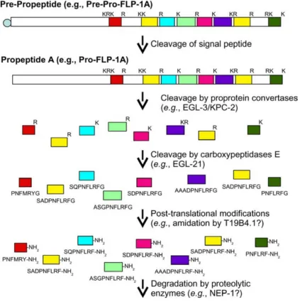
Neuropeptides in C. elegans
Neuropeptides are small signaling molecules that modulate synaptic connectivity throughout the animal kingdom (Kow and Pfaff, 1988; Li and Kim, 2008). These are short sequences of amino acids derived from large precursor genes as a result of
post-translational processing, and sometimes modification such as amidation, to become fully active (Figure 2.3, Li and Kim, 2008). A neuropeptide precursor gene may give rise to
multiple identical or different mature neuropeptides, which are then packaged inside dense core vesicles as they are transported to the nerve terminal (Strand, 1999). Increase of calcium level throughout the nerve terminal triggers release of neuropeptides from
dense core vesicles (Strand, 1999; Salio et al., 2006). Neuropeptides act through G-protein coupled receptors (GPCRs), and one neuropeptide may bind to multiple receptors,
making it difficult to discern the function of specific neuropeptide (Bargmann, 1998). The existence of neuropeptides in C. elegans was inferred by the observation of dense core vesicles in the nervous system in electron microscope images (White et al.,
The function of neuropeptide in promoting wakefulness was observed in mammals (Sakurai, 2007) and in zebrafish (Prober et al., 2006). Neuropeptide Y (NPY) possesses duo functions in promoting animal sleep and wakefulness, depending on the
site and vehicle of NPY introduction (Antonijevic et al., 2000; Dyzma et al., 2010). The
Drosophila NPY ortholog, sNPF neuropeptide, plays a role in regulating sleep homeostat (Shang et al., 2013). Despite all this, a conserved sleep promoting neuropeptide system has not been reported.
In C. elegans, it is proposed that the syntaxin regulator unc-13 and the calcium dependent activator protein for serotonin (CAPS) unc-31 mediate movement of dense core vesicle to the cell membrane (Richmond et al., 1999; Sieburth et al., 2007; Renden
et al., 2001; Grishanin et al., 2002). Mutants of unc-13 and unc-31 do not induce sleep, presumably due to their failure to package and transport sleep-promoting neuropeptides and neurotransmitters in ALA (Van Buskirk and Sternberg, 2007).
The only known neuropeptide in C. elegans ALA is a FMRFamide-like neuropeptide encoded by, flp-7, which does not induce sleep (Van Buskirk and Sternberg, 2007). It is estimated that at least hundreds if not thousands of genes are expressed in a neuron to define the specific functionality of a neuron (Hobert, 2012). There are 8,011 protein-coding genes expressed in the somatic male linker cell (Schwarz et al., 2012),
Development of ALA
Transgenic reporter analysis and imaging showed that ALA expresses three transcription factors, including ceh-10 (Wu et al, 2011) and ceh-17 (Pujol et al, 2000)in the Paired-like homeodomain class, and ceh-14 (Cassata et al, 2000) in the LIM homeodomain class families. Van Buskirk and Sternberg (2010) showed that these
transcription factors are essential for the generation and differentiation of ALA in a combinatorial and temporal fashion (Van Buskirk and Sternberg 2010). Moreover, absence or reduction of these regulatory genes abolishes expression of component genes
in the EGFR pathway, as well as other known ALA-expressed differentiation genes. As a result, these mutants lost their ability to respond to the ALA-induced sleep effect (Van
Buskirk and Sternberg 2010). But how might such genetic information translate into functional readout, i.e., what are the local genomic context and gene regulatory logic employed to elicit the sleep-inducing ability of the ALA neuron?
Differentiation genes, the genes that give rise to the functional characteristics of a cell, are often regulated by cell-specific motifs (Hobert, 2008). These motifs contain
regulatory binding sites of transcription factors that read them. Genes regulated by a common motif are often co-expressed, and may share the same function (Hobert, 2008). The C. elegans nervous system appears to fit into this model. Over the years, several motifs have been identified in C. elegans, including a cell type-specific motif (Wenick and Hobert, 2004), a neuronal subtype-specific motif (Kratsios et al, 2012; Zhang et al,
Connecting the dots towards the sleep state
The sleep-inducing property and the morphological features differentiate ALA
from the rest of the nervous system for single-neuron functional analysis. ALA induces C. elegans sleep through the EGFR signaling pathway (Van Buskirk and Sternberg, 2007) and I hypothesize that neuropeptides, as well as other differentiation genes, are involved in the sleep-inducing pathway. I propose to conduct an unbiased transcriptome profiling to catalog the protein-coding genes expressed in ALA. Deciphering their regulatory
interactions may shed light on understanding how genetic codes are translated into behavioral outputs, and provide an entry point to decode the genetic players in the
Reference
1. Altun, Z.F. and Hall, D.H. 2009. Introduction. In WormAtlas.
2. Antonijevic, I.A., Murck, H., Bohlhalter, S., Frieboes, R.M., Holsboer, F., and Steiger, A. (2000). Neuropeptide Y promotes sleep and inhibits ACTH and cortisol release in young men. Neuropharmacology 39(8), 1474-1481.
3. Doitsidou M, Flames N, Topalidou I, Abe N, Felton T, Remesal L, Popovitchenko T, Mann R, Chalfie M, Hobert O. 2013. A combinatorial regulatory signature controls terminal differentiation of the dopaminergic nervous system in C. elegans. Genes Dev27(12): 1391-1405.
4. Dupuy D, Bertin N, Hidalgo CA, Venkatesan K, Tu D, Lee D, Rosenberg J, Svrzikapa N, Blanc A, Carnec A, Carvunis AR, Pulak R, Shingles J, Reece-Hoyes J, Hunt-Newbury R, Viveiros R, Mohler WA, Tasan M, Roth FP, Le Peuch C, Hope IA, Johnsen R, Moerman DG, Barabási AL, Baillie D, Vidal M. 2007. Genome-scale analysis of in vivo spatiotemporal promoter activity in Caenorhabditis elegans. Nat Biotechnol 25(6): 663-668.
5. Dyzma, M., Boudjeltia, K.Z., Farut, B., and Kerkhofs M. (2010). Neuropeptide Y and sleep. Sleep Med. Rev. 14(3), 161-165.
6. Grishanin, R.N., Klenchin, V.A., Loyet, K.M., Kowalchyk, J.A., Ann, K., and Martin, T.F. (2002). Membrane association domains in Ca2+-dependent activator protein for secretion mediate plasma membrane and dense-core vesicle binding required for Ca2+-dependent exocytosis. J. Biol. Chem. 277, 22025–22034. 7. Hobert O. 2008. Regulatory logic of neuronal diversity: Terminal selector genes
and selector motifs. Proc Natl Acad Sci U.S.A. 105: 20067-20071.
8. Hobert, O. (2012). Neurogenesis in the nematode Caenorhabditis elegans.
WormBook ed. The C. elegans Research Community:doi/10.1895/wormbook,
http://www.wormbook.org.
9. Jarecki, J.L., Andersen, K., Konop, K.J., Knickelbine, J.J., Vestling, M.M., and Stretton, A.O. (2000). Mapping neuropeptide expression by mass spectrometry in single dissected identified neurons from the dorsal ganglion of the nematode
Ascaris suum. ACS Chem. Neurosci. 1, 505-519.
10. Kow, L.M., and D. W. (1988). Pfaff, D.W. Neuromodulatory actions of peptides.
Ann. Rev. Pharmacol. Toxico.28, 163
11. Kratsios P, Stolfi A, Levine M, Hobert O. 2011. Coordinated regulation of cholinergic motor neuron traits through a conserved terminal selector gene. Nat Neurosci15(2): 205-214.
12. Li, C. (2005). The ever-expanding neuropeptide gene families in the nematode Caenorhabditis elegans. Parasitology 131 Suppl. S109–S127.
13. Li, C., and Kim, K. (2008). Neuropeptides. WormBook ed. The C. elegans Research Community:doi/10.1895/wormbook, http://www.wormbook.org.
15. Pujol N, Torregrossa P, Ewbank JJ, Brunet JF. 2000. The homeodomain protein CePhox2/CEH-17 controls antero-posterior axonal growth in C. elegans. Development 127: 3361-3371.
16. Raizen, D.M., Zimmerman, J.E., Maycock, M.H., Ta, U.D., You, Y.J. Sundaram, M.V., and Pack, A.I. (2008). Lethargus is a Caenorhabditis elegans sleep-like state. Nature 451(7178), 569-572.
17. Renden, R., Berwin, B., Davis, W., Kyoungsook, A., Chin, C.-T., Kreber, R., Ganetzky, B, Martin, T.F.J., and Broadie, K. (2001). Drosophila CAPS is an essential gene that regulates dense-cre vesicle release and synaptic vesicle fusion. Neuron 31, 421–437.
18. Richmond, J.E., Davis, W.S, and Jorgensen, E.M. (1999). UNC-13 I srequired for synaptic vesicle fusion in C. elegans. Nat. Neurosci. 2, 959–964.
19. Sakurai, T. (2007). The neural circuit of orexin (hypocretin): maintaining sleep and wakefulness. Nat. Rev. Neurosci. 8(3),171-181.
20. Salio, C., Lossi, L., Ferrini, F., and Merighi, A. (2006). Neuropeptides as synaptic transmitters. Cell Tissue Res. 326, 583–598.
21. Schwarz EM, Kato M, Sternberg PW. 2012. Functional transcriptomics of a migrating cell in Caenorhabditis elegans. Proc Natl Acad Sci U S A. 109(40):
16246-16251.
22. Shang, Y., Donelson, N.C., Vecsey, C.G, Guo, F., Rosbash, M., and Griffith, L.C. (2013). Short neuropeptide F is a sleep-promoting inhibitory modulator. Neuron
80(1), 171-183.
23. Sieburth, D., Madison, J.M., and Kaplan, J.M. (2007). PKC-1 regulates secretion of neuropeptides. Nat. Neurosci.10, 49–57.
24. Strand, F.L. (1999). Neuropeptides. MIT Press, Cambridge, MA.
25. Troemel, E.R., Chou, J.H., Dwyer, N.D., Colbert, H.A., and Bargmann, C.I. (1995). Divergent seven transmembrane receptors are candidate chemosensory receptors in C. elegans. Cell 83(2), 207-218.
26. Van Buskirk C, Sternberg PW. 2007. Epidermal growth factor signaling induces behavioral quiescence in Caenorhabditis elegans. Nat Neurosci 10(10): 1300-1307.
27. Van Buskirk C, Sternberg PW. 2010. Paired and LIM class homeodomain proteins coordinate differentiation of the C. elegans ALA neuron. Development
137: 2065-2074.
28. Wenick AS, Hobert O. 2004. Genomic cis-regulatory architecture and trans-acting regulators of a single interneuron-specific gene battery in C. elegans. Dev Cell
6(6): 757-770.
29. White, J.G., Southgate, E., Thomson, J.N. and Brenner, S. (1986). The structure of the nervous system of the nematode C. elegans. Philos. Trans. R. Soc. Lond. Series B. Biol. Sci. 314, 1-340.
Chapter 3
Sleep
Abstract
General understanding of sleep is that it is a prolonged period of motor inactivity and slow response to external environmental changes such as chemical and mechanical
stimulation. Because of this seemingly unproductive and vulnerable state, animals are more prone to the danger of being caught by predators, and yet sleep is a behavior
observed across the animal kingdom, spanning from worms to humans (Allada and Siegal, 2008; Zimmerman et al, 2008). Such phenomenon highlights the importance of sleep, in that the benefits it offers outweighs the risk of death, and indicates that the need of sleep
is an internal drive and is perhaps indispensable for survival. Despite a growing community in sleep research, there remain two fundamental questions regarding sleep
that go unanswered: i) What is the function of sleep? ii) What is the molecular regulation of sleep? This chapter introduces current understandings of sleep, defines the definition of sleep, summarizes known molecular players regulating sleep, and discusses
approaches to facilitate sleep research.
Definition of sleep
Sleep was initially defined on the basis of electroencephalograms (EEGs), recordings that reflect cortical electrical activity alterations (Sehgal and Mignot, 2011).
During the sleep period, three states of behavior are found in EEG: wake, rapid eye movement (REM) sleep, and non-REM (NREM) sleep. In humans, a full night’s sleep is
mammalian and avian models, it precludes sleep research in other models such as fish, reptile, and bees, all of which do not present a well-defined cortex but exhibit a sleep-like state (Campbell and Tobler, 1984). Moreover, this approach is not practical for
high-throughput screening and is laborious for day-to-day experiments (Sehgal and Mignot, 2011). A more efficient and economic approach is to use simple animal models and
behavioral assays to measure a sleep-like state originally proposed by Campbell and Tobler (1984). This shifted the paradigm of sleep definition. The current definition of sleep behavior consists of four criteria: i) a prolonged but reversible period of voluntary
movement inactivity, ii) increased arousal threshold for response to sensory stimulants, iii) regulation by homeostasis, and iv) a circadian clock control (Sehgal and Mignot,
2011).
Function of sleep
Knowledge and recognition of the need of sleep come from complaints of sleep disorders, which are extremely common (Mahowald and Schenck, 2005). There are more
than 100 identified sleep disorders, and most of them fall into four categories: hypersomnia (i.e. excessive day-time sleep without obvious explanation, e.g. narcolepsy, obstructive sleep apnoea), insomnia (i.e. trouble falling and staying asleep, e.g. restless
leg syndrome), circadian rhythm disorders (e.g. delayed sleep syndrome), and parasomnias (i.e. complex behaviors arising from the sleep period, e.g. sleep walking,
sleep terrors).
plasticity and supports cognitive function (Diekelmann and Born, 2010; Poe et al., 2010). Others observed association of regulation of sleep and wakefulness with regulation of cerebral energy stores (Benington, et la., 1995). Insights from studies in sleep-deprived
mice and flies suggest that sleep has a role in curbing stress, and that the need for sleep is influenced by cellular stress (Naidoo et al., 2007). More recently, using real-time
two-photon imaging in mice, Lulu et al., (2013) showed that a critical function of sleep is to clear metabolite from the brain and to maintain metabolic homeostasis.
Molecules regulating sleep
Research in model organisms revealed that sleep is genetically regulated and is
evolutionarily conserved among animals (Sehgal and Mignot, 2011). Much discoveries in sleep regulation come from the identification of neuropeptides and neurotransmitters, as well as characterization of intracellular signaling molecules that are essential for
regulating sleep/wakefulness. Other molecules include ion channels and channel-regulating proteins, circadian clock genes, metabolic factors, and immune genes (Sehgal
and Mignot, 2011). This study will focus on the roles of neuropeptides and intracellular signaling pathway on sleep regulation.
Neuropeptide/receptor systems and neurotransmitters regulating sleep
In mammals and other vertebrates, numerous neuropeptide/receptor systems have
narcolepsy (Taheri et al., 2002; Sehgal and Mignot, 2011). Orthologs of hypocretins in flies and worms have yet to be reported. However, the neuropeptide pigment-dispersing factor (PDF) is a wake-promoting peptide and functions in an analogous fashion in fles
(Drosophila melanogaster) by inhibiting sleep-promoting neurotransmitters such as gamma-aminobutyric acid (GABA; Parisky et al., 2008). Consistent with this view, the
wake promoting property of C. elegans PDF-1/PDFR system is shown by its ability in governing exit of locomotor quiescence in lethargus (Choi et al., 2013). Other neurotransmitters implicated in sleep regulation are the wake-promoting histamine,
dopamine, acetylcholine, and norepinephrine, as well as sleep-promoting serotonin and adenosine (Sehgal and Mignot, 2011).
Intracellular signaling molecules regulating sleep
As the downstream target of neuropeptides and neurotransmitters, intracellular
signaling pathways also take part in regulating sleep and wakefulness. The mammalian CREB pathway promotes wakefulness (Graves et al., 2003; Hendricks et al., 2001), whereas Drosophila protein kinase A (PKA)/CREB pathway plays duo roles in a site-dependent manner. Pan-neuronal expression of PKA promotes wakefulness, but PKA expression in specific subsets of neurons promotes sleep (Joiner et al., 2006). The cyclic
guanosine monophosphate (cGMP) kinase regulates sleep in mammals (Langmesser et al., 2009) and promotes sleep in C. elegans and Drosophila (Raizen et al., 2008). Lastly, the epidermal growth factor receptor (EGFR) signaling pathway promotes sleep in worms,
(Kramer et al., 2001). Overexpression of the EGFR ligand in Drosophila increases sleep, and this effect is dependent on a functional EGFR and, at least in part, mediated by the extracellular signal-regulated kinases/mitogen-activated protein kinase (ERK/MAPK)
pathway (Foltenyi et al., 2007). C. elegans has one EGFR, LET-23, and reduction of this receptor as a result of loss-of-function mutation increases locomotor activity in the
sleep-like state of worms (Van Buskirk and Sternberg, 2007). On the contrary, overexpression of the sole EGFR ligand, LIN-3C, activates EGFR signaling through diacylglycerol and
phospholipase C-γ to induce a sleep-like state where worms cease locomotor and feeding
activities (Van Buskirk and Sternberg, 2007).
What remains to be explored?
Research on sleep had made enormous progress in the past decades due to the utilization of simple animal model organisms (Sehgal and Mignot, 2011). Whether the function of sleep is for building synaptic plasticity or for metabolite clearance, the genetic
basis of sleep regulation remains elusive. With the paradigm of sleep definition shifted from measuring EEGs to directly measuring rest/active behavior, research using model
References
1. Allada, R., and Siegel, J.M. (2008). Unearthing the phylogenetic roots of sleep. Curr. Biol. 18, R670-R679.
2. Benington, J.H., and H.C. Heller. (1995). Restoration of brain energy metabolism as the function of sleep. Prog. Neurobiol. 45, 347-360.
3. Campbell, S.S., and Tobler, I. (1984). Animal sleep: a review of sleep duration across phyogeny. Neurosci. Biobehav. Rev. 8, 269-300.
4. Choi, S., Chatzigeorgiou, M., Taylor, K.P., Schafer, W.R., and Kaplan, J.M. (2013). Analysis of NPR-1 reveals a circuit mechanism for behavioral quiescence in C. elegans. Neuron 78(5), 869-880.
5. Diekelmann, S., and Born, J. (2010). The memory function of sleep. Nat. Rev. Neurosci. 11, 114-126.
6. Graves, L.A., Hellman, K., Veasey, S., Blendy, J.A., Pack, A.I., and Abel, T. (2003). Genetic evidence for a role of CREB in sustained cortical arousal. K. Neurophysiol. 90, 1152-1159.
7. Hendricks, J.C., Williams, J.A., Panckeri, K., Kirk, D., Tello, M., Yin, J.C., and Sehgal, A. (2001). A non-circadian role for camp signaling and CREB activity in Drosophila rst homeostasis. Nat. Neurosci. 4, 1108-1115.
8. Joiner, W.J., Crocker, A., White, B.H., Sehgal, A. (2006). Sleep in Drosophila is regulated by adult mushroom bodies. Nature 441(7094), 757-760.
9. Kramer, A., Yang, F.C., Snodgrass, O., Li, X., Scammell, T.E., Davis, F.C., Weitz, C.J. (2001). Regulation of daily locomotor activity and sleep by hypothalamic EGF receptor signaling. Science 294(5551), 2511-2515.
10. Kushikata, T., Fang, J., Chen, Z., Wang, Y., and Krueger, J.M. (1998). Epidermal growth factor enhances spontaneous sleep in rabbits. Am. J. Physiol. 275(2 Pt 2), R509-514.
11. Langmesser, S., Franken, P., Feil, S., Emmenegger, Y., Albrecht, U., and Feil, R. (2009). cGMP-dependent protein kinase type I is implicated in the regulation of the timing and quality of sleep and wakefulness. PLoS One 4(1), e4238.
12. Mahowald, M.W., and Schenck., C.H. (2005). Insights from studying human sleep disorders. Nature 437, 1279-1285.
13. Naidoo, N., Casiano, V., Cater, J., Zimmerman, J., and Pack., A.I. (2007). A role for the molecular chaperone protein BiP/GRP78 in Drosophila sleep homeostasis. Sleep 30, 557-565.
14. Parisky, K.K., Agosto, J., Pulver, S.R., Shang, Y., Kuklin, E., Hodge, J.J., Kang, K., Liu, X., Garrity, P.A., Rosbash, M., and Griffith, L.C. (2008). PDF cells are a GABA-responsive wake-promoting component of the Drosophila sleep circuit. Neuron 60, 672-682.
15. Poe, G.R., Walsh, C.M., and Bjorness, T.E. (2010). Cognitive neuroscience of sleep. Prog. Brain Res. 185, 1-19.
16. Prober, D.A., Rihel, J., Onah, A.A., Sung, R.J., and Schier, A.F. (2006). Hypocretin/orexin overexpression induces an insomnia-like phenotype in zebrafish. J. Neurosci. 26(51), 13400-13410.
18. Raizen, D.M., Zimmerman, J.E., Maycock, M.H., Ta, U.D., You, Y.J. Sundaram, M.V., and Pack, A.I. (2008). Lethargus is a Caenorhabditis elegans sleep-like state. Nature 451(7178), 569-572.
19. Sakurai, T. (2007). The neural circuit of orexin (hypocretin): maintaining sleep and wakefulness. Nat. Rev. Neurosci. 8(3),171-181.
20. Sehgal, A., and Mignot, E. (2011). Genetics of sleep and sleep disorders. Cell 146,
194-207.
21. Taheri, S., Zeitzer, J.M., and Mignot, E. (2002). The role of hypocretins (orexins) in sleep regulation and narcolepsy. Annu. Rev. Neurosci. 25, 283–313.
22. Van Buskirk, C., and Sternberg, P.W. (2007). Epidermal growth factor signaling induces behavioral quiescence in Caenorhabditis elegans. Nat. Neurosci. 10(10),
1300-1307.
23. Xie, L., Kang, H., Xu, Q., Chen, M.J., Liao, Y., Thiyagarajan, M., O'Donnell, J., Christensen, D.J., Nicholson, C., Iliff, J.J., Takano, T., Deane, R., and Nedergaard, M. (2013). Sleep drives metabolite clearance from the adult brain. Science
342(6156), 373-377.
24. Zimmerman, J.E., Naidoo, N., Raizen D.M., and Pack, A.I. (2008). Conservation of sleep: Insights from non-mammalian model systems. Trends Neurosci. 31, 371-376.
Chapter 4
Sleep in
Caenorhabditis elegans
and
Abstract
A sleep-like state was recently characterized in two non-mammalian model organisms: the roundworm Caenorhabditis elegans and the zebrafish Danio rerio
(Zhdanova et al., 2001; Prober et al., 2006; Yokogawa et al., 2007; Van Buskirk and Sternberg, 2007; Raizen et al., 2008). Both of these model organisms employ regulatory
molecules similar to the mammalian models for sleep and wake promotion (see also Chapter 3). In addition, they are genetically traceable and are excellent for high-throughput behavioral screens. More importantly, the well-established genetic tools
enable easy manipulation on genes for functional analysis. Herein I introduce sleep in C. elegans and zebrafish, as well as the behavioral essays for analyzing sleep in each organism, and discuss the potential advantages of corroborating insights gained from these two models for elucidating genes involved in sleep behavior.
Worm sleep
A sleep-like state in C. elegans was first described in lethargus, a quiescence behavioral state at the end of each larval stage (Raizen et al., 2008). Lethargus in worms possesses specific characteristics that fulfill the criteria for sleep, i.e. reversibility of quiescence, elevated arousal threshold to sensory stimuli, and homeostasis. Although
C. elegans sleep is not restricted to the larval stage (Figure 4.1). In fact, sleep-like states are observed in adults and can be induced by intracellular signaling pathways such as anachronistic expression of the EGFR signaling (Figure 4.2; Van Buskirk and
Sternberg, 2007), and by satiety, through the TGFβ and cGMP pathways, in the presence
of high-nutrient food or full feeding after a long period of starvation (You et al., 2008; Gallagher et al., 2013). More evidence on EGFR signaling or satiety induced sleep-like
state comes from neuronal modulation analysis. Using optogenetic tools, Cho and Sternberg (2014) showed that a sleep-like state of C. elegans in response to both EGFR signaling activation and satiety share the same neuronal circuit modulation as in lethargus.
[image:48.612.180.438.347.582.2]Figure 4.2. Schematic diagram of sleep assay to assess locomotion, feeding and sensory arousal behaviors. Worms were placed in a water bath for heat shock treatment (30 min at 33°C) and recovered at their growing temperature for 2 hr. Worms were examined for the
sinusoidal movement as an indication for locomotion, pharyngeal pumping as an indicator for feeding, and rapid reversal movement when it withdraws from noxious smell like 1-octanol as an indication for sensory arousal.
Zebrafish sleep
Zebrafish is a vertebrate that has a sleep-like state, which is regulated by circadian
rhythm and homeostasis, and has reduced sensory responsiveness (Chiu and Prober, 2013). Zebrafish rest-activity rhythm is synchronized with the day-night cycle. Similar to their diurnal cousins like human, zebrafish are active in the daytime and quiescent at
night (Zimmerman et al., 2008, and references therein). Also, arousal state in zebrafish larvae can be characterized by changes in frequency and intensity of voluntary locomotor
using high-speed infrared video capture in conjunction with computational analysis for locomotor behaviors (Figure 4.3; Chiu and Prober, 2013).
Figure 4.3. Monitoring larval zebrafish sleep and wake behavior. (A) Zebrafish larval locomotor activity assay. Individual zebrafish larva are placed in each well of a 96-well-plate on the 5th day of development. The plate is placed in a temperature-controlled chamber that is illuminated by white lights during the day and continuously illuminated by infrared lights. The larvae are monitored by an infrared camera and the locomotor activity of each larva is recorded by a computer. (B) Representative locomotor activity data for each of 20 individual wild-type larvae (gray traces) and their mean locomotor activity (blue trace, ± standard error bar of the mean) is shown. Black and white bars
[image:50.612.160.419.147.563.2]although there is considerable variability among individuals. (C) An example of typical larval zebrafish behavior at the end of the day is shown. A rest bout is defined as a period of at least 1 min of inactivity, which is associated with an increase in arousal threshold (Prober et al., 2006). Rest latency indicates the time between lights-off at night and initiation of the first rest bout. Adopted and unmodified from Chiu and Prober, 2013.
Zebrafish also show physiological and pharmacological characteristics of
mammalian sleep (Chiu and Prober, 2013, and references therein). The hypothalamus-expressed neuropeptide hypocretin is the best-known and characterized sleep and arousal
regulator in zebrafish. Overexpression of hypocretin using a heat shock promoter induces an insomnia-like behavior in zebrafish (Prober et. al., 2006), where the animals have increased wakefulness, longer latency to sleep after lights off, decreased frequency and
length of sleep bouts at night, and are hyperaroused. Today, a sleep promoting neuropeptide has yet to be found in zebrafish.
What happens when a worm meets a fish?
The route to better understand the need of sleep is to find out its functions and regulations, which highlights the importance of uncovering essential genetic molecules
involved in the process. Despite intensive research in sleep, little is known about the fundamental molecules and their specific roles in regulating sleep and arousal. Identification of novel genes has been challenging due to the complex genome and a long
life cycle of mammalian models.
To understand the role of a component in an assemble, there is no better way than
genome, making it a good model for genetic analysis (WormBook, www.wormbook.org). One of the most attractive features of C. elegans is the ease of generating mutants (see Chapter 1). More importantly, the wealth of existing mutant collections provides an
References
1. Chiu, C.N., and Prober, D.A. (2013). Regulation of zebrafish sleep and arousal states: current and prospective approaches. Frontiers in Neural Circuits 7:58.
2. Cho, J.Y., and Sternberg P.W. (2014). Multilevel modulation of a sensory motor circuit during C. elegans sleep and arousal. Cell 156, 249-260.
3. Gallagher, T., Kim, J., Oldenbroek, M., Kerr, R, and You, Y.J. (2013). ASI regulates satiety quiescence in C. elegans. J. Neurosci. 33(23), 9716-9724.
4. Jeon, M., Gardner, H.F., Miller, E.A., Deshler, J., and Rougvie, A.E. (1999). Similarity of the C. elegans developmental timing protein LIN-42 to circadian rhythm proteins. Science 286(5442), 1141-1146.
5. Pfaff, D., Ribeiro, A., Matthews, J., and Kow, L.-M. (2008). Concepts and mechanism of generalized central nervous system arousal. Ann. N.Y. Acad. Sci
1129, 11-25.
6. Prober, D.A., Rihel, J., Onah, A.A., Sung, R.J., and Schier, A.F. (2006). Hypocretin/orexin overexpression induces an insomnia-like phenotype in zebrafish. J. Neurosci. 26(51), 13400-13410.
7. Raizen, D.M., Zimmerman, J.E., Maycock, M.H., Ta, U.D., You, Y.J. Sundaram, M.V., and Pack, A.I. (2008). Lethargus is a Caenorhabditis elegans sleep-like state. Nature 451(7178), 569-572.
8. Sehgal, A., and Mignot, E. (2011). Genetics of sleep and sleep disorders. Cell 146,
194-207.
9. TALE nuclease architecture for efficient genome editing. Nat. Biotechnol. 29:
143–148.
10. Van Buskirk, C., and Sternberg, P.W. (2007). Epidermal growth factor signaling induces behavioral quiescence in Caenorhabditis elegans. Nat. Neurosci. 10(10),
1300-1307.
11. White, J.G., Southgate, E., Thomson, J.N., Brenner, F.R.S. (1996). The structure of the nervous system of the nematode Caenorhabditis elegans. Philos Trans R Soc. Lond. B Biol. Sci. 314, 1-340.
12. Yokogawa, T., Marin, W., Faraco, J., Pezeron, G., Appelbaum, L., Zhang, J., Rosa, F., Mourrain, P., and Mignot, E. (2007). Charactrization of sleep in zebrafish and insomnia in hypocretin receptor mutants. PLoS Biol. 5(10), e277. 13. You, Y.J., Kim, J., Raizen, D.M., and Avery L. (2008). Insulin, cGMP, and
TGF-beta signals regulate food intake and quiescence in C. elegans: a model for satiety. Cell Metab. 7(3), 249-257.
14. Zhdanova, I.V., Wang, S.Y., Leclair, O.U., and Danilova, N.P. (2001). Melantonin promotes sleep-like state in zebrafish. Brain Res. 903, 263-268. 15. Zimmerman, J.E., Naidoo, N., Raizen D.M., and Pack, A.I. (2008). Conservation