5,5
000-Bis[(1
H
-imidazol-1-yl)methyl]-2,2
000-bipyridine methanol disolvate
Suk-Hee Moon,aTae Ho Kimband Ki-Min Parkb*
aDepartment of Food & Nutrition, Kyungnam College of Information and
Technology, Busan 616-701, Republic of Korea, andbDepartment of Chemistry and Research Institute of Natural Sciences, Gyeongsang National University, Jinju 660-701, Republic of Korea
Correspondence e-mail: kmpark@gnu.ac.kr
Received 25 January 2011; accepted 28 January 2011
Key indicators: single-crystal X-ray study;T= 173 K; mean(C–C) = 0.002 A˚;
Rfactor = 0.041;wRfactor = 0.144; data-to-parameter ratio = 16.7.
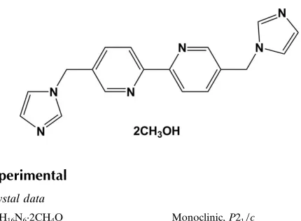
The title compound, C18H16N62CH3OH, was prepared by the
reaction of 5,50-bis(bromomethyl)-2,20-bipyridine with imida-zole. The main molecule lies on an inversion center located at the mid-point of the C—C bond joining the two pyridine rings. The asymmetric unit therefore contains one half-molecule and one methanol solvent molecule. The dihedral angle between the pyridine and imidazole rings is 72.32 (5). In the crystal, weak intermolecular O—H N, C—H N and C—H O hydrogen bonds contribute to the stabilization of the packing.
Related literature
For related syntheses, see: Sambrooket al.(2006); Zanget al.
(2010). For a related structure, see: Zang et al. (2010). For reference bond lengths, see: Allenet al.(1987).
Experimental
Crystal data C18H16N62CH4O
Mr= 380.45
Monoclinic,P21=c a= 4.5653 (4) A˚
b= 14.7886 (12) A˚
c= 14.5378 (11) A˚
= 93.805 (2) V= 979.35 (14) A˚3
Z= 2
MoKradiation
= 0.09 mm1
T= 173 K
0.350.300.10 mm
Data collection Bruker APEXII CCD
diffractometer
Absorption correction: multi-scan (SADABS; Sheldrick, 1996)
Tmin= 0.970,Tmax= 0.991
5948 measured reflections 2136 independent reflections 1619 reflections withI> 2(I)
Rint= 0.053
Refinement
R[F2> 2(F2)] = 0.041
wR(F2) = 0.144
S= 1.09 2136 reflections
128 parameters
H-atom parameters constrained
max= 0.21 e A˚
3 min=0.24 e A˚
[image:1.610.57.272.510.668.2]3
Table 1
Hydrogen-bond geometry (A˚ ,).
D—H A D—H H A D A D—H A
O1—H1A N3i
0.84 1.93 2.7662 (19) 171 C3—H3 O1 0.95 2.44 3.360 (2) 162 C8—H8 N1ii 0.95 2.57 3.422 (2) 149 C9—H9 O1iii
0.95 2.54 3.470 (2) 168
Symmetry codes: (i) xþ1;yþ1;z; (ii) x;yþ1 2;z
1 2; (iii) xþ1;y1
2;zþ 1 2.
Data collection:APEX2(Bruker, 2006); cell refinement:SAINT
(Bruker, 2006); data reduction:SAINT; program(s) used to solve structure: SHELXTL (Sheldrick, 2008); program(s) used to refine structure: SHELXTL; molecular graphics: SHELXTL and
DIAMOND(Brandenburg, 1998); software used to prepare material for publication:SHELXTL.
This research was supported by the National Nuclear R&D Program through the National Research Foundation (NRF) funded by the Ministry of Education, Science and Technology (2010–0018586)
Supplementary data and figures for this paper are available from the IUCr electronic archives (Reference: SJ5094).
References
Allen, F. H., Kennard, O., Watson, D. G., Brammer, L., Orpen, A. G. & Taylor, R. (1987).J. Chem. Soc. Perkin Trans. 2, pp. S1–19.
Brandenburg, K. (1998).DIAMOND. Crystal Impact GbR, Bonn, Germany. Bruker (2006).APEX2andSAINT. Bruker AXS Inc., Madison, Wisconsin,
USA.
Sambrook, M. R., Curiel, D., Hayes, E. J., Beer, P. D., Pope, S. J. A. & Faulkner, S. (2006).New J. Chem.30, 1133–1136.
Sheldrick, G. M. (1996).SADABS. University of Go¨ttingen, Germany. Sheldrick, G. M. (2008).Acta Cryst.A64, 112–122.
Zang, H.-Y., Lan, Y.-Q., Yang, G.-S., Wang, X.-L., Shao, K.-Z., Xu, G.-J. & Su, Z.-M. (2010).CrystEngComm,12, 434–445.
Acta Crystallographica Section E Structure Reports
Online
supporting information
Acta Cryst. (2011). E67, o554 [doi:10.1107/S1600536811003643]
5,5
′
-Bis[(1
H
-imidazol-1-yl)methyl]-2,2
′
-bipyridine methanol disolvate
Suk-Hee Moon, Tae Ho Kim and Ki-Min Park
S1. Comment
The title compound was prepared to use as a multi-dentate ligand in the formation of metallosupramolecules in line with
similar previously reported compounds (Sambrook et al., 2006; Zang et al., 2010).
In the title compound (Scheme 1, Fig. 1), two pyridine rings are coplanar because the title compound lies on a
crystallographic inversion center. The dihedral angle between the pyridine and imidazole rings is 72.32 (5)°. All the bond
lengths are within normal values (Allen et al., 1987).
In the crystal structure, as shown in Fig. 2, weak intermolecular O–H···N, C–H···N and C–H···O hydrogen bonds are
observed (Table 1). These intermolecular interactions may be contribute to the stabilization of the packing.
S2. Experimental
A mixture of imidazole (0.120 g, 1.76 mmol) and potassium hydroxide (0.440 g, 7.84 mmol) in DMSO (10 ml) was
stirred for 1 h. A DMSO solution (20 ml) of 5,5′-bis(bromomethyl)-2,2′-bipyridine (0.30 g, 0.88 mmol) was slowly added
and the solution stirred for 6 h at room temperature. After water (100 ml) was added, the reaction mixture was extracted
with chloroform (3×100 ml), washed with water and then dried over anhydrous MgSO4. The solvent was removed to give
the title compound in 63% yield. X-ray quality single crystals were obtained by slow evaporation of a solution in MeOH.
S3. Refinement
All H-atoms were positioned geometrically and refined using a riding model with d(C—H) = 0.95 Å, Uiso = 1.2Ueq(C) for
Figure 1
The molecular structure of the title compound, showing displacement ellipsoids drawn at the 50% probability level.
Hydrogen bonds are shown as dashed lines. (Symmetry code: i) -x + 2, -y + 1, -z + 1)
Figure 2
Crystal packing of the title compound with intermolecular O–H···N, C–H···N and C–H···O hydrogen bonds shown as
dashed lines. H atoms not involved in intermolecular interactions have been omitted for clarity (Symmetry codes: i) -x +
1, -y + 1, -z; ii) x, -y + 1/2, z - 1/2; iii) -x + 1, y - 1/2, -z + 1/2; iv) x, -y + 1/2, z + 1/2; v) -x + 2, -y + 1, -z + 1).
5,5′-Bis[(1H-imidazol-1-yl)methyl]-2,2′-bipyridine methanol disolvate
Crystal data
C18H16N6·2CH4O
Mr = 380.45
Monoclinic, P21/c
Hall symbol: -P 2ybc a = 4.5653 (4) Å
c = 14.5378 (11) Å β = 93.805 (2)° V = 979.35 (14) Å3
[image:3.610.126.487.362.531.2]Mo Kα radiation, λ = 0.71073 Å Cell parameters from 2441 reflections θ = 2.8–28.2°
µ = 0.09 mm−1
T = 173 K Plate, colorless 0.35 × 0.30 × 0.10 mm
Data collection
Bruker APEXII CCD diffractometer
Radiation source: fine-focus sealed tube Graphite monochromator
φ and ω scans
Absorption correction: multi-scan (SADABS; Sheldrick, 1996) Tmin = 0.970, Tmax = 0.991
5948 measured reflections 2136 independent reflections 1619 reflections with I > 2σ(I) Rint = 0.053
θmax = 27.0°, θmin = 2.0°
h = −4→5 k = −18→18 l = −16→18
Refinement
Refinement on F2
Least-squares matrix: full R[F2 > 2σ(F2)] = 0.041
wR(F2) = 0.144
S = 1.09 2136 reflections 128 parameters 0 restraints
Primary atom site location: structure-invariant direct methods
Secondary atom site location: difference Fourier map
Hydrogen site location: inferred from neighbouring sites
H-atom parameters constrained w = 1/[σ2(F
o2) + (0.0772P)2 + 0.1442P]
where P = (Fo2 + 2Fc2)/3
(Δ/σ)max < 0.001
Δρmax = 0.21 e Å−3
Δρmin = −0.24 e Å−3
Special details
Geometry. All e.s.d.'s (except the e.s.d. in the dihedral angle between two l.s. planes) are estimated using the full covariance matrix. The cell e.s.d.'s are taken into account individually in the estimation of e.s.d.'s in distances, angles and torsion angles; correlations between e.s.d.'s in cell parameters are only used when they are defined by crystal symmetry. An approximate (isotropic) treatment of cell e.s.d.'s is used for estimating e.s.d.'s involving l.s. planes.
Refinement. Refinement of F2 against ALL reflections. The weighted R-factor wR and goodness of fit S are based on F2,
conventional R-factors R are based on F, with F set to zero for negative F2. The threshold expression of F2 > σ(F2) is used
only for calculating R-factors(gt) etc. and is not relevant to the choice of reflections for refinement. R-factors based on F2
are statistically about twice as large as those based on F, and R- factors based on ALL data will be even larger.
Fractional atomic coordinates and isotropic or equivalent isotropic displacement parameters (Å2)
x y z Uiso*/Ueq
N1 0.8022 (3) 0.40136 (8) 0.45831 (9) 0.0287 (3)
N2 0.4002 (3) 0.36956 (9) 0.17390 (9) 0.0281 (3)
N3 0.5877 (4) 0.36182 (11) 0.03818 (10) 0.0441 (4)
C1 0.5974 (4) 0.37871 (10) 0.39254 (11) 0.0301 (4)
H1 0.5329 0.3176 0.3901 0.036*
C2 0.4722 (3) 0.43832 (10) 0.32748 (10) 0.0254 (4)
C3 0.5628 (4) 0.52770 (11) 0.33297 (11) 0.0302 (4)
H3 0.4831 0.5710 0.2900 0.036*
C4 0.7704 (4) 0.55308 (10) 0.40161 (11) 0.0290 (4)
H4 0.8328 0.6143 0.4070 0.035*
C5 0.8868 (3) 0.48825 (9) 0.46263 (10) 0.0230 (3)
H6B 0.1273 0.3589 0.2774 0.037*
H6A 0.1260 0.4573 0.2313 0.037*
C7 0.4342 (4) 0.41095 (12) 0.09281 (12) 0.0370 (4)
H7 0.3563 0.4689 0.0770 0.044*
C8 0.6558 (4) 0.28448 (12) 0.08778 (13) 0.0408 (5)
H8 0.7665 0.2353 0.0663 0.049*
C9 0.5437 (4) 0.28818 (11) 0.17102 (12) 0.0357 (4)
H9 0.5604 0.2436 0.2181 0.043*
O1 0.2950 (3) 0.64181 (9) 0.14603 (8) 0.0371 (3)
H1A 0.3447 0.6360 0.0917 0.056*
C10 −0.0113 (4) 0.65194 (15) 0.14519 (15) 0.0484 (5)
H10B −0.0687 0.6604 0.2084 0.073*
H10A −0.1069 0.5977 0.1186 0.073*
H10C −0.0717 0.7048 0.1080 0.073*
Atomic displacement parameters (Å2)
U11 U22 U33 U12 U13 U23
N1 0.0345 (8) 0.0239 (7) 0.0274 (7) −0.0042 (5) 0.0002 (6) 0.0016 (5) N2 0.0318 (8) 0.0278 (7) 0.0244 (7) 0.0002 (5) 0.0000 (6) −0.0046 (5) N3 0.0533 (10) 0.0507 (9) 0.0289 (8) 0.0131 (8) 0.0063 (7) −0.0033 (7) C1 0.0367 (9) 0.0246 (8) 0.0290 (8) −0.0063 (6) 0.0016 (7) −0.0023 (6) C2 0.0250 (8) 0.0304 (8) 0.0215 (7) 0.0000 (6) 0.0065 (6) −0.0043 (6) C3 0.0337 (9) 0.0288 (8) 0.0279 (8) 0.0038 (7) −0.0008 (7) 0.0034 (6) C4 0.0351 (9) 0.0219 (8) 0.0296 (8) −0.0011 (6) −0.0009 (7) 0.0008 (6) C5 0.0256 (8) 0.0240 (7) 0.0201 (7) 0.0001 (6) 0.0065 (6) −0.0015 (5) C6 0.0267 (9) 0.0379 (9) 0.0283 (9) −0.0004 (6) 0.0034 (7) −0.0065 (7) C7 0.0471 (11) 0.0361 (9) 0.0280 (9) 0.0088 (8) 0.0030 (8) 0.0007 (7) C8 0.0483 (11) 0.0390 (10) 0.0345 (10) 0.0132 (8) −0.0015 (8) −0.0112 (8) C9 0.0456 (11) 0.0273 (8) 0.0337 (9) 0.0037 (7) −0.0014 (8) −0.0043 (7)
O1 0.0342 (7) 0.0493 (7) 0.0277 (6) 0.0074 (5) 0.0024 (5) 0.0017 (5)
C10 0.0350 (10) 0.0590 (13) 0.0515 (12) 0.0067 (9) 0.0053 (9) −0.0012 (9)
Geometric parameters (Å, º)
N1—C1 1.335 (2) C4—H4 0.9500
N1—C5 1.3420 (19) C5—C5i 1.490 (3)
N2—C7 1.346 (2) C6—H6B 0.9900
N2—C9 1.372 (2) C6—H6A 0.9900
N2—C6 1.4653 (19) C7—H7 0.9500
N3—C7 1.313 (2) C8—C9 1.346 (3)
N3—C8 1.377 (2) C8—H8 0.9500
C1—C2 1.388 (2) C9—H9 0.9500
C1—H1 0.9500 O1—C10 1.406 (2)
C2—C3 1.386 (2) O1—H1A 0.8400
C2—C6 1.506 (2) C10—H10B 0.9800
C3—C4 1.382 (2) C10—H10A 0.9800
C4—C5 1.388 (2)
C1—N1—C5 117.36 (14) N2—C6—H6B 109.3
C7—N2—C9 106.80 (14) C2—C6—H6B 109.3
C7—N2—C6 126.80 (14) N2—C6—H6A 109.3
C9—N2—C6 126.30 (14) C2—C6—H6A 109.3
C7—N3—C8 104.71 (15) H6B—C6—H6A 108.0
N1—C1—C2 124.46 (14) N3—C7—N2 112.04 (16)
N1—C1—H1 117.8 N3—C7—H7 124.0
C2—C1—H1 117.8 N2—C7—H7 124.0
C3—C2—C1 117.31 (15) C9—C8—N3 110.55 (15)
C3—C2—C6 121.56 (15) C9—C8—H8 124.7
C1—C2—C6 121.11 (14) N3—C8—H8 124.7
C4—C3—C2 119.24 (15) C8—C9—N2 105.89 (15)
C4—C3—H3 120.4 C8—C9—H9 127.1
C2—C3—H3 120.4 N2—C9—H9 127.1
C3—C4—C5 119.28 (14) C10—O1—H1A 109.5
C3—C4—H4 120.4 O1—C10—H10B 109.5
C5—C4—H4 120.4 O1—C10—H10A 109.5
N1—C5—C4 122.32 (14) H10B—C10—H10A 109.5
N1—C5—C5i 116.18 (16) O1—C10—H10C 109.5
C4—C5—C5i 121.50 (16) H10B—C10—H10C 109.5
N2—C6—C2 111.44 (13) H10A—C10—H10C 109.5
C5—N1—C1—C2 1.6 (2) C9—N2—C6—C2 73.5 (2)
N1—C1—C2—C3 −1.5 (2) C3—C2—C6—N2 94.07 (17)
N1—C1—C2—C6 176.79 (15) C1—C2—C6—N2 −84.11 (18)
C1—C2—C3—C4 0.1 (2) C8—N3—C7—N2 −0.1 (2)
C6—C2—C3—C4 −178.13 (14) C9—N2—C7—N3 0.3 (2)
C2—C3—C4—C5 1.0 (2) C6—N2—C7—N3 177.02 (15)
C1—N1—C5—C4 −0.3 (2) C7—N3—C8—C9 −0.1 (2)
C1—N1—C5—C5i 179.33 (15) N3—C8—C9—N2 0.3 (2)
C3—C4—C5—N1 −0.9 (2) C7—N2—C9—C8 −0.3 (2)
C3—C4—C5—C5i 179.44 (16) C6—N2—C9—C8 −177.10 (16)
C7—N2—C6—C2 −102.61 (19)
Symmetry code: (i) −x+2, −y+1, −z+1.
Hydrogen-bond geometry (Å, º)
D—H···A D—H H···A D···A D—H···A
O1—H1A···N3ii 0.84 1.93 2.7662 (19) 171
C3—H3···O1 0.95 2.44 3.360 (2) 162
C8—H8···N1iii 0.95 2.57 3.422 (2) 149
C9—H9···O1iv 0.95 2.54 3.470 (2) 168