2,2
000-{1,1
000-[Butane-1,4-diylbis(oxy-nitrilo)]diethylidyne}di-1-naphthol
Wen-Kui Dong,* Jian-Chao Wu, Yin-Xia Sun, Jian Yao and Jun-Feng Tong
School of Chemical and Biological Engineering, Lanzhou Jiaotong University, Lanzhou 730070, People’s Republic of China
Correspondence e-mail: dongwk@mail.lzjtu.cn Received 28 April 2009; accepted 2 May 2009
Key indicators: single-crystal X-ray study;T= 298 K; mean(C–C) = 0.003 A˚;
Rfactor = 0.054;wRfactor = 0.173; data-to-parameter ratio = 13.2.
The title compound, C28H28N2O4, was synthesized by the reaction of 2-acetyl-1-naphthol with 1,4-bis(aminooxy)butane in ethanol. The molecule, which lies about an inversion centre, adopts a linear structure, in which the oxime groups and naphthalene ring systems assume an anticonformation. The intramolecular interplanar distance between parallel naphtha-lene rings is 1.054 (3) A˚ . Intramolecular O—H N hydrogen bonds are formed between the oxime nitrogen and hydroxy groups.
Related literature
For salen-type compounds, see: Atwood & Harvey (2001); Okabe & Oya (2000). For related structures, see: Dong et al.
(2007, 2008).
Experimental
Crystal data
C28H28N2O4 Mr= 456.52
Triclinic,P1
a= 6.9590 (15) A˚
b= 8.6598 (18) A˚
c= 10.596 (2) A˚ = 105.841 (2)
= 105.940 (2)
= 91.689 (1)
V= 586.9 (2) A˚3 Z= 1
MoKradiation = 0.09 mm1 T= 298 K
0.500.430.20 mm
Data collection
Bruker SMART 1000 CCD area-detector diffractometer Absorption correction: multi-scan
(SADABS; Sheldrick, 1996)
Tmin= 0.958,Tmax= 0.983
3020 measured reflections 2026 independent reflections 1350 reflections withI> 2(I)
Rint= 0.025
Refinement
R[F2> 2(F2)] = 0.054
wR(F2) = 0.173 S= 1.02 2026 reflections
154 parameters
H-atom parameters constrained
max= 0.23 e A˚
3 min=0.25 e A˚
3
Table 1
Hydrogen-bond geometry (A˚ ,).
D—H A D—H H A D A D—H A
O2—H2 N1 0.82 1.84 2.557 (2) 145
Data collection:SMART(Siemens, 1996); cell refinement:SAINT
(Siemens, 1996); data reduction:SAINT; program(s) used to solve structure:SHELXS97(Sheldrick, 2008); program(s) used to refine structure: SHELXL97 (Sheldrick, 2008); molecular graphics:
SHELXTL(Sheldrick, 2008); software used to prepare material for publication:SHELXTL.
The authors acknowledge finanical support from the ‘Jing Lan’ Talent Engineering Funds of Lanzhou Jiaotong Univer-sity.
Supplementary data and figures for this paper are available from the IUCr electronic archives (Reference: HG2505).
References
Atwood, D. A. & Harvey, M. J. (2001).Chem. Rev.101, 37–52.
Dong, W.-K., He, X.-N., Dong, C.-M., Wang, L., Zhong, J.-K., Chen, X. & Yu, T.-Z. (2007).Z. Kristallogr. New Cryst. Struct.222, 289–290.
Dong, W.-K., He, X.-N., Sun, Y.-X., Xu, L. & Guan, Y.-H. (2008).Acta Cryst.
E64, o1917.
Okabe, N. & Oya, N. (2000).Acta Cryst.C56, 1416–1417.
Sheldrick, G. M. (1996).SADABS. University of Go¨ttingen, Germany. Sheldrick, G. M. (2008).Acta Cryst.A64, 112–122.
Siemens (1996).SMARTandSAINT. Siemens Analytical X-ray Instruments Inc., Madison, Wisconsin, USA.
Acta Crystallographica Section E
Structure Reports Online
supporting information
Acta Cryst. (2009). E65, o1248 [doi:10.1107/S160053680901647X]
2,2
′
-{1,1
′
-[Butane-1,4-diylbis(oxynitrilo)]diethylidyne}di-1-naphthol
Wen-Kui Dong, Jian-Chao Wu, Yin-Xia Sun, Jian Yao and Jun-Feng Tong
S1. Comment
Salen-type compounds and their analogues are one of the most prevalent multidentate ligands in the field of modern
coordination chemistry (Atwood & Harvey, 2001), which can coordinate to transition or rare earth ions yielding
complexes with interesting properties that are useful in materials science and in biological systems (Okabe & Oya, 2000).
Herein, we report on the crystal structure of 2,2′-[(butane-1,4-diyldioxy)bis(nitriloethylidyne)]dinaphthol, shown in Fig.
1. The structure of the title compound consists of discrete C28H28N2O4 molecule, in which all bond lengths and angles are
in normal ranges. The molecule is disposed about a crystallographic centre of symmetry with the (–CH═N—O-(CH)4—O
—N═CH–) bridge adopting a linear-shaped structure. The oxime groups and naphthalene rings assume an anti
conformation. This structure is not similar to what was observed in our previously reported series salen-type compounds
containing four-methene bridge, which often adopt an E configuration (Dong et al., 2007, 2008).
The two naphthalene rings in each molecule of the title compound are parallel and the distance between them is
1.054 (3) Å. Two intramolecular O—H···N hydrogen bonds are formed between the oxime nitrogen and hydroxy groups.
S2. Experimental
2,2′-[(Butane-1,4-diyldioxy)bis(nitriloethylidyne)]dinaphthol was synthesized according to our previous work (Dong et
al., 2007). To an ethanol solution (5 ml) of 2-acetyl-1-naphthol (760.0 mg, 2.02 mmol) was added dropwise an ethanol
solution (3 ml) of 1,4-bis(aminooxy)butane (243.0 mg, 1.01 mmol). The mixture solution was stirred at 328–333 K for 75
h. After cooling to room temperature, the precipitate was filtered off, and washed successively three times with ethanol.
The product was dried in vacuo and purified by recrystallization from ethanol to yield 618.6 mg (yield 67.0%) of powder.
Single crystals were obtained by slow evaporation from a solution of ethanol/dichloromethane (1:2) of
2,2′-[(butane-1,4-diyldioxy)bis(nitriloethylidyne)]dinaphthol at room temperature for several weeks. Anal. Calcd. for C28H28N2O4: C, 73.66;
H, 6.18; N, 6.14; Found: C, 73.61; H, 6.23; N, 6.09%.
S3. Refinement
Non-H atoms were refined anisotropically. H atoms were treated as riding atoms with distances C—H = 0.97 (CH2), C—
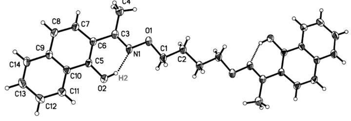
Figure 1
The molecule structure of the title compound with atom numbering [Symmetry codes: -x + 1, -y + 1, -z]. Displacement
ellipsoids for non-hydrogen atoms are drawn at the 30% probability level.
2,2′-{1,1′-[Butane-1,4-diylbis(oxynitrilo)]diethylidyne}di-1-naphthol
Crystal data
C28H28N2O4 Mr = 456.52
Triclinic, P1 Hall symbol: -P 1
a = 6.9590 (15) Å
b = 8.6598 (18) Å
c = 10.596 (2) Å
α = 105.841 (2)°
β = 105.940 (2)°
γ = 91.689 (1)°
V = 586.9 (2) Å3
Z = 1
F(000) = 242
Dx = 1.292 Mg m−3
Mo Kα radiation, λ = 0.71073 Å Cell parameters from 1246 reflections
θ = 2.5–27.6°
µ = 0.09 mm−1 T = 298 K
Block, pale-yellow 0.50 × 0.43 × 0.20 mm
Data collection
Bruker SMART 1000 CCD area-detector diffractometer
Radiation source: fine-focus sealed tube Graphite monochromator
φ and ω scans
Absorption correction: multi-scan (SADABS; Sheldrick, 1996)
Tmin = 0.958, Tmax = 0.983
3020 measured reflections 2026 independent reflections 1350 reflections with I > 2σ(I)
Rint = 0.025
θmax = 25.0°, θmin = 2.1° h = −8→8
k = −9→10
l = −12→6
Refinement
Refinement on F2
Least-squares matrix: full
R[F2 > 2σ(F2)] = 0.054 wR(F2) = 0.173 S = 1.02 2026 reflections 154 parameters 0 restraints
Primary atom site location: structure-invariant direct methods
Secondary atom site location: difference Fourier map
Hydrogen site location: inferred from neighbouring sites
H-atom parameters constrained
w = 1/[σ2(F
o2) + (0.096P)2 + 0.0813P]
where P = (Fo2 + 2Fc2)/3
(Δ/σ)max < 0.001
Δρmax = 0.23 e Å−3
Special details
Geometry. All e.s.d.'s (except the e.s.d. in the dihedral angle between two l.s. planes) are estimated using the full covariance matrix. The cell e.s.d.'s are taken into account individually in the estimation of e.s.d.'s in distances, angles and torsion angles; correlations between e.s.d.'s in cell parameters are only used when they are defined by crystal symmetry. An approximate (isotropic) treatment of cell e.s.d.'s is used for estimating e.s.d.'s involving l.s. planes.
Refinement. Refinement of F2 against ALL reflections. The weighted R-factor wR and goodness of fit S are based on F2,
conventional R-factors R are based on F, with F set to zero for negative F2. The threshold expression of F2 > σ(F2) is used
only for calculating R-factors(gt) etc. and is not relevant to the choice of reflections for refinement. R-factors based on F2
are statistically about twice as large as those based on F, and R- factors based on ALL data will be even larger.
Fractional atomic coordinates and isotropic or equivalent isotropic displacement parameters (Å2)
x y z Uiso*/Ueq
N1 0.4299 (3) 0.3289 (2) 0.31934 (16) 0.0435 (5) O1 0.3505 (2) 0.37340 (18) 0.19981 (14) 0.0515 (5) O2 0.7182 (2) 0.30688 (18) 0.52155 (15) 0.0505 (5)
H2 0.6665 0.3326 0.4519 0.076*
C1 0.5112 (3) 0.4251 (3) 0.1562 (2) 0.0455 (6)
H1A 0.5966 0.3396 0.1410 0.055*
H1B 0.5923 0.5189 0.2259 0.055*
C2 0.4211 (3) 0.4667 (3) 0.0252 (2) 0.0449 (6)
H2A 0.3277 0.5459 0.0400 0.054*
H2B 0.3460 0.3705 −0.0449 0.054*
C3 0.2958 (3) 0.2877 (2) 0.3702 (2) 0.0402 (5) C4 0.0759 (3) 0.2939 (3) 0.3091 (2) 0.0627 (7)
H4A 0.0578 0.3501 0.2408 0.094*
H4B 0.0198 0.3496 0.3798 0.094*
H4C 0.0091 0.1859 0.2673 0.094*
C5 0.5732 (3) 0.2467 (2) 0.5630 (2) 0.0372 (5) C6 0.3690 (3) 0.2344 (2) 0.49419 (19) 0.0365 (5) C7 0.2306 (3) 0.1639 (2) 0.5453 (2) 0.0433 (5)
H7 0.0937 0.1554 0.5009 0.052*
C8 0.2912 (3) 0.1088 (2) 0.6565 (2) 0.0438 (6)
H8 0.1961 0.0608 0.6851 0.053*
C9 0.4976 (3) 0.1238 (2) 0.7294 (2) 0.0386 (5) C10 0.6397 (3) 0.1961 (2) 0.6838 (2) 0.0370 (5) C11 0.8449 (3) 0.2185 (3) 0.7606 (2) 0.0506 (6)
H11 0.9400 0.2668 0.7321 0.061*
C12 0.9047 (4) 0.1696 (3) 0.8761 (3) 0.0600 (7)
H12 1.0397 0.1872 0.9270 0.072*
C13 0.7640 (4) 0.0933 (3) 0.9182 (2) 0.0564 (7)
H13 0.8068 0.0574 0.9955 0.068*
C14 0.5661 (3) 0.0711 (3) 0.8475 (2) 0.0479 (6)
H14 0.4743 0.0207 0.8770 0.057*
Atomic displacement parameters (Å2)
O1 0.0585 (10) 0.0695 (11) 0.0386 (8) 0.0082 (8) 0.0167 (7) 0.0329 (7) O2 0.0468 (9) 0.0664 (10) 0.0556 (10) 0.0120 (7) 0.0240 (7) 0.0362 (8) C1 0.0570 (13) 0.0519 (13) 0.0402 (12) 0.0112 (10) 0.0242 (10) 0.0234 (10) C2 0.0573 (14) 0.0482 (12) 0.0360 (12) 0.0094 (11) 0.0180 (10) 0.0188 (9) C3 0.0498 (12) 0.0394 (11) 0.0345 (11) 0.0036 (9) 0.0159 (9) 0.0121 (9) C4 0.0544 (15) 0.0897 (19) 0.0525 (14) 0.0030 (13) 0.0111 (11) 0.0397 (13) C5 0.0447 (12) 0.0363 (11) 0.0393 (11) 0.0080 (9) 0.0219 (9) 0.0152 (9) C6 0.0443 (12) 0.0380 (11) 0.0313 (10) 0.0048 (9) 0.0155 (9) 0.0124 (8) C7 0.0434 (12) 0.0523 (13) 0.0374 (11) 0.0003 (10) 0.0131 (9) 0.0178 (10) C8 0.0487 (13) 0.0495 (13) 0.0404 (12) −0.0015 (10) 0.0203 (10) 0.0182 (10) C9 0.0505 (13) 0.0354 (11) 0.0375 (11) 0.0105 (9) 0.0202 (9) 0.0149 (9) C10 0.0438 (12) 0.0367 (11) 0.0379 (11) 0.0138 (9) 0.0177 (9) 0.0157 (9) C11 0.0466 (13) 0.0589 (14) 0.0597 (14) 0.0164 (11) 0.0218 (11) 0.0315 (11) C12 0.0498 (14) 0.0742 (17) 0.0654 (16) 0.0220 (12) 0.0132 (12) 0.0376 (13) C13 0.0643 (16) 0.0638 (15) 0.0534 (14) 0.0232 (12) 0.0170 (12) 0.0356 (12) C14 0.0621 (15) 0.0493 (13) 0.0455 (13) 0.0138 (11) 0.0239 (11) 0.0262 (10)
Geometric parameters (Å, º)
N1—C3 1.285 (3) C5—C10 1.427 (3)
N1—O1 1.398 (2) C6—C7 1.423 (3)
O1—C1 1.425 (2) C7—C8 1.357 (3)
O2—C5 1.350 (2) C7—H7 0.9300
O2—H2 0.8200 C8—C9 1.415 (3)
C1—C2 1.503 (3) C8—H8 0.9300
C1—H1A 0.9700 C9—C10 1.413 (3)
C1—H1B 0.9700 C9—C14 1.414 (3)
C2—C2i 1.509 (4) C10—C11 1.414 (3)
C2—H2A 0.9700 C11—C12 1.365 (3)
C2—H2B 0.9700 C11—H11 0.9300
C3—C6 1.475 (3) C12—C13 1.400 (3)
C3—C4 1.498 (3) C12—H12 0.9300
C4—H4A 0.9600 C13—C14 1.354 (3)
C4—H4B 0.9600 C13—H13 0.9300
C4—H4C 0.9600 C14—H14 0.9300
C5—C6 1.394 (3)
C3—N1—O1 113.72 (17) C5—C6—C7 117.71 (18)
N1—O1—C1 109.26 (15) C5—C6—C3 122.01 (18)
C5—O2—H2 109.5 C7—C6—C3 120.26 (18)
O1—C1—C2 107.96 (18) C8—C7—C6 122.4 (2)
O1—C1—H1A 110.1 C8—C7—H7 118.8
C2—C1—H1A 110.1 C6—C7—H7 118.8
O1—C1—H1B 110.1 C7—C8—C9 120.59 (19)
C2—C1—H1B 110.1 C7—C8—H8 119.7
H1A—C1—H1B 108.4 C9—C8—H8 119.7
C1—C2—C2i 112.2 (2) C10—C9—C14 118.88 (19)
C2i—C2—H2A 109.2 C14—C9—C8 122.24 (19)
C1—C2—H2B 109.2 C9—C10—C11 118.88 (18)
C2i—C2—H2B 109.2 C9—C10—C5 119.50 (18)
H2A—C2—H2B 107.9 C11—C10—C5 121.61 (19)
N1—C3—C6 116.56 (18) C12—C11—C10 120.5 (2)
N1—C3—C4 122.53 (18) C12—C11—H11 119.8
C6—C3—C4 120.91 (18) C10—C11—H11 119.8
C3—C4—H4A 109.5 C11—C12—C13 120.4 (2)
C3—C4—H4B 109.5 C11—C12—H12 119.8
H4A—C4—H4B 109.5 C13—C12—H12 119.8
C3—C4—H4C 109.5 C14—C13—C12 120.6 (2)
H4A—C4—H4C 109.5 C14—C13—H13 119.7
H4B—C4—H4C 109.5 C12—C13—H13 119.7
O2—C5—C6 122.92 (17) C13—C14—C9 120.8 (2)
O2—C5—C10 116.22 (18) C13—C14—H14 119.6
C6—C5—C10 120.86 (18) C9—C14—H14 119.6
C3—N1—O1—C1 176.83 (17) C7—C8—C9—C14 178.56 (19)
N1—O1—C1—C2 178.10 (15) C14—C9—C10—C11 −2.1 (3)
O1—C1—C2—C2i 176.3 (2) C8—C9—C10—C11 176.85 (18)
O1—N1—C3—C6 178.15 (15) C14—C9—C10—C5 178.64 (16)
O1—N1—C3—C4 −2.3 (3) C8—C9—C10—C5 −2.4 (3)
O2—C5—C6—C7 177.98 (17) O2—C5—C10—C9 −176.58 (16)
C10—C5—C6—C7 −2.5 (3) C6—C5—C10—C9 3.8 (3)
O2—C5—C6—C3 −0.4 (3) O2—C5—C10—C11 4.2 (3)
C10—C5—C6—C3 179.16 (17) C6—C5—C10—C11 −175.34 (18)
N1—C3—C6—C5 7.8 (3) C9—C10—C11—C12 0.5 (3)
C4—C3—C6—C5 −171.77 (19) C5—C10—C11—C12 179.74 (19) N1—C3—C6—C7 −170.53 (17) C10—C11—C12—C13 1.5 (4)
C4—C3—C6—C7 9.9 (3) C11—C12—C13—C14 −2.0 (4)
C5—C6—C7—C8 −0.4 (3) C12—C13—C14—C9 0.3 (3)
C3—C6—C7—C8 178.04 (18) C10—C9—C14—C13 1.7 (3)
C6—C7—C8—C9 1.8 (3) C8—C9—C14—C13 −177.2 (2)
C7—C8—C9—C10 −0.4 (3)
Symmetry code: (i) −x+1, −y+1, −z.
Hydrogen-bond geometry (Å, º)
D—H···A D—H H···A D···A D—H···A