Determination of the diversity and
antibiotic resistance
profiles
of
Staphylococcus
species from dogs with otitis
externa and examination of
mecA
gene occurrence
K. Metiner, A.F. Bagcigil, A. Ilgaz
Veterinary Faculty, University of Istanbul, Avcilar, Istanbul, Turkey
ABSTRACT: The aim of this study was to determine the distribution of Staphylococci from swab samples of dogs with otitis externa and to determine their antibiotic resistance profiles, particularly methicillin resistance. For this purpose 116 ear swab samples were collected from 100 dogs and examined for the presence of Staphylococ-cus species by conventional culture methods. Antibiotic susceptibility of the isolates was determined by the disk diffusion test and for methicillin resistance, by PCR. Forty Staphylococci were isolated from 37 (31.9%) of the 116 ear swabs. Among the 40 isolates, 30 of them were coagulase-positive Staphylococcus species (CPS), while 10 (25%) were coagulase-negative Staphylococcus spp. (CNS). S. pseudintermedius (n = 11), S. aureus (n = 8), other not determined Staphylococcus spp. (n = 7), S. chromogenes (n = 7), S. schleiferi coagulans (n = 3), S. hyicus (n = 1), S. hominis subsp. hominis (n = 1), S. simulans (n = 1), S. saprophyticus (n = 1) were isolated. Results of the anti-biotic susceptibility tests have shown that 60% of the isolates were resistant to sulfamethoxazole/trimethoprim, 32.5% of them were resistant to erythromycin, 25% were resistant to clindamycin, and all isolates (100%) were sensitive to amoxicillin/clavulanic acid and cephazolin. The majority of isolates (97.5%) were sensitive to cipro-floxacin and gentamicin which are frequently used in otitis externa treatment. It was determined that only one (2.5%) (S. hominis subsp. hominis) of the 40 isolates was resistant to methicillin and carried the mecA gene. We found 77% of Staphylococcus spp. to be resistant to one or more antimicrobial drugs, and 25% of Staphylococcus species were found to be resistant to three or more antimicrobial classes. Thus, multidrug-resistance as detected in our study should always be taken into account and close attention should be given to the antimicrobial therapy protocols of pet animals.
Keywords: dog; otitis externa; Staphylococcus spp.; methicillin resistance; mecA gene
Staphylococci persist on mucosal surfaces and on skin in a commensal form. However, they also represent the main factor in many animal diseases. Staphylococci are divided into coagulase-positive and negative subclasses. The coagulase-positive Staphylococci (CPS) are related with more chronic and sub-acute infections, while the coagu-lase-negative Staphylococci (CNS) have long been neglected in terms of pathogenic research (Mouney et al. 2013).
S. intermedius has been reported to be the most frequently isolated species among the CPS both in healthy and infected dogs (Sasaki et al. 2007). It has also been reported that S. aureus and S. intermedius are responsible for many skin problems and otitis externa infections in pet animals (Guardabassi et al.
dogs, the pathogenic potential of these microor-ganisms in animals has not yet been fully accepted (Van Duijkeren et al. 2004; Epstein et al. 2009). The transfer resistance among dogs, cats, horses and other domestic animals is possible, and these car-rier animals are reported to act as reservoirs both for other animals and humans (Weese et al. 2010; Loeffler et al. 2010a).
In this study, the purpose was to determine the distribution of Staphylococcus species isolated from dogs with otitis externa, to establish their antibiotic resistance profiles, to determine the Staphylococcus species that are resistant to me-thicillin and to examine the presence of the mecA genewith PCR.
MATERIAL AND METHODS
In this study, 116 ear swab samples collected from 100 dogs with ear canal complaints were examined. The samples were collected from dogs brought to private veterinary clinics located in different dis-tricts of Istanbul or to clinics of Istanbul University, Faculty of Veterinary Medicine. In addition, some of the dogs were living in dog shelters.
The swab samples were inoculated into Nutrient Broth (Oxoid Ltd, Hampshire, England) supple-mented with 1% horse serum, and incubated at 37 °C under aerobic conditions. After a 24–48 h incubation period, passages were performed onto mannitol-salt agar (MSA, Oxoid), and onto blood agar base (Oxoid) containing 5% sheep blood.
Pure Gram-positive cultures were examined for Staphylococcus spp. identification, by performing coagulase, catalase, oxidase, DNase, haemolysis, pigment production, urease activity, mannitol fermentation, esculin hydrolysis, novobiocin and polymyxin B antibiotic susceptibility tests (Holt et al. 1994). The results were confirmed by API ID32 STAPH (Bio-Merieux Co. Ltd., France). In order to discriminate 18 isolates which were identified as Staphylococcus intermedius group (SIG; S. inter-medius, S. delphini or S. pseudintermedius) pheno-typically, a multiplex-PCR was performed. Bacterial DNA was extracted using the QIAamp DNA extrac-tionkit (Qiagen, Cat. No.69506). The PCR reac-tion mixture consisted of 2 µl of DNA extract in a total volume of 50 µl and was performed accord-ing to Sasaki et al. (2010). DNA fragments were analysed by electrophoresis in 1 ×
Tris-acetate-EDTA on a 1% agarose gel stained with ethidium bromide. Staphylococcus intermedius: (CCM5729), Staphylococcus pseudintermedius: (NVAV-6045), S. delphini group B, (H-2C) and S. delphini group A; (F-13b) were used as positive controls.
The antibiotic resistance of the isolates was ex-amined according to the standards of the Clinical and Laboratory Standards Institute (CLSI) using the following antibiotic disks: amikacin (30 µg), amoxicillin/clavulanic acid (20/10 µg), ampicillin (10 µg), cefazolin (30 µg), ceftriaxone (30 µg), cip-rofloxacin (5 µg), clindamycin (2 µg), encip-rofloxacin (5 µg), erythromycin (15 µg), gentamicin (10 µg), kanamycin (30 µg), lincomycin (2 µg), neomycin (30 µg), penicillin G (10 µg), rifampicin (5 µg), sul-famethoxazole/trimethoprim (1.25/23.75 µg), tet-racycline (30 µg), tobramycin (10 µg), vancomycin (30 µg), oxacillin (1 µg), (CLSI 2010). Methicillin resistance was confirmed by determining the pres-ence of the mecA gene in a PCR assay carried out according to Jonas et al. (2002).
RESULTS
A total of 116 swab samples were examined from 100 dogs with otitis externa. In 37 samples bacte-rial growth was observed; 40 Staphylococcus spe-cies were isolated from those 37 ear swabs. It was determined that 30 of these isolates (75%) were CPS, while 10 (25%) were CNS (Table 1). Eight S. aureus (20%), seven S. chromogenes (17.5%), three S. schleiferi coagulans (7.5%), one S. hyicus (2.5%),one S. hominis subsp. hominis (2.5%), one S. simulans (2.5%), one S. saprophyticus (2.5%) and eighteen SIG (42.5%) isolates were recovered. Eleven out of the 18 SIG isolates were determined to be S. pseudintermedius after PCR analysis. In the remaining seven isolates which were identi-fied as S. intermedius by biochemical tests and API no bands were observed; these were idenfied as Staphylococcus spp. (Figure 1).
was also multidrug resistant; it was resistant to ampicillin, ciprofloxacin, enrofloxacin, kanamy-cin, gentamikanamy-cin, tetracycline, tobramykanamy-cin, erythro-mycin, sulfamethoxazole/trimethoprim (Table 1). Other than this, an additional 10 Staphylococci isolates were determined to be multidrug
[image:3.595.97.515.96.282.2]resist-ant (resistresist-ant to three or more resist-antibiotic groups). Additionally, among eight isolates the same resist-ance pattern - resistresist-ance to clindamycin, erythro-mycin and sulfamethoxazole/trimethoprim, was observed (Table 1). Results of antibiotic suscepti-bility tests are shown in Table 2.
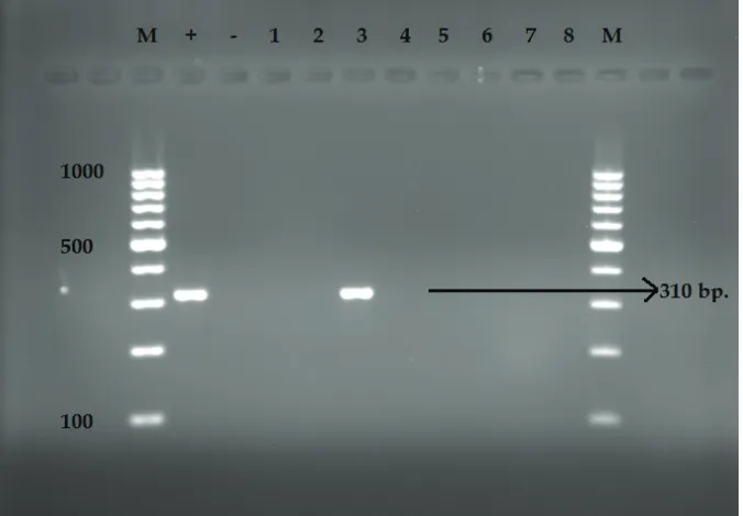
[image:3.595.132.470.461.696.2]Figure 2. The 310 bp amplicon of the mecA gene; 2% agarose gel. PCR analysis for the mecA gene in staphylococci isolated from dogs with external otitis. M = DNA molecular weight marker (100 bp); + = S. aureus (mecA, positive, 310 bp); – = negative control; line 3 = mecA positive isolate (Staphylococcus hominis subsp. hominis, 310 bp); lines 1, 2, 4, 5, 6, 7, 8 = mecA, negative isolates
Figure 1. Multiplex-PCR products on a 1% agarose gel. M = DNA molecular weight marker (100 bp); line 1, 2 =
Staphylococcus spp.; lines 3–13 = Staphylococcus pseudintermedius 926 bp band; line 14 = negative control; line 15 =
Staphylococcus intermedius (CCM5729), positive control, 430 bp; line 16 = Staphylococcus pseudintermedius (NVAV-6045), positive control,926 bp; line 17 = S. delphini (H-2C ), group B, positive control, 1135 bp; line 18 = S. delphini
DISCUSSION
Otitis externa is a frequent problem in dogs and there have been many studies conducted on the
ae-tiology of this disease. S. aureus and S. intermedius group were the most common strains identified in those studies (Keskin et al. 1999; Sarierler and Kirkan 2004; Strommenger et al. 2006; Boost et al. 2008; Ozturk et al. 2010).
Some researchers have reported that the isolation rate of members of the Staphylococcus intermedius
group is higher than other staphylococci, while it has also been shown that among SIG, S. pseudoint-ermedius isolation is higher than other SIG mem-bers(Futagawa-Saito et al. 2006; Jones et al. 2007; Sasaki et al. 2007; Vanni et al. 2009).Similarly, in our study S. pseudintermedius was the most com-mon staphylococcus detected from ear swabs.
[image:4.595.64.534.113.322.2]Studies on methicillin-resistant staphylococci are focused mainly on farm animals, in Turkey. There have also been a few studies in dogs (Findik et al. 2009; Bagcigil et al. 2012). Findik et al. (2009) re-ported the isolation of S. aureus from 80 (20.5%) swab samples from 390 dogs and detected the mecA gene only in three (3.8%) isolates. Bagcigil et al. (2012) isolated methicillin-resistant coagulase negative staphylococci (MRCNS) from five dogs (23.8%) out of 21 dogs. In the current study, only one methicillin-resistant S hominis subsp. hominis isolate was detected both phenotypically and geno-typically. Although the rate of S. pseudintermedius and S. aureus isolation was relatively high, the ab-sence of the mecA gene was reassuring.
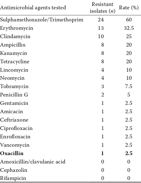
Table 2. Results of antimicrobial susceptibility tests
Antimicrobial agents tested isolates (Resistant n) Rate (%) Sulphamethoxazole/Trimethoprim 24 60
Erythromycin 13 32.5
Clindamycin 10 25
Ampicillin 8 20
Kanamycin 8 20
Tetracycline 8 20
Lincomycin 4 10
Neomycin 4 10
Tobramycin 3 7.5
Penicillin G 2 5
Gentamicin 1 2.5
Amicacin 1 2.5
Ceftriaxone 1 2.5
Ciprofloxacin 1 2.5
Enrofloxacin 1 2.5
Vancomycin 1 2.5
Oxacillin 1 2.5
Amoxicillin/clavulanic acid 0 0
Cephazolin 0 0
[image:4.595.63.290.463.758.2]Rifampicin 0 0
Table 1. Resistance pattern of strains of staphylococci species isolated from otitis externa in dogs
Resistant
isolates Antimicrobial resistance pattern (n isolates) CPS organism (n)
S. aureus (8) 5/8, 62.5% CD + E + K + N + P + SXT + T + TB (1), CD + E + K + SXT + P (1), SAM + AM (1), AM + G (1), SXT (1)
S. pseudintermedius (11) 7/11, 63.6% AM + CD + E + K + L + SXT + T (1), CD + E + K + N + SXT (1), CD + E + K + SXT (1), SXT + T (2), AM + T (1), AM (1), SXT (2) Staphylococcus spp. (SIG) (7) 7/7, 100% CD + E + L + SXT (1), CD + E + SXT (1), E + SXT (1), T + RD (1), AM (1), SXT (2) S. schleiferi coagulans (3) 2/3, 66. 7% SXT + T (1), SXT (1)
S. hyicus (1) 1/1, 100% SXT (1)
CNS organism (n)
S. chromogenes (7) 4/7, 57.1% AM + CD + E + K + N + SXT + T + TB (1), CD + E + L + N + P + RD + SXT + V (1), E + N (1), CD (1)
S. hominis subsp. hominis (1) 1/1, 100% A + CRO + CF + ENR + E + G + K + SXT + T + TB + OX (1)
S. simulans (1) 1/1, 100% SXT (1)
S. saprophyticus (1) 1/1, 100% E + SXT (1)
Ozturk et al. (2010) reported that only five (9.3%) of the 54 CPS isolated from 96 dogs with otitis externa, pyoderma, and skin wounds were methicil-lin-resistant phenotypically, and they did not carry the mecA gene. We did not detect any methicillin-resistant CPS isolates. Some studies performed in dogs have also reported a similarly low prevalence (Vanderhaeghen et al. 2012; Wedley 2012).
It has been reported that resistance rates to peni-cillin G, erythromycin, tetracycline and fusidic acid among S. aureus isolates is high, and it was pro-posed that the acquisition of resistance to these antibiotics was due to their higher usage by veteri-narians (Boost et al. 2008). Staphylococcus species isolated from dogs with pyoderma were found to be resistant to streptomycin, kanamycin, neomy-cin and erythromyneomy-cin (28%), to clindamyneomy-cin (22%) and to gentamicin and enrofloxacin (2%; Boost et al. 2008).
Keskin et al. (1999) reported that 82.5% of the bacteria isolated from the dogs with otitis externa were resistant to enrofloxacin, 65.5% to cepha-losporins, 44.4% to gentamicin and tetracycline, 34.9% to spiramycin, 26.9 to ampicillin, while 20.6% were resistant to lincomycin. Sarierler and Kirkan (2004) reported that bacteria isolated from dogs with otitis externa were resistant to oxytetracycline (100%), ciprofloxacin (100%), kanamycin (87.5%), penicillin G (72.5%), erythromycin (57.5%), gen-tamicin (55%), ampicillin (50%) and cefoperazone (50.0%). In our study, 2.5% of the isolates were re-sistant to gentamicin and ciprofloxacin which are frequently used in the treatment of dogs with otitis externa. The susceptibility rates of the isolates to ciprofloxacin were close to the rates obtained in the study by Sarierler and Kirkan (2004). In a study conducted in our country on dogs with otitis ex-terna it was reported that Staphylococcus species were highly resistant (63.1%) to ampicillin (Keskin et al. 1999). In our study, we determined that the resistance rate to ampicillin was 20%. The differ-ence between these results is striking. In our study, the highest resistance rates we detected were to sul-famethoxazole/trimethoprim (60%), erythromycin (32.5%), clindamycin (25%), and penicillin G (5%), respectively. We consider that in recent years the use of penicillin G in cats and dogs has decreased, while that of sulfamethoxazole/trimethoprim and erythromycin has increased, and therefore re-sistance has developed over time. The rere-sistance rates to clindamycin and gentamicin detected in
our study are in accordance with those detected by Ganiere et al. (2005).
Van Duijkeren et al. (2004) reported that all 10 mecA-positive Staphylococci strains bar one were multidrug-resistant and warned that if they were resistant to all antibiotic types used in dogs, the infections would be difficult to treat. We de-termined in our study that the isolate which was detected as mecA-positive was multi-resistant. The importance of multidrug-resistance among bac-teria is increasing in both human and veterinary medicine. Ganiere et al. (2005) found 41 of the 50 S. intermedius strains (82%) to be resistant to one or more antimicrobial drugs and 21 strains (42%) to be resistant to three or more antimicrobial classes. We found 31 of the 40 Staphylococcus spp. (77.7%) to be resistant to one or more antimicrobial drugs and 10 Staphylococcus species (25%) were observed to be resistant to three or more antimicrobial classes. This multidrug-resistance should not be ignored and the necessary importance and attention should be given to the antimicrobial therapy protocols of pet animals.
Acknowledgements
Thanks to Prof. Dr. Baris Sareyyupoglu (Ankara University, Diskapi, Ankara, Turkey) for his pre-cious help in providing S. pseudintermedius refer-ence strains (S. pseudintermedius: NVAV-6045), S. delphini group A reference strains (S. delphini: F 13b), S. delphini group B reference strains (S. del-phini H-2C) and to Prof. Dr. Ali Aydin (University of Istanbul, Avcilar, Istanbul, Turkey) for his pre-cious help in providing S. intermedius reference strains (CCM 5729).
REFERENCES
Bagcigil FA, Ikiz S, Guzel O, Parkan Yaramis C, Ilgaz A (2012): Species distribution of methicillin resistant Staph-ylococci isolated from animals, environmental samples and staffs. The Journal of Faculty of Veterinary Medicine, Istanbul University 38, 151–160.
CLSI – Clinical and Laboratory Standards Institute (2010): Performance standards for antimicrobial susceptibility testing: Twentieth informational supplement M100-S20. CLSI, Wayne, PA, USA.
Duquette RA, Nuttall TJ (2004): Methicillin-resistant Staphylococcus aureus in dogs and cats: an emerging problem? Journal of Small Animal Practice 45, 591–597. Epstein CR, Yam WC, Peiris, JSM, Epstein RJ (2009): Me-thicillin-resistant commensal staphylococci in healthy dogs as a potential zoonotic reservoir for community-acquired antibiotic resistance. Infection, Genetics and Evolution 9, 283–285.
Findik A, Akan N, Onuk EE, Cakiroglu D, Ciftci A (2009): Methicillin resistance profile and molecular typing of Staphylococcus aureus strains isolated from noses of the healthy dogs. Journal of the Faculty of Veterinary Medi-cine, Kafkas University 15, 925–930.
Futagawa-Saito K, Ba-Thein W, Sakurai N, Fukuyasu T (2006): Prevalence of virulence factors in Staphylococcus intermedius isolates from dogs and pigeons. BMC Vet-erinary Research 26, 2–4.
Ganiere JP, Medaille C, Mangion C (2005): Antimicrobial drug susceptibility of Staphylococcus intermedius clini-cal isolates from canine pyoderma. Journal of Veterinary Medicine B, Infectious Diseases and Veterinary Public Health 52, 25–31.
Guardabassi L, Loeber ME, Jacobson A (2004a): Transmis-sion of multiple antimicrobial-resistant Staphylococcus intermedius between dogs affected by deep pyoderma and their owners. Veterinary Microbiology 98, 23–27. Guardabassi L, Schwarz S, Lloyd DH (2004b): Pet animals
as reservoirs of antimicrobial-resistant bacteria. Journal of Antimicrobial Chemotherapy 54, 321–332.
Holt JG, Krieg NR, Sneath PHA, Staley JT, Williams ST (1994): Bergey’s Manual of Determinative Bacteriology. 9th ed. Lippincott Williams & Wilkins, UK. 532, 544–551.
Jonas D, Speck M, Daschner FD, Grundmann H (2002): Rapid PCR based identification of methicillin-resistant Staphylococcus aureus from screening swabs. Journal of Clinical Microbiology 40, 1821–1823.
Jones RD, Kania SA, Rohrbach BW Frank LA, Bemis DA (2007): Prevalence of oxacillin- and multidrug-resistant staphylococci in clinical samples from dogs: 1772 samples (2001–2005). Journal of the American Veterinary Medi-cal Association 230, 221–227.
Keskin O, Kokcu L, Akan M (1999): Identification and an-timicrobial sensitivity of microorganisms isolated from otitic dogs. Veterinary Journal of Ankara University 46, 163–168.
Loeffler A, Pfeiffer DU, Lloyd DH, Smith H, Soares-Magal-haes R, Lindsay JA (2010a): Methicillin-resistant
Staphy-lococcus aureus carriage in UK veterinary staff and owners of infected pets: new risk groups. Journal of Hos-pital Infection 74, 282–288.
Loeffler A, Pfeiffer DU, Lindsay JA, Soares-Magalhaes R, Lloyd DH (2010b): Lack of transmission of methicillin-re-sistant Staphylococcus aureus (MRSA) between apparently healthy dogs in a rescue kennel. Veterinary Microbiology 141, 178–181.
Mouney MC, Stiles J, Townsend WM, Guptill L, Weese JS (2013): Prevalence of methicillin-resistant Staphylococ-cus spp. in the conjunctival sac of healthy dogs. Veteri-nary Ophthalmology doi: 10.1111/vop. 12130, 1–4. O’Mahony R, Abbott Y, Leonard FC, Markey BK, Quinn PJ,
Pollock PJ, Fanning S, Rossney AS (2005): Methicillin-resistant Staphylococcus aureus (MRSA) isolated from animals and veterinary personnel in Ireland. Veterinary Microbiology 109, 285–296.
Ozturk D, Avki S, Turutoglu H, Yigitarslan K, Sagnak S (2010): Methicillin resistance among coagulase-positive staphylococci isolated from dogs with otitis externa, skin wounds and pyoderma. Journal of the Faculty of Veteri-nary Medicine, Kafkas University 6, 651–656.
Sarierler M, Kirkan S (2004): Microbiological diagnosis and therapy of canine otitis externa. Veteriner Cerrahi Dergisi 10, 11–15.
Sasaki T, Kikuchi K, Tanaka Y, Takahashi N, Kamata S, Hi-ramatsu K (2007): Methicillin-resistant Staphylococcus pseudintermedius in a veterinary teaching hospital. Jour-nal of Clinical Microbiology 45, 1118–1125.
Sasaki T, Tsubakishita S, Tanaka Y, Sakusabe A, Ohtsuka M, Hirotaki S, Kawakami T, Fukata T, Hiramatsu K (2010): Multiplex-PCR method for species identification of co-agulase positive staphylococci. Journal of Clinical Micro-biology 48, 765–769.
Strommenger B, Kehrenberg C, Kettlitz C, Cuny C, Verspohl J, Witte W, Schwarz S (2006): Molecular characterization of methicillin-resistant Staphylococcus aureus strains from pet animals and their relationship to human isolates. Journal of Antimicrobial Chemotherapy 57, 461–465. Tanner MA, Everett CL, Youvan DC (2000): Molecular
phy-logenetic evidence for noninvasive zoonotic transmission of Staphylococcus intermedius from a canine pet to a human. Journal of Clinical Microbiology 38, 1628–1631. Van Duijkeren E, Box ATA, Heck MEOC, Wannet WJB,
Vanni M, Tognetti R, Pretti C, Crema F, Soldani G, Meucci V, Intorre L (2009): Antimicrobial susceptibility of Staph-ylococcus intermedius and StaphStaph-ylococcus schleiferi isolated from dogs. Research in Veterinary Science 87, 192–195.
Wedley AL (2012): Prevalance of Staphylococcus spp. Car-riage in dogs. Department of Epidemiology and Popula-tion Health, Institute of InfecPopula-tion and Global Health, University of Liverpool.
Weese JS, Rousseau J, Willey BM, Archambault M, McGeer A, Low DE (2010): Methicillin-resistant Staphylococcus
aureus in horses at a veterinary teaching hospital: fre-quency, characterization, and association with clinical
disease. Journalof Veterinary Internal Medicine 20,
182–186.
Received: 2014–06–03 Accepted after corrections: 2015–04–16
Corresponding Author: