Evaluation of platelet function in horses undergoing
colic surgery using the PFA-100 platelet function
analyser
A. Iwaszko-Simonik, S. Graczyk
Department of Immunology, Pathophysiology and Veterinary Preventive Medicine, Wroclaw University of Environmental and Life Sciences, Wroclaw, Poland
ABSTRACT: Acute colic in horses, especially presentations requiring surgical correction, such as large colon volvulus, very often involves defective or excess platelet activation. The PFA-100 is a new point-of-care analyser that evaluates platelet function by measuring closure time (CT) in the whole blood of healthy horses in a stan-dardised manner. However, there are no reports on platelet function in horses with colic measured by the PFA-100. The aim of the present study was to investigate platelet function in a group of horses sufferingascending colon displacement (ACD) and who underwent surgery compared to a group of control (C) horses. Thirty ACD cases and twenty untreated clinically healthy control horses were included. Blood samples were collected from the ACD horses prior to and 24, 48 and 72 h after laparotomy. Red blood cell count (RBC), haematocrit (HCT), hae-moglobin (Hb) concentration, white blood cell count (WBC), total plasma protein (TP) concentration, platelet count (PLT), platelet volume (MPV) and ADP closure time (CT-ADP) were measured. CT was determined on a PFA-100® device using collagen/ADP cartridges. There were no significant differences in RBC, PLT and MPV between C and ACD horses pre-operatively. Reduced PLT was observed in the post-operative period. The mean CT-ADP of C horses was 96.43 ± 12.53 and 91.43 ± 2.51 seconds in the ACD group prior to surgery. CT-ADP was changed after surgery whereby CT-ADP time was more than doubled. We conclude that the PFA-100® can be used to detect platelet function defects in horses.
Keywords: colic; closure time; ascending colon displacement; PFA-100; platelets; horse
Supported by the Faculty of Veterinary Medicine, Wroclaw University of Environmental and Life Sciences, Wroclaw, Poland.
Platelets play a pivotal role in haemostasis by forming haemostatic plugs, and are also involved in inflammatory and immune processes (Segura et al. 2005; Segura et al. 2006; Dallap Schaer and Epstein 2009; Piccione et al. 2010; Yeaman 2010). Their function can be seen as a succession of over-lapping events involving adhesion, aggregation, secretion and promotion of pro-coagulant activ-ity (McMichael 2005; Dallap Schaer and Epstein 2009). Platelets can be activated by several agonists such as thrombin, collagen, adenosine diphosphate (ADP) or lipopolysaccharide (LPS) (Piccione et al. 2010). However, the response of platelets to ago-nists varies significantly between species (Segura
et. 2005; Brooks et al. 2007). For many years, ag-gregometry and bleeding time have been the only standard clinical tests used to evaluate primary haemostasis in humans and animals. Both tests have major limitations in assessing platelet func-tion. The major drawback of aggregometry is that this in vitro technique does not mimic the in vivo
new accurate tests that provide parameters such as closure time (CT). This parameter can be obtained using the platelet function analyser (PFA-100®),
which is a new point-of-care platelet function ana-lyser that evaluates human platelet function in a small volume of whole blood. The device assesses the process of primary haemostasis by stimulating platelet adhesion and aggregation under high shear stress. Whole, citrate blood is aspirated through an aperture in a collagen membrane coated with plate-let agonists (ADP or epinephrine EPI). This design mimics in vivo endothelial damage and platelet plug organization. Time until plug formation is measured in seconds and is called closure time (Jandrey et al. 2008; Clancey et al. 2009). The PFA-100 permits rapid detection of inherited, acquired, or drug-in-duced platelet dysfunction. Two different cartridges containing ADP or EPI can be used to differentiate haemostatic disorders caused by administration of acetylsalicylic acid (ASA) and other functional platelet disorders (Salat et al. 2002; Harrison 2005a; Harrison 2005b; Hayward et al. 2006; Jandrey 2012).
Recent studies described the use of the PFA-100 for evaluation of primary haemostasis in humans, dogs, cats, pigs and horses (Callan and Giger 2001; Wuillemin et al. 2002; Jandrey et al. 2008; Brooks et al. 2009; Clancey et al. 2009). In horses, this tech-nique has been standardised and has been used to assess platelet function (Segura et al. 2005; Jandrey et al. 2008). Use of cartridges coated with the agonists collagen and adenosine diphosphate (COL/ADP) is recommended over the cartridges coated with col-lagen and epinephrine (COL/EPI), as epinephrine does not consistently induce platelet aggregation in horses (Segura et al. 2005; Segura et al. 2006).
Colic is an important diagnostic and therapeutic problem in equine practice (Nelson et al. 2013). Acute colic causes such as large colon volvulus can induce changes in the coagulation system (Dallap et al. 2003). These changes are secondary to the haemodynamic and functional disorders of the gas-trointestinal tract and are also a consequence of an inflammatory process (Levi et al. 1997; Dallap et al. 2003; Levi and Opal 2006; Dallap Schaer and Epstein 2009; Levi et al. 2011). Therefore, haem-orrhage and prolonged bleeding after surgery or thromboembolic complications are realistic risks (Dallap et al. 2003; Segura et al. 2005; Segura et al. 2006; Alsaad and Nori 2009). Most previous studies on haemostatic changes in the course of colic were mainly focused on secondary haemostasis (Dallap
et al. 2003; Dallap Schaer and Epstein 2009).Only a few studies assessed platelet function in horses with gastrointestinal disease using aggregometry or thromboelastography (Epstein et al. 2011; Brooks et al. 2007; Mendez-Angulo et al. 2010; Epstein et al. 2013); however, to our knowledge, there have been no reports evaluating platelet function in horses with colic using the PFA-100. Considering the po-tential usefulness of the PFA-100 in the assessment of equine platelet function, it was decided to in-vestigate platelet function in the course of acute surgical colic. We hypothesised that both the sys-temic disorders underlying the causes of the colic as well as medical treatment and surgery induce alterations in platelet function. Taking into account the prognostic importance of platelet function the observations were made prior to and in the first three days following the laparotomy procedure.
MATERIAL AND METHODS
This study was approved by the II Local Ethics Committee at the Wroclaw University of Environ- mental and Life Sciences, Poland.
Animals. The study included thirty horses that underwent abdominal surgery for disorders in which the anatomical position of the ascend-ing colon was abnormal. In many cases this was a non-strangulating volvulus (torsion of < 360°); in other cases the displaced position of colon parts could not be exactly classified during manipulation. The study population, therefore, included all horses with any deviation in part of or in the entire ascend-ing colon relative to the normal anatomical posi-tion. For this study, the cases were categorized as ascending colon displacement (ACD). Cases were collected between January 2009 and October 2010.
The procedures were performed at a clinic of the Department of Surgery of the Faculty of Veterinary Medicine, Wroclaw University of Environmental and Life Sciences, Poland. Client-owned horses were included in the study after signed owner consent. Horses belonged to the following breeds: Polish halfbreds n = 23, Thoroughbreds n = 3, Silesians n = 2, Great-Polish horses n = 2; 13 were female and 17 were male. Ages ranged from five to 15 years old with a mean of 8.5 ± 2.9 years.
rectal examination. The decision for surgical inter-vention was based on clinical signs. The criteria for explorative laparotomy were: heart rate (> 60 bpm), capillary refill time (CRT) > 2 s), colour of mu-cous membranes (normal/abnormal-hyperaemic or cyanotic), abdominal auscultation (no sounds, decreased, normal or increased activity), a rectal examination indicating intestinal displacement and the presence of nasogastric reflux.
Horses were hospitalised for 10–14 days and dis-charged in good general condition.
The untreated control group (C) comprised twenty healthy Polish halfbred horses (eight geldings, five stallions and seven mares), with a mean age of 7.5 ± 2.1 years (range 5–12 years) and mean body weight of 550 ± 50 kg. All horses were deemed healthy for inclusion in the study if they had no history of bleed-ing accordbleed-ing to the owner, had no abnormalities at physical examination (body temperature, mucous membranes, heart and respiratory rates, capillary re-fill time) and showed no evidence of platelet clumps in blood smears. The animals were housed in pad-docks and fed on grass, hay and received a balanced ration of oat. Water was available ad libitum. The horses were regularly dewormed and vaccinated against influenza and tetanus and did not receive any other drugs within the two months prior to the study. The control horses were included in the study after signed owner consent.
Experimental design. The study was a compara-tive study between two groups and a longitudinal study for treated horses. The observation period for the latter extended from the day of the surgery to the third post-operative day. The first blood sample was taken at admission, i.e., pre-operatively (Day 0). Thereafter, the samples were collected 24 hours (Day 1), 48 hours (Day 2) and 72 hours (Day 3) post-operatively both from horses with colic and from the control animals.
This blood sampling schedule was based on the assumption that the first three days after the sur-gery are critical for further therapy and prognosis.
Blood samples. All blood samples were collected by direct jugular venipuncture using a 19-G nee-dle (S-Monovette neenee-dle, Sarstedt, Numbrecht, Germany) and vacuum tubes (S-Monovette; Sarstedt, Numbrecht, Germany). Samples were obtained in the following order: one EDTA tube and one citrate tube and one tube to obtain the serum. Blood (total volume: 2 ml) was collected into the tube containing K3EDTA for haematology;
WBC count, RBC count, HCT, HGB concentration, PLT and MPV were determined. The anticoagulant tubes were gently turned several times immedi-ately after collection to allow for adequate mixing. Haematology was performed just after blood col-lection using an automated haematology analyser (PE-6800 vet; Procan Electronics INC, Shenzhen, China), equipped with multispecies software. The applied methods were recommended by the manu-facturer. Peripheral blood smears were examined for visual judgement of platelet morphology and platelet clumping (Hinchcliff et al. 1993). The total protein (TP) concentration was measured in the serum using an automated biochemical analyser (Pointe 180; Pointe Scientific INC., Canton, USA).
For determination of the closure time (CT), blood samples from each horse were collected into 10 ml plastic tubes containing 3.8% sodium citrate (0.129 M) in a ratio 1 : 9 (anticoagulant to blood). The closure time was determined using the PFA-100 (PFA-100; Siemens Healthcare, Malvern, USA) ac-cording to the manufacturer’s instructions. Citrated whole blood samples used for CT analysis were stored at room temperature (23 °C) and analysed within 30–60 min after collection. Cartridges coated with the agonists COL/ADP (Dade PFA Collagen/ ADP test cartridge; Siemens Healthcare, Malvern, USA) were used for all measurements. All blood samples from all horses were run in duplicate. The mean of the duplicates was taken into account. The COL/ADP cartridges were stored at 4 °C and pre-warmed at room temperature for at least 10 min before analysis. Immediately before analysis, blood was mixed by gentle rotating and tilting movements of the tubes on a mixer. Once at room tempera-ture, test cartridges were inserted into the housing carousel of the analyser and loaded with 800 µl of mixed citrated whole blood. The analyser measures the time from initial blood flow until flow arrest in seconds and this result is recorded as the CT.
RESULTS
All thirty ACD horses survived until the end of the study. No horses had to be excluded due to confounding disorders.
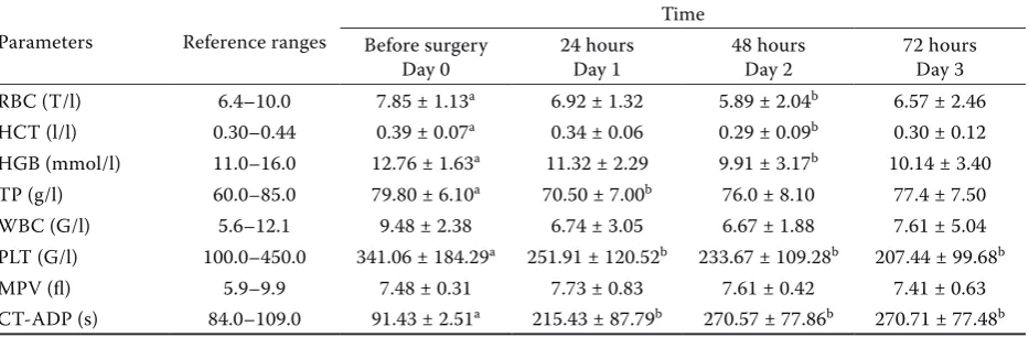
The results of the RBC, HCT, Hb, WBC, TPand CT-ADP in the C group and the ACD group in the pre-operative period (Day 0) are shown in Table 1. The results of the same parameters over three days in the ACD group are presented in Table 2. RBC was sig-nificantly higher in the ACD group compared to the C group; the RBC of the ACD group was significantly elevated prior to surgery (Day 0) compared to Day 2 after surgery (P < 0.05). The changes in RBC count were accompanied by the associated changes in HCT and HGB concentrations. There were mild but non-significant increases in HCT and HGB in the ACD group prior to surgery compared to the controls. After surgery HCT had decreased significantly by Day 2.
No significant differences in WBC and TP were found between the C and the ACD groups prior to surgery (Day 0) (Table 1), as well as in the ACD group pre- and post-operatively (Table 2), except for TP, which was significantly higher (P < 0.05) on Day 0 than on Day 1.
[image:4.595.63.531.140.278.2]There were no significant differences in PLT be-tween the C and the ACD groups prior to surgery (Table 1). In the ACD group, a gradual decrease of the PLT was observed throughout the entire post-operative period (Table 2). PLT decreased from a mean of 341.1 ± 184.3 G/l at Day 0 to 207.4 ± 99.7 G/L at Day 3. Compared to the prior to surgery values (Day 0) the counts were statistically signifi-cantly reduced (P < 0.05), at 24, 48 and 72 h after the surgery. The PLT values in the post-operative period were not significantly different. The reduced PLT count in the horses from the surgery group was accompanied by a statistically significant reduction
Table 2. RBC count, HCT, HGB concentration, total WBC count, PLT, MPV and CT-ADP inhorses with large colon volvulus, including horses before colic surgery (Day 0), 24 hours (Day 1), 48 hours (Day 2), 72 hours (Day 3) after the operation. Results are presented as mean ± SD. Different lowercase letters indicate significant differences (P < 0.05)
Parameters Reference ranges
Time
Before surgery
Day 0 24 hoursDay 1 48 hoursDay 2 72 hoursDay 3
RBC (T/l) 6.4–10.0 7.85 ± 1.13a 6.92 ± 1.32 5.89 ± 2.04b 6.57 ± 2.46
HCT (l/l) 0.30–0.44 0.39 ± 0.07a 0.34 ± 0.06 0.29 ± 0.09b 0.30 ± 0.12
HGB (mmol/l) 11.0–16.0 12.76 ± 1.63a 11.32 ± 2.29 9.91 ± 3.17b 10.14 ± 3.40
TP (g/l) 60.0–85.0 79.80 ± 6.10a 70.50 ± 7.00b 76.0 ± 8.10 77.4 ± 7.50
WBC (G/l) 5.6–12.1 9.48 ± 2.38 6.74 ± 3.05 6.67 ± 1.88 7.61 ± 5.04
PLT (G/l) 100.0–450.0 341.06 ± 184.29a 251.91 ± 120.52b 233.67 ± 109.28b 207.44 ± 99.68b
MPV (fl) 5.9–9.9 7.48 ± 0.31 7.73 ± 0.83 7.61 ± 0.42 7.41 ± 0.63
[image:4.595.65.532.603.756.2]CT-ADP (s) 84.0–109.0 91.43 ± 2.51a 215.43 ± 87.79b 270.57 ± 77.86b 270.71 ± 77.48b
Table 1. Red blood cell (RBC) count, hematocrit (HCT), haemoglobin (HGB) concentration, total white blood cell (WBC) count, platelet count (PLT), mean platelet volume (MPV) and closure time (CT-ADP) in control horses (C) and horses with large colon volvulus in the pre-operative period (Day 0). Results are presented as mean ± SD
Parameters Reference ranges Control horsesControl Colic horses before surgeryDay 0 P-value
RBC (T/l) 6.4–10.0 6.89 ± 1.31 7.85 ± 1.13 0.04
HCT (l/l) 0.30–0.44 0.35 ± 0.04 0.39 ± 0.07 0.07
HGB (mmol/l) 11.0–16.0 13.60 ± 1.67 12.76 ± 1.63 0.28
TP (g/l) 60.0–85.0 74.80 ± 2.10 79.80 ± 6.10 0.04
WBC (G/l) 5.6–12.1 8.99 ± 2.28 9.48 ± 2.38 0.85
PLT (G/l) 100.0–450.0 417.36 ± 199.66 341.06 ± 184.29 0.26
MPV (fl) 5.9–9.9 9.83 ± 1.57 7.48 ± 0.31 0.00
in MPV. MPV was significantly lower (P < 0.05) in the ACD group prior to surgery (7.48 ± 0.31) than in the controls (9.83 ± 1.57). MPV remained low throughout the entire post-operative period and MPV values did not differ significantly during this time (Table 2).
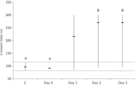
The reference range for CT-ADP (ADP closure time) in the C group was 81–116 s (mean 96.4 ± 12.5 s). In the ACD group CT-ADP prior to surgery (Day 0) was nearly 5 s shorter than in the C group, but this difference was not significant (P = 0.26). After surgery, mean CT-ADP values were doubled compared to the controls (C) and Day 0. Mean CT-ADP values were significantly (P < 0.05) prolonged at 24, 48 and 72 h post-surgery in comparison with pre-operative values (Table 2, Figure 1).
DISCUSSION
The goal of the present study was to investigate platelet function in horses undergoing large colon surgery prior to and for up to three days after opera-tion. Platelet function as well as platelet count and volume, was included too. The capacity of platelets to adhere and aggregate was determined using the PFA-100 device. Mean PLT and MPV gradually de-creased due to surgery for intestinal displacement. Various authors have indicated that after surgical intervention thrombocytopenia is more likely to occur than after conservative treatment (Chang 1996; Dallap et al. 2003; Yaguchi et al. 2004). Post-operative reduction of platelet counts may be due to platelet activation and subsequent consumption in the process of clot formation, sequestration in the spleen or adhesion to endothelial surfaces, as well
as destruction and exhaustion of thrombopoiesis (Levi et al. 1997; Salat et al. 1999; Nunez et al. 2001; Kimberley et al. 2004; Levi and Opal 2006; Alsaad and Nori 2009; Levi et al. 2011). Several authors indicate that pseudothromocytopenia may occur in horse blood collected in EDTA (Hinchliff et al. 1993), but visual evaluation of blood smears can exclude its presence. Therefore, in our study every blood sample was assessed by microscopic observa-tion of blood film for platelet clumps. Because we did not observe platelet clumps in any blood smears, we could rule out pseudothrombocytopenia.
Systemic inflammation is characterised by pro-duction and release of extra leukocytes and acute phase proteins (El-Ashker et al. 2014). WBC counts are used to judge the severity of inflammation. In our study total the WBC of the ACD group was not changed and TP was only marginally increased compared to the controls. Our results are con-sistent with findings obtained by Fagliary et al. (2008), who demonstrated that marked leucocy-tosis only occurs in colic horses with septic shock symptoms which did not survive. Apparently, our cases had not yet deteriorated to the point of il-eus. Dehydration and shock symptoms were not observed as expected based on the normal HCT and TP concentrations. We observed a marked reduction in MPV over the entire follow-up pe-riod. Inflammatory processes triggered by the ileus may cause damage to the integrity of the intestinal mucosal barrier which subsequently may impair thrombopoiesis and contribute to reduced platelet volume (MPV) (Kapsoritakis et al. 2001; Danese et al. 2007; Kaushansky 2009). WBC in our cases did not indicate clear inflammation; however, the surgery, anaesthesia and medical therapy with
crys-50 100 150 200 250 300 350
C Day 0 Day 1 Day 2 Day 3
C
lo
su
re
ti
m
e
(s
)
A A
[image:5.595.66.343.96.272.2]B B
talloids and NSAID may have exerted an effect on platelet volume. These factors need to be studied in more detail. Our study shows that gastro-intestinal surgery of relative healthy horses still causes a re-markable effect on the coagulation system.
This study showed that a reduced platelet count, accompanied by lower mean platelet volume in the surgically treated horses, occurs concomitantly with a prolonged closure time (CT). CT measurement with the PFA-100® (Platelet Function Analyzer-100) using ADP as a platelet agonist allows rapid as-sessment of the adhesion and aggregation capac-ity of platelets in horses. Our finding confirms the results of Segura et al. (2005) who evaluated the usefulness of the PFA-100® for platelet func-tion assessment in healthy and acetyl-salicylic acid (ASA)-treated horses. Our reference interval for CT-ADP in clinically healthy horses is 81–116 s and is similar to Segura et al. (2005). However, our data are not in agreement with those of Monreal et al. (2000) showing activation of coagulation and the fibrinolytic system in the course of acute horse colic. This can be explained by the different gas-trointestinal (GI) status of the animals in the two studies. Sampling time relative to surgery has a marked effect on subsequent findings. Therefore, comparison of studies is only possible after stand-ardisation of sampling time and clear definition of the GI pathologies. Furthermore, interpretation of observed CT changes after colic surgery should account for possible effects of other therapeutic agents used concurrently, especially NSAID as well as the progressive reduction in platelet count.
Acknowledgements
The authors are grateful to Dr Radomir Henklewski and Dr Anna Biazik (Wroclaw University of Environmental and Life Sciences, Wroclaw, Poland) for the help in data collection and Prof. Janusz Orda (Wroclaw University of Environmental and Life Sciences, Wroclaw, Poland) for his statistical expertise.
REFERENCES
Alsaad KM, Abid-albar A. Nori (2009): Equine colic and coagulation disorders. Journal of Animal and Veterinary Advances 8, 2675–2679.
Brooks AC, Menzies-Gow NJ, Wheeler-Jones C, Bailey SR, Cunningham FM, Elliott J (2007): Endotoxin-induced activation of equine platelets: evidence for direct activa-tion of p38 MAPK pathways and vasoactive mediator production.Inflammation Research56, 154–161. Brooks MB, Randolph J, Warner K, Center S (2009):
Evalu-ation of platelet function screening tests to detect plate-let procoagulant deficiency in dogs with Scott syndrome. Veterinary Clinical Pathology 38, 306–315.
Callan MB, Giger U (2001): Assessment of point of care instru-ment for identification of primary hemostatic disorders in dogs. American Journal of Veterinary Research 62, 652–658. Chang JC (1996): Postoperative thrombocytopenia: with
etiologic, diagnostic, and therapeutic consideration. American Journal of the Medical Sciences 311, 96–105. Clancey N, Burton S, Horney B, MacKenzie A, Nicastro A,
Cote E (2009): Evaluation of platelet function in dogs with cardiac disease using the PFA-100 platelet function ana-lyzer. Veterinary Clinical Pathology 38, 299–305.
Dallap-Schaer BL, Epstein K (2009): Coagulopathy of the critically ill equine patient. Journal of Veterinary Emer-gency and Critical Care 19, 53–65.
Dallap B, Dolente B, Boston R (2003): Coagulation profiles in 27 horses with large colon volvulus. Journal of Vet-erinary Emergency and Critical Care 13, 215–225. Danese S, Papa A, Saibeni S, Repici A, Malesci A, Vecchi
M (2007): Inflammation and coagulation in inflammatory bowel disease: The clot thickens. American Journal of Gastroenterology 102, 174–186.
El-Ashker M., Salama M., Rizk A., El-Bosh M (2014): The use of inflammatory markers as a prognostic aid for trau-matic reticuloperitonitis in water buffalo (Bubalus buba-lis). Veterinarni Medicina 59, 239–246
Epstein KL, Brainard BM, Giguere S, Vrono Z, Moore JN (2013): Serial Viscoelastic and traditional coagulation testing with gastrointestinal disease. Journal of Veterinary Emergency and Critical Care 23, 504–516.
Epstein KL, Brainard BM, Gomez-Ibanez MAF, Lopes MA, Barton MH, Moore JN (2011): Thromboelastography in horses with acute gastrointestinal disease. Journal of Vet-erinary Emergency and Critical Care 25, 307–314. Fagliary JJ, Silva SL, Silva PC, Pereira GT (2008): Leukocytes
and plasma levels of acute phase proteins in horses with acute abdomen underwent laparotomy (Leucograma e teores plas-maticos de proteinas de fase aguda de equinos portadores de abdomen agudo e submetidos a laparotomia). Arquivo Bra-sileiro de Medicina Veterinaria e Zootecnia 60, 322–328. Harrison P (2005a): Platelet function analysis. Blood 19,
111–123.
children and adults. British Journal of Haematology 130, 3–10.
Hayward CPM, Harrison P, Cattaneo M, Ortel TL, Rao AK (2006): Platelet function analyzer (PFA)-100 closure time in the evaluation of platelet disorders and platelet func-tion. Journal ofThrombosis and Haemostasis4, 312–319. Hinchcliff KW, Kociba GJ, Mitten LA (1993): Diagnosis of
EDTA-dependent pseudothrombocytopenia in a horse. Journal of the American Veterinary Medical Association 203, 1715-1716.
Jandrey KE (2012): Assessment of platelet function. Journal of Veterinary Emergency and Critical Care 22, 81–98. Jandrey KE, Norris JW, MacDonald KA, Kittleson MD,
Ta-blin F (2008): Platelet function in clinically healthy cats and cats with hypertriphic cardiomyopathy: analysis us-ing the Platelet Function Analyzer-100. Veterinary Clin-ical Pathology 37, 385–388.
Kapsoritakis AN, Koukourakis MI, Sfiridaki A, Potamianos SP, Kosmadaki MG, Koutroubakis IE, Kouroumalis EA (2001): Mean platelet volume: A useful maker of inflam-matory bowel disease activity. American Journal of Gas-troenterology 96, 776–781.
Kaushansky K (2009): Determinants of platelet number and regulation of thrombopoiesis. Hematology 1, 147–152. Kimberley M, McGurrin J, Arroyo LG, Bienzle D (2004): Flow
cytometric detection of platelet-bound antibody in three horses with immune-mediated thrombocytopenia. Journal of the American Veterinary Medical Association224, 83–87. Levi M, Opal SM (2006): Coagulation abnormalities in
critically ill patients. Critical Care Clinics 10, 222–230. Levi M, Poll T, Cate H, Deventer JH (1997): The
cytokine-mediated imbalance between coagulant and anticoagu-lant mechanisms in sepsis and endotoxaemia. European Journal of Clinical Investigation 27, 3–9.
Levi M, Schultz M, Poll T (2011): Coagulation biomarkers in critically ill patents. Critical Care Clinics 27, 281–297. McMichael M (2005): Primary hemostasis. Journal of
Vet-erinary Emergency and Critical Care 15, 1–8.
Mendez-Angulo JL, Mudge MC, Vilar-Saavedra P, Stingle N, Couto CG (2010): Thromboelastography in healthy horses and horses with inflammatory gastrointestinal disorders and suspected coagulopathies. Journal of Vet-erinary Emergency and Critical Care 20, 488–493. Monreal L, Angles A, Espada Y, Monasterio J, Monreal M
(2000): Hypercoagulation and hypofibrinolysis in horses
with colic and DIC. Equine Veterinary Journal Supple-ment 32, 19–25.
Nelson BB, Lordan LL, Hassel DM (2013): Risk factors as-sociated with gastrointestinal dysfunction in horses un-dergoing elective procedures under general anaesthesia. Equine Veterinary Journal Supplement 45, 8–14. Nunez R, Gomes-Keller MA, Schwarzwald C, Feige K (2001):
Assessment of Equine Autoimmune Thrombocytopenia (EAT) by flow cytometry. Blood Disorders 1, 1–13. Piccione G, Casella S, Gianetto C, Assenza A, Caola G
(2010): Effect of different storage conditions on platelet aggregation in horse. Journal of Equine Veterinary Sci-ence 30, 371–375.
Salat A, Bodingbauer G, Boehm D, Murabito M, Tochkow E, Sautner T, Mueller MR, Fuegger R (1999): Changes of platelet surface antigens in patients suffering from ab-dominal septic shock. Thrombosis Research 95, 289–294. Salat A, Kroess S, Felfernig-Boehm D., Felfernig M, Fleck T,
Schmidt D, Pulaki S, Mueller MR (2002): Comparison of in vitro closure time (PFA-100) with whole blood electri-cal aggregometry and platelet surface antigen expression in healthy volunteers. Thrombosis Research105, 205–208. Segura D, Monreal L, Espada Y, Pastor J, Mayós I, Homedes J (2005): Assessment of platelet function analyser in horses. References range and influence of a platelet ag-gregation inhibitor. Veterinary Journal170, 108–112. Segura D, Monreal L (2008): Poor reproducibility of
tem-plate bleeding time in horses. Journal of Veterinary In-ternal Medicine 22, 238–241.
Segura D, Monreal L, Perez-Pujol S, Pino M, Ordinas A, Brugues R, White JG, Escolar G (2006): Assessment of platelet function in horses: ultrastructure, flow cytom-etry and perfusion techniques. Journal of Veterinary In-ternal Medicine 20, 581–588.
Wuillemin WA, Gasser KM, Zeerleder SS, Lammle B (2002): Evaluation of a platelet function analyser (PFA-100) in patients with a bleeding tendency. Swiss Medical Weekly 132, 443–448.
Yaguchi A, Lobo FL, Pradier O (2004): Platelet function in sep-sis. Journal ofThrombosis and Haemostasis2, 2096–2102. Yeaman MR (2010): Platelets in defense against bacterial
path-ogens. Cellular and Molecular Life Sciences 67, 525–544.
Received: 2014–09–24 Accepted after corrections: 2015–08–11
Corresponding Author:
Alicja Iwaszko-Simonik, Wroclaw University of Environmental and Life Sciences, Department of Immunology, Pathophysiology and Veterinary Preventive Medicine, C.K. Norwida 31, 50 375 Wroclaw, Poland