5,8-Dimethoxy-2-phenyl-1,4-dihydro-quinoline-3-carbonitrile
Souheila Ladraa,aAbdelmalek Bouraiou,aSofiane Bouacida,b* Thierry Roisnelcand Ali Belfaitaha
aLaboratoire des Produits Naturels d’Origine Ve´ge´tale et de Synthe`se Organique, PHYSYNOR, Universite´ Mentouri-Constantine, 25000 Constantine, Algeria,bUnite´ de Recherche de Chimie de l’Environnement, et Mole´culaire Structurale, CHEMS, Universite´ Mentouri-Constantine, 25000 Algeria, andcCentre de Difractome´trie X, UMR 6226 CNRS Unite´ Sciences Chimiques de Rennes, Universite´ de Rennes I, 263 Avenue du Ge´ne´ral Leclerc, 35042 Rennes, France
Correspondence e-mail: bouacida_sofiane@yahoo.fr
Received 2 August 2010; accepted 6 August 2010
Key indicators: single-crystal X-ray study;T= 150 K; mean(C–C) = 0.003 A˚; Rfactor = 0.049;wRfactor = 0.133; data-to-parameter ratio = 16.4.
The crystal structure of the title molecule, C18H16N2O2, can be
described as two types of crossed layers parallel to the (110) and (110) planes. An intramolecular N—H O hydrogen bond occurs.
Related literature
For our previous work on the preparation of quinoline deri-vatives see: Benzerkaet al.(2008); Ladraaet al.(2009, 2010); Moussaouiet al.(2002); Menasraet al.(2005); Belfaitahet al. (2006); Bouraiouet al.(2006, 2007, 2008). For more details of quinoline reduction, see: Dauphinee & Forrest (1978); Srik-rishna et al. (1996); Vierhapper & Eliel (1975); Lim et al. (1995).
Experimental
Crystal data
C18H16N2O2 Mr= 292.33
Monoclinic,P21=c a= 3.9952 (3) A˚
b= 20.4544 (15) A˚
c= 17.7313 (13) A˚ = 95.976 (5)
V= 1441.12 (18) A˚3
Z= 4
MoKradiation = 0.09 mm 1
T= 150 K
0.270.070.05 mm
Data collection
Bruker APEXII diffractometer Absorption correction: multi-scan
(SADABS: Sheldrick, 2002)
Tmin= 0.702,Tmax= 0.996
12491 measured reflections 3292 independent reflections 1975 reflections withI> 2(I)
Rint= 0.051
Refinement
R[F2> 2(F2)] = 0.049 wR(F2) = 0.133 S= 1.03 3292 reflections
201 parameters
H-atom parameters constrained
max= 0.17 e A˚ 3
min= 0.29 e A˚ 3
Table 1
Hydrogen-bond geometry (A˚ ,).
D—H A D—H H A D A D—H A
N1—H1 O1 0.88 2.29 2.649 (2) 104
Data collection:APEX2(Bruker, 2001); cell refinement:SAINT
(Bruker, 2001); data reduction:SAINT; program(s) used to solve structure: SIR2002 (Burla et al., 2003); program(s) used to refine
structure: SHELXL97 (Sheldrick, 2008); molecular graphics:
ORTEP-3 for Windows (Farrugia, 1997) and DIAMOND (Bran-denburg & Berndt, 2001); software used to prepare material for publication:WinGX(Farrugia, 1999).
We are grateful to all personel of the PHYSYNOR laboratory, Universite´ Mentouri-Constantine, Alge´rie for their assistance.
Supplementary data and figures for this paper are available from the IUCr electronic archives (Reference: HG2695).
References
Belfaitah, A., Ladraa, S., Bouraiou, A., Benali-Cherif, N., Debache, A. & Rhouati, S. (2006).Acta Cryst.E62, o1355–o1357.
Benzerka, S., Bouraiou, A., Bouacida, S., Rhouati, S. & Belfaitah, A. (2008).
Acta Cryst.E64, o2089–o2090.
Bouraiou, A., Belfaitah, A., Bouacida, S., Benard-Rocherulle, P. & Carboni, B. (2007).Acta Cryst.E63, o1626–o1628.
Bouraiou, A., Debache, A., Rhouati, S., Carboni, B. & Belfaitah, A. (2008).J. Heterocycl. Chem.45, 329–333.
Bouraiou, A., Menasra, H., Debache, A., Rhouati, S. & Belfaitah, A. (2006).J. Soc. Alger. Chim.16, 171–183.
Brandenburg, K. & Berndt, M. (2001).DIAMOND. Crystal Impact, Bonn, Germany.
Bruker (2001). APEXII andSAINT. Bruker AXS Inc., Madison, Wisconsin, USA.
Burla, M. C., Caliandro, R., Camalli, M., Carrozzini, B., Cascarano, G. L., De Caro, L., Giacovazzo, C., Polidori, G. & Spagna, R. (2003).J. Appl. Cryst.38, 381–388.
Dauphinee, G. A. & Forrest, T. P. (1978).Can. J. Chem.56, 632–634. Farrugia, L. J. (1997).J. Appl. Cryst.30, 565.
Farrugia, L. J. (1999).J. Appl. Cryst.32, 837–838.
Ladraa, S., Bouraiou, A., Bouacida, S., Roisnel, T. & Belfaitah, A. (2009).Acta Cryst.C65, o475–o478.
Ladraa, S., Bouraiou, A., Bouacida, S., Roisnel, T. & Belfaitah, A. (2010).Acta Cryst.E66, o693.
Lim, C. L., Pyo, S. H., Kim, T. Y., Yim, E. S. & Han, B. H. (1995).Bull. Korean Chem. Soc.16, 374–377.
Menasra, H., Kedjadja, A., Rhouati, S., Carboni, B. & Belfaitah, A. (2005).
Synth. Commun.35, 2779–2788.
organic compounds
o2312
Ladraaet al. doi:10.1107/S1600536810031685 Acta Cryst.(2010). E66, o2312–o2313Acta Crystallographica Section E
Structure Reports Online
Moussaoui, F., Belfaitah, A., Debache, A. & Rhouati, S. (2002).J. Soc. Alger. Chim.12, 71–78.
Sheldrick, G. M. (2002).SADABS. Bruker AXS Inc., Madison, Wisconsin, USA
Sheldrick, G. M. (2008).Acta Cryst.A64, 112–122.
Srikrishna, A., Reddy, T. J. & Viswajanani, R. (1996).Tetrahedron,52, 1631– 1636.
supporting information
sup-1 Acta Cryst. (2010). E66, o2312–o2313
supporting information
Acta Cryst. (2010). E66, o2312–o2313 [https://doi.org/10.1107/S1600536810031685]
5,8-Dimethoxy-2-phenyl-1,4-dihydroquinoline-3-carbonitrile
Souheila Ladraa, Abdelmalek Bouraiou, Sofiane Bouacida, Thierry Roisnel and Ali Belfaitah
S1. Comment
In quinoline and its derivatives it is usually the pyridine ring which is reduced first. Sodium in liquid ammonia converted
quinoline to 1,2-dihydroquinoline (Dauphinee et al., 1978). 1,2,3,4-tetrahydroquinoline was obtained by catalytic
hydrogenation and by reduction with borane and sodium cyanoborohydride (Srikrishna et al., 1996),
5,6,7,8-tetrhydro-quinoline by catalytic hydrogenation over platinum oxide or 5% palladium or rhodium on carbon in triflouroacetic acid
(Vierhapper et al., 1975). Vigorous hydrogenation gave cis and trans-decahydroquinoline. The reducing proprieties of
hydrazine are due to its thermal decomposition to hydrogen and nitrogen. The heating of hydrazine with aromatic
hydro-carbons at 160–280°C effected complete hydrogenation of the aromatic ring. On the other hand, zinc is used to a limited
extent for reductions of double bonds conjugated with strongly polar groups and partial reduction of some aromatics. The
majority of reductions with zinc are carried out in acids: hydrochloric, sulfuric, formic and especially acetic. In previous
works, we were interested in the design and synthesis of new molecules that contain a quinolyl moiety (Benzerka et al.,
2008; Ladraa et al., 2009, 2010, Moussaoui et al., 2002; Menasra et al., 2005; Belfaitah et al., 2006 and Bouraiou et
al.,2006, 2007, 2008). In this paper, we report the structure determination of new compound that result from an unwanted
reduction of the pyridine ring of 3-cyano-5,8-dimethoxy-2-phenylquinoline. Our attempt to create a tetrazine ring linked
quinolyl moiety, using hydrazine in the presence of Cu(NO3)2.3H2O-Zn, was failed and led to
1,4-dihydro-5,8-dimeth-oxy-2-phenylquinoline-3-carbonitrile (I).
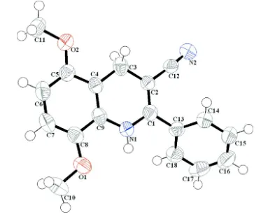
The molecular geometry and the atom-numbering scheme of (I) are shown in Fig. 1. The asymmetric unit of title
compound contains a 1,4-dihydroquinolyl unit bearing a phenyl ring at position C-2, nitril group at C-3 and two methoxy
at C-5 and C-8. The two rings of 1,4-dihydroquinolyl moiety are fused in an axial fashion and form a dihedral angle of
0.17 (5)°. The phenyl ring form also with quinolyl plane a dihedral angle of 45.38 (6)°. The crystal packing can be
described by two types of crossed layers which 1,4-dihydroquinolyl ring is parallel to (110) and (-110)planes respectively
(Fig. 2). The crystal packing is stabilized by intramolecular hydrogen bond (N—H···O) and Van der Waals interactions,
resulting in the formation of a three-dimensional network and reinforcing a cohesion of structure. Hydrogen-bonding
parameters are listed in table 1.
S2. Experimental
Compound (I) was obtained by modification of reported procedure (Lim et al., 1995). Refluxing a mixture of 1 eq. of
3-cyano-5,8-dimethoxy-2-phenylquinoline, 2 eq. of zinc and 1 eq. of Cu(NO2)2. 3H2O in the presence of 4 eq. of hydrazine
monohydrate for 3 days lead to the corresponding 1,4-dihydro-5,8-dimethoxy-2-phenylquinoline-3-carbonitrile I. The
product was purified by column chromatography. Single crystals suitable for X-ray diffraction analysis were obtained by
dissolving the corresponding compound in CH2Cl2/Petroleum ether mixture and letting the solution for slow evaporation
S3. Refinement
All non-H atoms were refined with anisotropic atomic displacement parameters. All H atoms were localized on Fourier
[image:4.610.112.485.121.434.2]maps but introduced in calculated positions and treated as riding on their parent C atom.
Figure 1
(Farrugia, 1997) the structure of the title compound with the atomic labelling scheme. Displacement are drawn at the
supporting information
sup-3 Acta Cryst. (2010). E66, o2312–o2313
Figure 2
(Brandenburg & Berndt, 2001) A diagram of the layered crystal packing of (I) viewed down the c axis.
5,8-Dimethoxy-2-phenyl-1,4-dihydroquinoline-3-carbonitrile
Crystal data
C18H16N2O2
Mr = 292.33
Monoclinic, P21/c
Hall symbol: -P 2ybc
a = 3.9952 (3) Å
b = 20.4544 (15) Å
c = 17.7313 (13) Å
β = 95.976 (5)°
V = 1441.12 (18) Å3
Z = 4
F(000) = 616
Dx = 1.347 Mg m−3
Mo Kα radiation, λ = 0.71073 Å Cell parameters from 2693 reflections
θ = 2.3–25.3°
µ = 0.09 mm−1
T = 150 K Stick, colourless 0.27 × 0.07 × 0.05 mm
Data collection
Bruker APEXII diffractometer
Graphite monochromator
CCD rotation images, thin slices scans Absorption correction: multi-scan
(SADABS: Sheldrick, 2002)
Tmin = 0.702, Tmax = 0.996 12491 measured reflections
3292 independent reflections 1975 reflections with I > 2σ(I)
Rint = 0.051
θmax = 27.6°, θmin = 2.5°
h = −3→5
k = −26→24
l = −22→22
Refinement
Refinement on F2 Least-squares matrix: full
R[F2 > 2σ(F2)] = 0.049
wR(F2) = 0.133
S = 1.03 3292 reflections 201 parameters 0 restraints
Primary atom site location: structure-invariant direct methods
Secondary atom site location: difference Fourier map
Hydrogen site location: inferred from neighbouring sites
H-atom parameters constrained
w = 1/[σ2(F
o2) + (0.0462P)2 + 0.6371P] where P = (Fo2 + 2Fc2)/3
(Δ/σ)max < 0.001 Δρmax = 0.17 e Å−3 Δρmin = −0.29 e Å−3
Special details
Geometry. All e.s.d.'s (except the e.s.d. in the dihedral angle between two l.s. planes) are estimated using the full covariance matrix. The cell e.s.d.'s are taken into account individually in the estimation of e.s.d.'s in distances, angles and torsion angles; correlations between e.s.d.'s in cell parameters are only used when they are defined by crystal symmetry. An approximate (isotropic) treatment of cell e.s.d.'s is used for estimating e.s.d.'s involving l.s. planes.
Refinement. Refinement of F2 against ALL reflections. The weighted R-factor wR and goodness of fit S are based on F2, conventional R-factors R are based on F, with F set to zero for negative F2. The threshold expression of F2 > σ(F2) is used only for calculating R-factors(gt) etc. and is not relevant to the choice of reflections for refinement. R-factors based on F2 are statistically about twice as large as those based on F, and R- factors based on ALL data will be even larger.
Fractional atomic coordinates and isotropic or equivalent isotropic displacement parameters (Å2)
x y z Uiso*/Ueq
C2 0.0694 (5) 0.18185 (9) 0.72449 (11) 0.0252 (4) C3 0.2040 (5) 0.17402 (10) 0.80517 (11) 0.0302 (5) H3A 0.0148 0.1763 0.8370 0.036* H3B 0.3573 0.2110 0.8198 0.036* C4 0.3890 (5) 0.11164 (9) 0.82153 (11) 0.0258 (4) C5 0.5299 (5) 0.09642 (10) 0.89555 (11) 0.0280 (5) C6 0.7054 (5) 0.03884 (10) 0.91013 (12) 0.0329 (5) H6 0.7993 0.0289 0.9603 0.040* C7 0.7448 (5) −0.00496 (10) 0.85068 (12) 0.0329 (5) H7 0.8668 −0.0444 0.8609 0.040* C8 0.6090 (5) 0.00849 (9) 0.77773 (11) 0.0282 (5) C10 0.8195 (6) −0.09023 (10) 0.72813 (14) 0.0402 (6) H10A 1.0483 −0.0807 0.7509 0.060* H10B 0.8296 −0.1130 0.6798 0.060* H10C 0.7053 −0.1180 0.7626 0.060* C11 0.6472 (6) 0.13355 (11) 1.02389 (11) 0.0377 (5) H11A 0.5699 0.0926 1.0450 0.056* H11B 0.5963 0.1701 1.0566 0.056* H11C 0.8906 0.1313 1.0211 0.056* C12 −0.1237 (5) 0.23988 (10) 0.70655 (11) 0.0273 (5) C13 0.0034 (5) 0.14369 (9) 0.58792 (11) 0.0268 (4) C14 0.0519 (5) 0.20237 (10) 0.55075 (11) 0.0319 (5) H14 0.1607 0.2378 0.5779 0.038* C15 −0.0572 (6) 0.20951 (11) 0.47439 (12) 0.0383 (6) H15 −0.0232 0.2498 0.4496 0.046* C16 −0.2153 (6) 0.15839 (12) 0.43412 (13) 0.0419 (6) H16 −0.2928 0.1637 0.3820 0.050* C17 −0.2606 (6) 0.09925 (12) 0.46989 (12) 0.0409 (6) H17 −0.3670 0.0639 0.4421 0.049* C18 −0.1503 (5) 0.09145 (11) 0.54663 (12) 0.0347 (5) H18 −0.1794 0.0507 0.5709 0.042* C9 0.4271 (5) 0.06718 (9) 0.76313 (11) 0.0265 (4) N1 0.2916 (4) 0.07985 (8) 0.68900 (9) 0.0294 (4) H1 0.3166 0.0506 0.6536 0.035* N2 −0.2790 (5) 0.28731 (9) 0.69710 (10) 0.0370 (5) O1 0.6360 (4) −0.02995 (7) 0.71508 (8) 0.0351 (4) O2 0.4784 (4) 0.14325 (7) 0.94924 (8) 0.0337 (4)
Atomic displacement parameters (Å2)
U11 U22 U33 U12 U13 U23
supporting information
sup-5 Acta Cryst. (2010). E66, o2312–o2313
C8 0.0294 (11) 0.0226 (10) 0.0335 (11) −0.0027 (8) 0.0070 (8) 0.0023 (8) C10 0.0401 (14) 0.0264 (12) 0.0548 (15) 0.0061 (10) 0.0088 (11) −0.0030 (10) C11 0.0463 (14) 0.0409 (13) 0.0245 (11) −0.0055 (10) −0.0022 (9) 0.0027 (9) C12 0.0341 (12) 0.0240 (11) 0.0240 (10) −0.0047 (9) 0.0044 (8) −0.0027 (8) C13 0.0271 (11) 0.0275 (11) 0.0258 (11) 0.0017 (8) 0.0033 (8) −0.0019 (8) C14 0.0389 (13) 0.0288 (11) 0.0287 (11) 0.0025 (9) 0.0061 (9) 0.0010 (9) C15 0.0514 (15) 0.0366 (13) 0.0279 (12) 0.0090 (11) 0.0092 (10) 0.0048 (9) C16 0.0462 (15) 0.0560 (16) 0.0233 (11) 0.0130 (12) 0.0022 (10) −0.0021 (10) C17 0.0403 (14) 0.0482 (15) 0.0332 (12) 0.0000 (11) −0.0009 (10) −0.0140 (11) C18 0.0380 (13) 0.0311 (12) 0.0347 (12) −0.0034 (9) 0.0030 (9) −0.0031 (9) C9 0.0262 (11) 0.0225 (10) 0.0309 (11) −0.0038 (8) 0.0043 (8) 0.0050 (8) N1 0.0406 (11) 0.0229 (9) 0.0246 (9) 0.0000 (7) 0.0025 (7) 0.0010 (7) N2 0.0475 (12) 0.0287 (10) 0.0348 (10) 0.0044 (9) 0.0045 (8) −0.0014 (8) O1 0.0422 (9) 0.0258 (8) 0.0378 (9) 0.0044 (6) 0.0071 (7) −0.0003 (6) O2 0.0436 (9) 0.0320 (8) 0.0244 (8) 0.0003 (6) −0.0019 (6) 0.0000 (6)
Geometric parameters (Å, º)
C1—N1 1.363 (3) C10—H10B 0.9800 C1—C2 1.378 (3) C10—H10C 0.9800 C1—C13 1.486 (3) C11—O2 1.435 (2) C2—C12 1.433 (3) C11—H11A 0.9800 C2—C3 1.484 (3) C11—H11B 0.9800 C3—C4 1.488 (3) C11—H11C 0.9800 C3—H3A 0.9900 C12—N2 1.154 (3) C3—H3B 0.9900 C13—C14 1.393 (3) C4—C9 1.398 (3) C13—C18 1.400 (3) C4—C5 1.408 (3) C14—C15 1.386 (3) C5—O2 1.381 (2) C14—H14 0.9500 C5—C6 1.381 (3) C15—C16 1.381 (3) C6—C7 1.405 (3) C15—H15 0.9500 C6—H6 0.9500 C16—C17 1.386 (3) C7—C8 1.377 (3) C16—H16 0.9500 C7—H7 0.9500 C17—C18 1.395 (3) C8—O1 1.374 (2) C17—H17 0.9500 C8—C9 1.413 (3) C18—H18 0.9500 C10—O1 1.441 (2) C9—N1 1.393 (2) C10—H10A 0.9800 N1—H1 0.8800
C2—C3—H3B 108.8 C18—C13—C1 120.61 (18) C4—C3—H3B 108.8 C15—C14—C13 120.5 (2) H3A—C3—H3B 107.7 C15—C14—H14 119.7 C9—C4—C5 118.87 (18) C13—C14—H14 119.7 C9—C4—C3 120.23 (17) C16—C15—C14 120.4 (2) C5—C4—C3 120.90 (18) C16—C15—H15 119.8 O2—C5—C6 124.87 (17) C14—C15—H15 119.8 O2—C5—C4 114.55 (17) C15—C16—C17 119.9 (2) C6—C5—C4 120.58 (19) C15—C16—H16 120.1 C5—C6—C7 119.86 (19) C17—C16—H16 120.1 C5—C6—H6 120.1 C16—C17—C18 120.2 (2) C7—C6—H6 120.1 C16—C17—H17 119.9 C8—C7—C6 120.88 (19) C18—C17—H17 119.9 C8—C7—H7 119.6 C17—C18—C13 120.0 (2) C6—C7—H7 119.6 C17—C18—H18 120.0 O1—C8—C7 126.04 (18) C13—C18—H18 120.0 O1—C8—C9 114.84 (17) N1—C9—C4 121.09 (17) C7—C8—C9 119.11 (19) N1—C9—C8 118.22 (18) O1—C10—H10A 109.5 C4—C9—C8 120.68 (18) O1—C10—H10B 109.5 C1—N1—C9 121.88 (17) H10A—C10—H10B 109.5 C1—N1—H1 119.1 O1—C10—H10C 109.5 C9—N1—H1 119.1 H10A—C10—H10C 109.5 C8—O1—C10 116.18 (16) H10B—C10—H10C 109.5 C5—O2—C11 116.77 (16)
supporting information
sup-7 Acta Cryst. (2010). E66, o2312–o2313
Hydrogen-bond geometry (Å, º)
D—H···A D—H H···A D···A D—H···A