Chloranilic acid: a redetermination at
100 K
Grzegorz Dutkiewicz,aH. S. Yathirajan,bQ. N. M. Hakim Al-arique,bB. Narayanacand Maciej Kubickia*
aDepartment of Chemistry, Adam Mickiewicz University, Grunwaldzka 6, 60-780
Poznan´, Poland,bDepartment of Studies in Chemistry, University of Mysore,
Manasagangotri, Mysore 570 006, India, andcDepartment of Studies in Chemistry,
Mangalore University, Mangalagangotri 574 199, India Correspondence e-mail: [email protected]
Received 9 January 2010; accepted 27 January 2010
Key indicators: single-crystal X-ray study;T= 100 K; mean(C–C) = 0.002 A˚;
Rfactor = 0.025;wRfactor = 0.056; data-to-parameter ratio = 13.1.
The crystal structure of chloranilic acid, C6H2Cl2O4, was first described by Andersen in 1967 [Andersen, E. K. (1967). Acta Cryst. 22, 188–191] at room temperature using visually estimated intensities. Taking into account the importance of the title compound, we have redetermined the structure at 100 (1) K. The approximately planar molecule [the maximum deviation from the mean plane through the ring is 0.0014 (9) A˚ for the ring atoms and 0.029 (3) A˚ for the other atoms] occupies a special position, lying across the center of symmetry. In the crystal structure, a two-dimensional hydrogen-bonded network sustained by O—H O inter-actions runs approximately parallel to [101]. The two-dimensional layers are further packed in a parallel fashion, stabilized by Cl Cl interactions [Cl Cl = 3.2838 (8) A˚ , C— Cl Cl = 152.96 (6)].
Related literature
For charge-transfer complexes of chloranilic acid, see: Gotoh
et al.(2006, 2007, 2008); Gotoh & Ishida (2009); Ishida (2004); Ishida & Kashino (1999). For a recent study of the formation of either salts or co-crystals by chloranilic acid with different organic bases, see: Molcˇanov & Kojic´-Prodic´ (2010). For the previous determination of the title structure, see: Andersen (1967a) and of its hydrate, see: Andersen (1967b). For hydrogen-bond motifs, see: Bernstein et al. (1995). For a description of the Cambridge Structural Database, see: (Allen, 2002).
Experimental
Crystal data
C6H2Cl2O4 Mr= 208.98
Monoclinic,P21=n a= 7.5338 (12) A˚ b= 5.5225 (10) A˚ c= 8.5720 (12) A˚
= 104.868 (11)
V= 344.70 (10) A˚3 Z= 2
MoKradiation
= 0.90 mm1 T= 100 K 0.30.10.1 mm
Data collection
Oxford Diffraction Xcalibur Eos diffractometer
Absorption correction: multi-scan (CrysAlis PRO; Oxford Diffraction (2009)) Tmin= 0.857,Tmax= 1.000
6154 measured reflections 774 independent reflections 698 reflections withI> 2(I) Rint= 0.032
Refinement
R[F2> 2(F2)] = 0.025 wR(F2) = 0.056 S= 1.09 774 reflections
59 parameters
All H-atom parameters refined max= 0.37 e A˚
3 min=0.27 e A˚
3
Table 1
Hydrogen-bond geometry (A˚ ,).
D—H A D—H H A D A D—H A
O2—H2 O3i
0.82 (2) 2.00 (2) 2.7516 (15) 152 (2)
Symmetry code: (i)xþ5 2;y
1 2;zþ
1 2.
Data collection: CrysAlis PRO(Oxford Diffraction, 2009); cell refinement:CrysAlis PRO; data reduction:CrysAlis PROprogram(s) used to solve structure:SIR92 (Altomare et al., 1993); program(s) used to refine structure: SHELXL97 (Sheldrick, 2008); molecular graphics:Stereochemical Workstation Operation Manual (Siemens, 1989); software used to prepare material for publication: SHELXL97.
QNMHA thanks the University of Mysore for research facilities.
Supplementary data and figures for this paper are available from the IUCr electronic archives (Reference: DS2017).
organic compounds
Acta Cryst.(2010). E66, o497–o498 doi:10.1107/S1600536810003387 Dutkiewiczet al.
o497
Acta Crystallographica Section EStructure Reports Online
Allen, F. H. (2002).Acta Cryst.B58, 380–388.
Altomare, A., Cascarano, G., Giacovazzo, C. & Guagliardi, A. (1993).J. Appl. Cryst.26, 343–350.
Andersen, E. K. (1967a).Acta Cryst.22, 188–191. Andersen, E. K. (1967b).Acta Cryst.22, 191–196.
Bernstein, J., Davis, R. E., Shimoni, L. & Chang, N.-L. (1995).Angew. Chem. Int. Ed. Engl.34, 1555–1573.
Gotoh, K., Asaji, T. & Ishida, H. (2007).Acta Cryst.C63, o17–o20. Gotoh, K., Asaji, T. & Ishida, H. (2008).Acta Cryst.C64, o550–o553.
Gotoh, K., Ishikawa, R. & Ishida, H. (2006).Acta Cryst.E62, o4738–o4740. Ishida, H. (2004).Acta Cryst.E60, o1900–o1901.
Ishida, H. & Kashino, S. (1999).Acta Cryst.C55, 1923–1926.
Molcˇanov, K. & Kojic´-Prodic´, B. (2010).CrystEngComm.In the press. doi: 10.1039/b908492d.
Oxford Diffraction (2009).CrysAlis PRO. Oxford Diffraction Ltd, Yarnton, England.
Sheldrick, G. M. (2008).Acta Cryst.A64, 112–122.
supporting information
sup-1 Acta Cryst. (2010). E66, o497–o498
supporting information
Acta Cryst. (2010). E66, o497–o498 [https://doi.org/10.1107/S1600536810003387]
Chloranilic acid: a redetermination at 100
K
Grzegorz Dutkiewicz, H. S. Yathirajan, Q. N. M. Hakim Al-arique, B. Narayana and Maciej
Kubicki
S1. Comment
The crystal structures of various charge-transfer complexes of chloranilic acid have been reported (Gotoh, Asaji et al.,
2008; Gotoh, Asaji et al., 2007; Gotoh & Ishida, 2009; Gotoh, Ishikawa, et al., 2006; Ishida, 2004; Ishida & Kashino,
1999). Very recently, a study on the formation of either salts or co-crystals by chloranilic acid with the different organic
bases was published (Molčanov & Kojić-Prodić, 2010).
There is a number of structures in the Cambridge Database (Allen, 2002) that contain the chloranilic acid
(2,5-di-chloro-3,6-dihydroxycyclohexa-2,5-diene-1,4-dione, I - Scheme 1), either as a neutral molecule or as an anion (mono- or
di-). Interestingly, the only determination of the structure of the acid itself dates back to 1967 (Andersen, 1967a;
hereinafter referred to as KA67). The structure was refined based on the visually estimated intensities of the diffraction
spots obtained by means of the Weissenberg equi-inclination method. The quality of this structure is excellent taking into
account the technology involved, but - having in mind the importance of this small molecule - thanks to the advancement
of the methodology it might be desirable to get the more accurate results. Here we report the results of the structure
determination of (I) at 100 (1) K. The unit cell parameters of the accompanying room temperature experiment are in an
excellent agreement with the data of KA67, but the model is much better, for instance in terms of R factors (8.9% in
1967, with 22 reflections omitted vs. 2.5% in the present determination), the only symmetry independent hydrogen atom
was found in the difference Fourier map in KA67 and left in the position found, while now it was isotropicaly refined,
etc. Nevertheless, the basic features of the structure are similar, and both the precision and depth of the analysis in KA67
and accompanying paper on the hydrate (Andersen, 1967b) are really remarkable.
We have chosen to describe the structure in the P21/n space group instead of P21/a used in KA67, in order to have
smaller β angle (104.87° instead of 122.77°); the transformation matrix is {-1 0 -1 0 1 0 1 0 0}. The molecule of I lies in
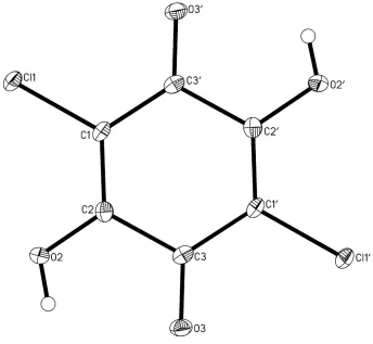
the special position, across the center of symmetry (Z′=1/2). The whole molecule is planar (Fig. 1); the maximum
deviation form the mean plane through 6 ring atom is 0.0014 (9) Å for the ring atom and 0.029 (3)Å for the other atoms.
The bond length pattern confirms the dominant double-bond character for the bonds C3—O3 (1.224 (2) Å) and C1—C2
(1.349 (2) Å) and single-bond for C2—C3 (1.507 (2) Å) and - to the lesser extent - for C1—C3′ (1.450 (2) Å).
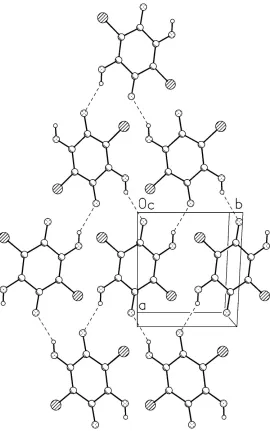
In the crystal structure the main packing motif arises as the result of relatively strong intermolecular O—H···O hydrogen
bonds, which make the antiparallel chains of molecules related by the 21 screw along y direction; using the graph-set
notation (Bernstein et al., 1995), these first-order chains will be described as C(5). The neighboring chains are
interconnected to give the centrosymmetric second-order rings R44(22) - cf. Fig. 2. These structures produce the
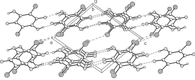
one-molecule thick layers of one-molecules which expand along [101] direction, and the neighboring chains are connected by
means of van der Waals interactions and probably also by weak halogen bonds, with Cl···Cl distance of 3.2838 (8)Å and
Chloranilic acid was purchased from Loba Chemie, Mumbai, India. X-ray quality crystals were obtained from methanol
solution after slow evaporation.
S3. Refinement
The position of the hydrogen atom was found in the difference Fourier map and both the positional and isotropic thermal
[image:4.610.133.477.175.492.2]parameters weres freely refined.
Figure 1
Anisotropic ellipsoid representation of the compound I together with atom labelling scheme. The ellipsoids are drawn at
supporting information
[image:5.610.171.441.69.500.2]sup-3 Acta Cryst. (2010). E66, o497–o498
Figure 2
The hydrogen bonded motifs in the crystal structure of I. Hydrogen bonds are shown as dashed lines. (a) the N—H···N
chain. Symmetry codex: (i) x,y,z; (ii) x,-3/2+y,3/2-z; (iii) 2-x,1/2+y,3/2-z; (iv) 2-x,-1/2+y,3/2-z; (v) 2-x,-3/2+y,3.2-z. (b)
Figure 3
Crystal packing as seen along y-direction. Hydrogen bonds and Cl···Cl contacts are shown as dashed lines.
Chloranilic acid
Crystal data
C6H2Cl2O4
Mr = 208.98 Monoclinic, P21/n
Hall symbol: -P 2yn a = 7.5338 (12) Å b = 5.5225 (10) Å c = 8.5720 (12) Å β = 104.868 (11)° V = 344.70 (10) Å3
Z = 2
F(000) = 208 Dx = 2.014 Mg m−3
Mo Kα radiation, λ = 0.71073 Å Cell parameters from 4539 reflections θ = 2.8–27.8°
µ = 0.90 mm−1
T = 100 K Prism, red
0.3 × 0.1 × 0.1 mm
Data collection
Oxford Diffraction Xcalibur Eos diffractometer
Radiation source: Enhance (Mo) X-ray Source Graphite monochromator
Detector resolution: 16.1544 pixels mm-1
ω–scan
Absorption correction: multi-scan
(CrysAlis PRO; Oxford Diffraction (2009)) Tmin = 0.857, Tmax = 1.000
6154 measured reflections 774 independent reflections 698 reflections with I > 2σ(I) Rint = 0.032
θmax = 27.9°, θmin = 3.2°
h = −9→9 k = −7→7 l = −11→10
Refinement
Refinement on F2
Least-squares matrix: full R[F2 > 2σ(F2)] = 0.025
wR(F2) = 0.056
S = 1.09 774 reflections 59 parameters 0 restraints
Primary atom site location: structure-invariant direct methods
Secondary atom site location: difference Fourier map
Hydrogen site location: inferred from neighbouring sites
All H-atom parameters refined w = 1/[σ2(F
o2) + (0.0204P)2 + 0.3502P]
where P = (Fo2 + 2Fc2)/3
(Δ/σ)max < 0.001
Δρmax = 0.37 e Å−3
supporting information
sup-5 Acta Cryst. (2010). E66, o497–o498
Special details
Geometry. All esds (except the esd in the dihedral angle between two l.s. planes) are estimated using the full covariance matrix. The cell esds are taken into account individually in the estimation of esds in distances, angles and torsion angles; correlations between esds in cell parameters are only used when they are defined by crystal symmetry. An approximate (isotropic) treatment of cell esds is used for estimating esds involving l.s. planes.
Refinement. Refinement of F2 against ALL reflections. The weighted R-factor wR and goodness of fit S are based on F2,
conventional R-factors R are based on F, with F set to zero for negative F2. The threshold expression of F2 > σ(F2) is used
only for calculating R-factors(gt) etc. and is not relevant to the choice of reflections for refinement. R-factors based on F2
are statistically about twice as large as those based on F, and R- factors based on ALL data will be even larger.
Fractional atomic coordinates and isotropic or equivalent isotropic displacement parameters (Å2)
x y z Uiso*/Ueq
C1 0.8496 (2) −0.1699 (3) 0.47066 (18) 0.0102 (3) Cl1 0.66487 (5) −0.36207 (7) 0.44138 (4) 0.01295 (13) C2 0.9798 (2) −0.1990 (3) 0.38967 (18) 0.0101 (3) O2 0.97189 (15) −0.3741 (2) 0.28296 (13) 0.0132 (3) H2 1.064 (3) −0.381 (4) 0.249 (3) 0.034 (7)* C3 1.14042 (19) −0.0274 (3) 0.41584 (17) 0.0095 (3) O3 1.25301 (15) −0.0641 (2) 0.33789 (13) 0.0126 (2)
Atomic displacement parameters (Å2)
U11 U22 U33 U12 U13 U23
C1 0.0081 (7) 0.0100 (8) 0.0122 (7) −0.0022 (6) 0.0020 (6) 0.0014 (6) Cl1 0.01094 (19) 0.0138 (2) 0.0150 (2) −0.00495 (14) 0.00488 (13) −0.00150 (14) C2 0.0113 (7) 0.0081 (7) 0.0102 (7) 0.0005 (6) 0.0016 (6) 0.0013 (6) O2 0.0121 (5) 0.0133 (6) 0.0163 (6) −0.0014 (4) 0.0075 (5) −0.0045 (4) C3 0.0084 (7) 0.0103 (7) 0.0098 (7) 0.0010 (6) 0.0021 (6) 0.0041 (6) O3 0.0121 (5) 0.0131 (6) 0.0147 (5) 0.0003 (4) 0.0072 (4) 0.0008 (4)
Geometric parameters (Å, º)
C1—C2 1.349 (2) C2—C3 1.508 (2) C1—C3i 1.450 (2) O2—H2 0.82 (2)
C1—Cl1 1.7164 (15) C3—O3 1.2240 (18) C2—O2 1.3217 (19)
C2—C1—C3i 121.02 (14) C1—C2—C3 120.71 (14)
C2—C1—Cl1 121.38 (12) C2—O2—H2 112.9 (17) C3i—C1—Cl1 117.59 (11) O3—C3—C1i 124.53 (14)
O2—C2—C1 122.23 (14) O3—C3—C2 117.19 (14) O2—C2—C3 117.05 (13) C1i—C3—C2 118.27 (13)
C3i—C1—C2—O2 −178.48 (14) O2—C2—C3—O3 −0.8 (2)
Cl1—C1—C2—C3 179.51 (11) C1—C2—C3—C1i −0.4 (2)
Symmetry code: (i) −x+2, −y, −z+1.
Hydrogen-bond geometry (Å, º)
D—H···A D—H H···A D···A D—H···A
O2—H2···O3ii 0.82 (2) 2.00 (2) 2.7516 (15) 152 (2)