N
,
N
000-Bis(3-chloro-2-fluorobenzylidene)-ethane-1,2-diamine
Hoong-Kun Fun* and Reza Kia‡
X-ray Crystallography Unit, School of Physics, Universiti Sains Malaysia, 11800 USM, Penang, Malaysia
Correspondence e-mail: hkfun@usm.my
Received 3 September 2008; accepted 4 September 2008
Key indicators: single-crystal X-ray study;T= 100 K; mean(C–C) = 0.001 A˚; Rfactor = 0.030;wRfactor = 0.089; data-to-parameter ratio = 38.2.
The molecule of the title centrosymmetric Schiff base compound, C16H12Cl2F2N2, adopts an E configuration with
respect to the azomethine C N bond. The imino groups are coplanar with the aromatic rings. Within the molecule, the planar units are parallel, but extend in opposite directions from the dimethylene bridge. An interesting feature of the crystal structure is the short intermolecular Cl F [3.1747 (5) A˚ ] interactions, which are shorter than the sum of the van der Waals radii of these atoms. These interactions link neighbouring molecules along the b axis. The crystal structure is further stabilized by – interactions, with a centroid–centroid distance of 3.5244 (4) A˚ .
Related literature
For bond-length data, see Allen et al. (1987). For related structures, see, for example: Fun & Kia (2008a,b): Fun, Kargar & Kia (2008); Fun, Kia & Kargar (2008). For information on Schiff base complexes and their applications, see, for example: Pal et al. (2005); Calligaris & Randaccio (1987); Hou et al.
(2001); Ren et al.(2002). For hydrogen-bonding motifs, see: Bernsteinet al.(1995).
Experimental
Crystal data
C16H12Cl2F2N2 Mr= 341.18
Monoclinic,P21=c a= 7.2249 (2) A˚
b= 11.3676 (2) A˚
c= 10.3368 (2) A˚
= 120.906 (1)
V= 728.42 (3) A˚3 Z= 2
MoKradiation
= 0.46 mm1 T= 100.0 (1) K 0.520.410.29 mm
Data collection
Bruker SMART APEXII CCD area-detector diffractometer Absorption correction: multi-scan
(SADABS; Bruker, 2005)
Tmin= 0.794,Tmax= 0.878
16842 measured reflections 3821 independent reflections 3403 reflections withI> 2(I)
Rint= 0.025
Refinement
R[F2> 2(F2)] = 0.030 wR(F2) = 0.088 S= 1.05 3821 reflections
100 parameters
H-atom parameters constrained
max= 0.45 e A˚
3
min=0.35 e A˚
3
Data collection:APEX2(Bruker, 2005); cell refinement:APEX2; data reduction: SAINT (Bruker, 2005); program(s) used to solve structure: SHELXTL (Sheldrick, 2008); program(s) used to refine structure:SHELXTL; molecular graphics:SHELXTL; software used to prepare material for publication:SHELXTLandPLATON(Spek, 2003).
HKF and RK thank the Malaysian Government and Universiti Sains Malaysia for the Science Fund grant No. 305/ PFIZIK/613312. RK thanks Universiti Sains Malaysia for the award of a postdoctoral research fellowship.
Supplementary data and figures for this paper are available from the IUCr electronic archives (Reference: SJ2537).
References
Allen, F. H., Kennard, O., Watson, D. G., Brammer, L., Orpen, A. G. & Taylor, R. (1987).J. Chem. Soc. Perkin Trans. 2, pp. S1–S19.
Bernstein, J., Davis, R. E., Shimoni, L. & Chang, N.-L. (1995).Angew. Chem. Int. Ed. Engl.34, 1555–1573.
Bruker (2005).APEX2,SAINTandSADABS. Bruker AXS Inc., Madison, Wisconsin, USA.
Calligaris, M. & Randaccio, L. (1987). Comprehensive Coordination Chemistry, Vol. 2, edited by G. Wilkinson, pp. 715–738. London: Pergamon. Fun, H.-K., Kargar, H. & Kia, R. (2008).Acta Cryst.E64, o1308.
Fun, H.-K. & Kia, R. (2008a).Acta Cryst.E64. submitted. [CV2444]. Fun, H.-K. & Kia, R. (2008b).Acta Cryst.E64, o1722–o1723. Fun, H.-K., Kia, R. & Kargar, H. (2008).Acta Cryst.E64, o1335.
Hou, B., Friedman, N., Ruhman, S., Sheves, M. & Ottolenghi, M. (2001).J. Phys. Chem. B,105, 7042–7048.
Pal, S., Barik, A. K., Gupta, S., Hazra, A., Kar, S. K., Peng, S.-M., Lee, G.-H., Butcher, R. J., El Fallah, M. S. & Ribas, J. (2005).Inorg. Chem.44, 3880– 3889.
Ren, S., Wang, R., Komatsu, K., Bonaz-Krause, P., Zyrianov, Y., McKenna, C. E., Csipke, C., Tokes, Z. A. & Lien, E. J. (2002).J. Med. Chem.45, 410–419. Sheldrick, G. M. (2008).Acta Cryst.A64, 112–122.
Spek, A. L. (2003).J. Appl. Cryst.36, 7–13.
Acta Crystallographica Section E
Structure Reports
Online
supporting information
Acta Cryst. (2008). E64, o1916 [doi:10.1107/S1600536808028419]
N
,
N
′
-Bis(3-chloro-2-fluorobenzylidene)ethane-1,2-diamine
Hoong-Kun Fun and Reza Kia
S1. Comment
Schiff bases are among the most prevalent mixed-donor ligands in the field of coordination chemistry in which there has
been growing interest, mainly because of their wide application in areas such as biochemistry, synthesis, and catalysis
(Pal et al., 2005; Hou et al., 2001; Ren et al., 2002). Many Schiff base complexes have been structurally characterized, but only a relatively small number of free Schiff bases have had their X-ray structures reported (Calligaris & Randaccio,
1987). As an extension of our work (Fun, Kargar & Kia 2008; Fun, Kia & Kargar 2008) on the structural characterization
of Schiff base ligands, the title compound (I), is reported here.
The molecule of the title compound (Fig. 1), adopts an E configuration with respect to the azomethine C═N bond. The bond lengths (Allen et al., 1987) and angles are within normal ranges and are comparable with the values found in related structures (Fun & Kia 2008a,b; Fun, Kargar & Kia 2008; Fun, Kia & Kargar 2008). The two planar units are parallel but extend in opposite directions from the dimethylene bridge. The interesting feature of the crystal structure is the short
intermolecular Cl···F interactions [symmetry code: x, -1/2 - y, -1/2 + z] with a distance of 3.1747 (5) Å, which is shorter than the sum of the van der Waals radii of these atoms. These interactions link neighbouring molecules along the b-axis. The crystal structure is further stabilized by π–π interactions with a centroid to centroid distance of 3.5244 (4) Å [Cg1–
Cg1; symmetry code, 2 - x, -y, -z; Cg1 is the centroid of the C1–C6 benzene ring].
S2. Experimental
The synthetic method has been described earlier (Fun, Kargar & Kia, 2008). Single crystals suitable for X-ray diffraction
were obtained by evaporation of an ethanol solution at room temperature.
S3. Refinement
All of the hydrogen atoms were positioned geometrically with C—H = 0.95 or 0.99 Å and refined in riding mode with
Figure 1
The molecular structure of (I) with atom labels and 50% probability ellipsoids for non-H atoms. The suffix A corresponds
to symmetry code (-x + 2, -y, -z + 1).
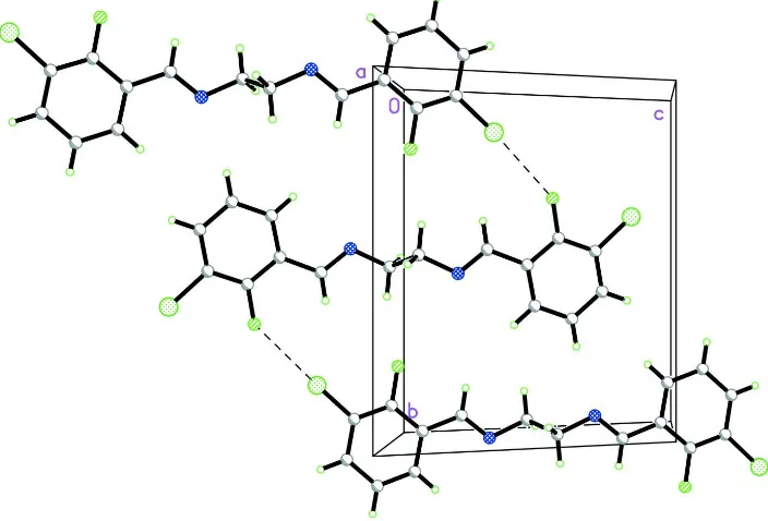
Figure 2
The crystal packing of (I), viewed approximately down the a-axis, showing the linking of the molecules by Cl···F contacts along the b-axis. Intermolecular interactions are shown as dashed lines.
N,N′-Bis(3-chloro-2-fluorobenzylidene)ethane-1,2-diamine
Crystal data
C16H12Cl2F2N2
Mr = 341.18
Monoclinic, P21/c
Hall symbol: -P 2ybc
a = 7.2249 (2) Å
b = 11.3676 (2) Å
c = 10.3368 (2) Å
β = 120.906 (1)°
V = 728.42 (3) Å3
F(000) = 348
Dx = 1.556 Mg m−3
Mo Kα radiation, λ = 0.71073 Å Cell parameters from 8904 reflections
θ = 2.9–41.0°
µ = 0.46 mm−1
[image:3.610.129.481.261.500.2]Data collection
Bruker SMART APEXII CCD area-detector diffractometer
Radiation source: fine-focus sealed tube Graphite monochromator
φ and ω scans
Absorption correction: multi-scan (SADABS; Bruker, 2005)
Tmin = 0.794, Tmax = 0.878
16842 measured reflections 3821 independent reflections 3403 reflections with I > 2σ(I)
Rint = 0.025
θmax = 37.5°, θmin = 2.9°
h = −11→12
k = −19→19
l = −17→17
Refinement
Refinement on F2
Least-squares matrix: full
R[F2 > 2σ(F2)] = 0.030
wR(F2) = 0.088
S = 1.05 3821 reflections 100 parameters 0 restraints
Primary atom site location: structure-invariant direct methods
Secondary atom site location: difference Fourier map
Hydrogen site location: inferred from neighbouring sites
H-atom parameters constrained
w = 1/[σ2(F
o2) + (0.0499P)2 + 0.131P]
where P = (Fo2 + 2Fc2)/3
(Δ/σ)max = 0.001
Δρmax = 0.45 e Å−3
Δρmin = −0.35 e Å−3
Special details
Experimental. The low-temperature data was collected with the Oxford Cyrosystem Cobra low-temperature attachment. Geometry. All e.s.d.'s (except the e.s.d. in the dihedral angle between two l.s. planes) are estimated using the full covariance matrix. The cell e.s.d.'s are taken into account individually in the estimation of e.s.d.'s in distances, angles and torsion angles; correlations between e.s.d.'s in cell parameters are only used when they are defined by crystal symmetry. An approximate (isotropic) treatment of cell e.s.d.'s is used for estimating e.s.d.'s involving l.s. planes.
Refinement. Refinement of F2 against ALL reflections. The weighted R-factor wR and goodness of fit S are based on F2,
conventional R-factors R are based on F, with F set to zero for negative F2. The threshold expression of F2 > σ(F2) is used
only for calculating R-factors(gt) etc. and is not relevant to the choice of reflections for refinement. R-factors based on F2
are statistically about twice as large as those based on F, and R- factors based on ALL data will be even larger.
Fractional atomic coordinates and isotropic or equivalent isotropic displacement parameters (Å2)
x y z Uiso*/Ueq
Cl1 0.65534 (3) −0.131316 (15) −0.342553 (18) 0.01852 (5) F1 0.71054 (8) −0.18319 (4) −0.04997 (5) 0.01952 (9) N1 0.83760 (10) 0.03597 (6) 0.29627 (6) 0.01620 (10) C1 0.72180 (10) −0.06703 (6) −0.07109 (7) 0.01350 (10) C2 0.69340 (10) −0.02919 (6) −0.20779 (7) 0.01388 (10) C3 0.70125 (11) 0.09019 (6) −0.23283 (7) 0.01577 (11)
H3 0.6802 0.1170 −0.3265 0.019*
C4 0.74016 (11) 0.17052 (6) −0.11975 (8) 0.01704 (11)
H4 0.7457 0.2523 −0.1363 0.020*
C5 0.77080 (11) 0.13130 (6) 0.01692 (8) 0.01554 (11)
H5 0.7977 0.1868 0.0934 0.019*
C6 0.76267 (10) 0.01123 (6) 0.04403 (7) 0.01320 (10) C7 0.80350 (10) −0.03353 (6) 0.19019 (7) 0.01486 (11)
H7 0.8043 −0.1160 0.2053 0.018*
H8A 0.8762 −0.1043 0.4279 0.021*
H8B 0.7805 0.0092 0.4659 0.021*
Atomic displacement parameters (Å2)
U11 U22 U33 U12 U13 U23
Cl1 0.02300 (8) 0.01901 (9) 0.01500 (8) −0.00281 (5) 0.01081 (6) −0.00347 (5) F1 0.0292 (2) 0.01227 (18) 0.01890 (19) −0.00308 (15) 0.01361 (17) 0.00007 (14) N1 0.0180 (2) 0.0181 (2) 0.0121 (2) 0.00091 (18) 0.00742 (18) 0.00082 (17) C1 0.0142 (2) 0.0125 (2) 0.0135 (2) −0.00087 (18) 0.00691 (18) 0.00018 (18) C2 0.0139 (2) 0.0153 (3) 0.0124 (2) −0.00011 (18) 0.00672 (18) −0.00027 (18) C3 0.0166 (2) 0.0165 (3) 0.0141 (2) 0.0015 (2) 0.0079 (2) 0.00275 (19) C4 0.0201 (3) 0.0140 (3) 0.0170 (2) 0.0024 (2) 0.0096 (2) 0.0025 (2) C5 0.0183 (3) 0.0131 (3) 0.0151 (2) 0.00170 (19) 0.0085 (2) 0.00006 (18) C6 0.0133 (2) 0.0140 (2) 0.0121 (2) 0.00054 (18) 0.00633 (18) 0.00056 (18) C7 0.0158 (2) 0.0161 (3) 0.0122 (2) −0.00085 (19) 0.00683 (19) 0.00053 (19) C8 0.0178 (2) 0.0212 (3) 0.0122 (2) −0.0012 (2) 0.0076 (2) 0.0008 (2)
Geometric parameters (Å, º)
Cl1—C2 1.7235 (7) C4—C5 1.3865 (10)
F1—C1 1.3476 (8) C4—H4 0.9500
N1—C7 1.2687 (9) C5—C6 1.4007 (9)
N1—C8 1.4568 (9) C5—H5 0.9500
C1—C2 1.3878 (9) C6—C7 1.4733 (9)
C1—C6 1.3906 (9) C7—H7 0.9500
C2—C3 1.3882 (9) C8—C8i 1.5267 (13)
C3—C4 1.3934 (10) C8—H8A 0.9900
C3—H3 0.9500 C8—H8B 0.9900
C7—N1—C8 116.79 (6) C4—C5—H5 119.5
F1—C1—C2 118.62 (6) C6—C5—H5 119.5
F1—C1—C6 119.48 (6) C1—C6—C5 117.68 (6)
C2—C1—C6 121.90 (6) C1—C6—C7 119.95 (6)
C1—C2—C3 119.61 (6) C5—C6—C7 122.33 (6)
C1—C2—Cl1 119.54 (5) N1—C7—C6 121.26 (6)
C3—C2—Cl1 120.84 (5) N1—C7—H7 119.4
C2—C3—C4 119.62 (6) C6—C7—H7 119.4
C2—C3—H3 120.2 N1—C8—C8i 109.32 (7)
C4—C3—H3 120.2 N1—C8—H8A 109.8
C5—C4—C3 120.12 (6) C8i—C8—H8A 109.8
C5—C4—H4 119.9 N1—C8—H8B 109.8
C3—C4—H4 119.9 C8i—C8—H8B 109.8
C4—C5—C6 121.07 (6) H8A—C8—H8B 108.3
F1—C1—C2—C3 179.08 (6) C2—C1—C6—C5 1.11 (9)
C6—C1—C2—C3 −1.35 (10) F1—C1—C6—C7 2.90 (9)
C6—C1—C2—Cl1 177.08 (5) C4—C5—C6—C1 −0.31 (10)
C1—C2—C3—C4 0.78 (10) C4—C5—C6—C7 177.39 (6)
Cl1—C2—C3—C4 −177.63 (5) C8—N1—C7—C6 −177.18 (6)
C2—C3—C4—C5 −0.01 (10) C1—C6—C7—N1 −178.79 (6)
C3—C4—C5—C6 −0.22 (11) C5—C6—C7—N1 3.55 (10)
F1—C1—C6—C5 −179.33 (6) C7—N1—C8—C8i 117.01 (8)