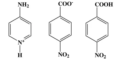
Aminopyridinium nitrobenzoate
4-nitrobenzoic acid
Ching Kheng Quah, Samuel Robinson Jebas‡ and Hoong-Kun Fun*
X-ray Crystallography Unit, School of Physics, Universiti Sains Malaysia, 11800 USM, Penang, Malaysia
Correspondence e-mail: hkfun@usm.my
Received 29 August 2008; accepted 29 August 2008
Key indicators: single-crystal X-ray study;T= 100 K; mean(C–C) = 0.001 A˚; Rfactor = 0.044;wRfactor = 0.133; data-to-parameter ratio = 23.4.
The asymmetric unit of the title compound, C5H7N2 +
-C7H4NO4
C7H5NO4, consists of an aminopyridinium cation, a 4-nitrobenzoate anion and a neutral 4-nitrobenzoic acid molecule. The pyridine ring forms dihedral angles of 64.70 (5)
and 70.37 (5), respectively, with the benzene rings of
4-nitrobenzoic acid and 4-nitrobenzoate. In the crystal structure, the cations, anions and the neutral 4-nitrobenzoic acid molecules are linked by O—H O and N—H O hydrogen bonds, forming a two-dimensional network parallel to (001). Adjacent networks are cross-linkedvia C—H O hydrogen bonds and – stacking interactions [centroid–centroid distances 3.6339 (6) and 3.6566 (6) A˚ ].
Related literature
For the biological activity of 4-aminopyridine, see: Judgeet al. (2006); Schwid et al.(1997); Struppet al.(2004). For related structures, see: Chao & Schempp (1977); Anderson et al. (2005); Andrau & White, (2003); Bhattacharya et al.(1994); Karleet al.(2003).
Experimental
Crystal data
C5H7N2+C7H4NO4
C7H5NO4 Mr= 428.36
Triclinic,P1
a= 6.4561 (1) A˚
b= 6.8598 (1) A˚
c= 20.9055 (3) A˚ = 85.826 (1)
= 87.975 (1)
= 86.188 (1) V= 920.92 (2) A˚3 Z= 2
MoKradiation = 0.12 mm1 T= 100.0 (1) K 0.400.360.29 mm
Data collection
Bruker SMART APEXII CCD area-detector diffractometer Absorption correction: multi-scan
(SADABS; Bruker, 2005)
Tmin= 0.952,Tmax= 0.965
24945 measured reflections 6647 independent reflections 5169 reflections withI> 2(I)
Rint= 0.031
Refinement
R[F2> 2(F2)] = 0.044 wR(F2) = 0.132 S= 1.05 6647 reflections 284 parameters 1 restraint
H atoms treated by a mixture of independent and constrained refinement
max= 0.43 e A˚ 3
[image:1.610.69.260.566.689.2]min=0.39 e A˚ 3
Table 1
Hydrogen-bond geometry (A˚ ,).
D—H A D—H H A D A D—H A
O3A—H1O3 O3Bi 0.82 1.63 2.4457 (11) 170 N3—H3A O3Bii
0.86 2.14 2.9977 (12) 172 N3—H3B O4Bi
0.86 2.07 2.8758 (12) 155 N2—H1N2 O4Aiii
0.85 (1) 1.99 (1) 2.7726 (12) 153 (1) C2B—H2BA O1Biv
0.93 2.52 3.2187 (13) 133 C8—H8A O3Av
0.93 2.56 3.4565 (13) 161 C12—H12A O1Avi
0.93 2.55 3.4427 (13) 162
Symmetry codes: (i) xþ1;yþ2;zþ1; (ii) xþ2;yþ2;zþ1; (iii)
xþ1;y1;z; (iv) xþ3;yþ1;zþ2; (v) x;y1;z; (vi)
xþ2;yþ1;zþ1.
Data collection:APEX2(Bruker, 2005); cell refinement:APEX2; data reduction: SAINT (Bruker, 2005); program(s) used to solve structure: SHELXTL (Sheldrick, 2008); program(s) used to refine structure:SHELXTL; molecular graphics:SHELXTL; software used to prepare material for publication:SHELXTLandPLATON(Spek, 2003).
HKF and SRJ thank the Malaysian Government and Universiti Sains Malaysia for Science Fund grant No. 305/ PFIZIK/613312. SRJ thanks Universiti Sains Malaysia for a post-doctoral research fellowship. CKQ thanks Universiti Sains Malaysia for a student assistanceship.
Supplementary data and figures for this paper are available from the IUCr electronic archives (Reference: CI2664).
References
Anderson, F. P., Gallagher, J. F., Kenny, P. T. M. & Lough, A. J. (2005).Acta Cryst.E61, o1350–o1353.
Andrau, L. & White, J. (2003).Acta Cryst.E59, o77–o79.
Bhattacharya, S., Dastidar, P. & Guru Row, T. N. (1994).Chem. Mater.6, 531– 537.
organic compounds
o1878
Quahet al. doi:10.1107/S1600536808027761 Acta Cryst.(2008). E64, o1878–o1879Acta Crystallographica Section E
Structure Reports Online
ISSN 1600-5368
Bruker (2005).APEX2,SAINTandSADABS. Bruker AXS Inc., Madison, Wisconsin, USA.
Chao, M. & Schempp, E. (1977).Acta Cryst.B33, 1557–1564. Judge, S. & Bever, C. (2006).Pharmacol. Ther.111, 224–259.
Karle, I., Gilardi, R. D., Chandrashekhar Rao, Ch., Muraleedharan, K. M. & Ranganathan, S. (2003).J. Chem. Crystallogr.33, 727–749.
Schwid, S. B., Petrie, M. D., McDermott, M. P., Tierney, D. S., Mason, D. H. & Goodman, A. D. (1997).Neurology,48, 817–821.
Sheldrick, G. M. (2008).Acta Cryst.A64, 112–122. Spek, A. L. (2003).J. Appl. Cryst.36, 7–13.
supporting information
sup-1
Acta Cryst. (2008). E64, o1878–o1879supporting information
Acta Cryst. (2008). E64, o1878–o1879 [doi:10.1107/S1600536808027761]
4-Aminopyridinium 4-nitrobenzoate 4-nitrobenzoic acid
Ching Kheng Quah, Samuel Robinson Jebas and Hoong-Kun Fun
S1. Comment
4-Aminopyridine (Fampridine) is used clinically in Lambert-Eaton myasthenic syndrome and multiple sclerosis because
by blocking potassium channels, it prolongs the action potentials thereby increasing transmitter release at the
neuromuscular junction (Judge et al., 2006; Schwid et al., 1997; Strupp et al., 2004). The crystal structure of
4-amino-pyridine has been reported (Chao & Schempp, 1977; Anderson et al., 2005). As an extension of our systematic study of
hydrogen bonding patterns of 4-aminopyridine with aromatic carboxylic acids, we report here the crystal structure of the
title compound.
The asymmetric unit of the title compound contains one 4-aminopyridinium cation, one 4-nitrobenzoate anion and one
4-nitrobenzoic acid molecule. A proton transfer from the carboxyl group of 4-nitrobenzoic acid to atom N2 of
4-amino-pyridine resulted in the formation of ions. This lead to the widening of C8—N2—C12 angle of the 4-amino-pyridine ring to
120.86 (9)°, compared to 115.25 (13)° in the unprotonated 4-aminopyridine (Anderson et al., 2005). This type of
protonation is observed in various 4-aminopyridine acid complexes (Bhattacharya et al., 1994; Karle et al., 2003). The
bond lengths and angles of the 4-aminopyridne are comparable to the values reported earlier for 4-aminopyridine (Chao
& Schempp, 1977; Anderson et al., 2005). The bond lengths and angles of the 4-nitrobenzoic acid is found to be
normal(Andrau & White, 2003).
The dihedral angle between the benzene rings of 4-nitrobenzoic acid (C1A-C6A) and 4-nitrobenzoate (C1B-C6B) units
is 6.62 (5)°. The pyridine (N2/C8—C12) ring forms dihedral angles of 64.70 (5)° and 70.37 (5)°, respectively, with the
C1A-C6A and C1B-C6B rings.
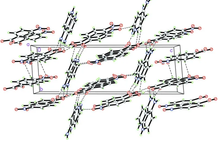
In the crystal structure, the cations, anions and the neutral 4-nitrobenzoic acid molecules are linked to form a
two-dimensional network (Fig. 2) parallel to the (0 0 1) by O—H···O and N—H···O hydrogen bonds (Table 1). The adjacent
networks are cross-linked via C—H···O hydrogen bonds. The crystal packing is further consolidated by π–π stacking
interactions between symmetry-related C1A-C6A (centroid Cg1) and C1B-C6B (centroid Cg2) rings, with Cg1···Cg1i and
Cg2···Cg2vii distances of 3.6566 (6) Å and 3.6339 (6) Å, respectively [symmetry codes: (i) 1-x, y, 1-z; (vii) x, y,
2-z].
S2. Experimental
4-Aminopyridine and 4-nitrobenzoic acid were mixed in equimolar ratio in methanol and warmed in a water bath for 2 h.
Colourless single crystals were obtained after a week on slow evaporation.
S3. Refinement
Atom H1N2 was located from a difference map and was refined with the N-H distance restrained to 0.85 (1) Å. The
remaining H atoms were positioned geometrically with C-H = 0.93 Å, N-H = 0.86 Å and O-H = 0.82Å, and refined using
Figure 1
The asymmetric unit of the title compound, showing 50% probability displacement ellipsoids and the atom-numbering
scheme.
Figure 2
The crystal packing of the title compound, viewed along the a axis. Hydrogen bonds are shown as dashed lines.
(I)
Crystal data
C5H7N2+·C7H4NO4−·C7H5NO4
Mr = 428.36
Triclinic, P1
Hall symbol: -P 1
a = 6.4561 (1) Å
b = 6.8598 (1) Å
c = 20.9055 (3) Å
α = 85.826 (1)°
β = 87.975 (1)°
γ = 86.188 (1)°
V = 920.92 (2) Å3
Z = 2
F(000) = 444
Dx = 1.545 Mg m−3
Mo Kα radiation, λ = 0.71073 Å
Cell parameters from 6200 reflections
θ = 2.2–29.2°
[image:4.610.128.483.304.536.2]supporting information
sup-3
Acta Cryst. (2008). E64, o1878–o1879T = 100 K
Block, colourless
0.40 × 0.36 × 0.29 mm
Data collection
Bruker SMART APEXII CCD area-detector diffractometer
Radiation source: fine-focus sealed tube Graphite monochromator
φ and ω scans
Absorption correction: multi-scan (SADABS; Bruker, 2005)
Tmin = 0.952, Tmax = 0.965
24945 measured reflections 6647 independent reflections 5169 reflections with I > 2σ(I)
Rint = 0.031
θmax = 32.5°, θmin = 1.0°
h = −9→9
k = −10→10
l = −31→31
Refinement
Refinement on F2
Least-squares matrix: full R[F2 > 2σ(F2)] = 0.044
wR(F2) = 0.132
S = 1.05
6647 reflections 284 parameters 1 restraint
Primary atom site location: structure-invariant direct methods
Secondary atom site location: difference Fourier map
Hydrogen site location: inferred from neighbouring sites
H atoms treated by a mixture of independent and constrained refinement
w = 1/[σ2(F
o2) + (0.0736P)2 + 0.1221P]
where P = (Fo2 + 2Fc2)/3
(Δ/σ)max = 0.001
Δρmax = 0.43 e Å−3
Δρmin = −0.39 e Å−3
Special details
Experimental. The data was collected with the Oxford Cyrosystem Cobra low-temperature attachment.
Geometry. All e.s.d.'s (except the e.s.d. in the dihedral angle between two l.s. planes) are estimated using the full covariance matrix. The cell e.s.d.'s are taken into account individually in the estimation of e.s.d.'s in distances, angles and torsion angles; correlations between e.s.d.'s in cell parameters are only used when they are defined by crystal symmetry. An approximate (isotropic) treatment of cell e.s.d.'s is used for estimating e.s.d.'s involving l.s. planes.
Refinement. Refinement of F2 against ALL reflections. The weighted R-factor wR and goodness of fit S are based on F2,
conventional R-factors R are based on F, with F set to zero for negative F2. The threshold expression of F2 > σ(F2) is used
only for calculating R-factors(gt) etc. and is not relevant to the choice of reflections for refinement. R-factors based on F2
are statistically about twice as large as those based on F, and R- factors based on ALL data will be even larger.
Fractional atomic coordinates and isotropic or equivalent isotropic displacement parameters (Å2)
x y z Uiso*/Ueq
O1A 0.61416 (13) 0.64153 (13) 0.61139 (4) 0.02534 (18)
O1B 1.43798 (12) 0.64460 (14) 1.07244 (4) 0.02613 (19)
O2A 0.89023 (12) 0.63198 (13) 0.54960 (4) 0.02492 (18)
O2B 1.15700 (13) 0.66730 (15) 1.13139 (4) 0.02794 (19)
O3A 0.31404 (11) 0.97209 (12) 0.29069 (4) 0.02024 (16)
H1O3 0.2342 1.0163 0.2628 0.030*
O3B 0.89111 (11) 0.90736 (11) 0.80183 (4) 0.01831 (15)
O4A 0.02402 (11) 0.98398 (11) 0.35318 (4) 0.01950 (16)
O4B 0.59084 (12) 0.90329 (13) 0.85892 (4) 0.02425 (18)
N1A 0.70215 (13) 0.66162 (13) 0.55851 (4) 0.01667 (17)
N1B 1.24848 (13) 0.67136 (13) 1.07904 (4) 0.01646 (17)
N3 0.73597 (13) 0.85350 (13) 0.20200 (5) 0.02020 (18)
H3A 0.8354 0.9309 0.2022 0.024*
H3B 0.6223 0.8922 0.1834 0.024*
C1A 0.24956 (15) 0.84572 (14) 0.46101 (5) 0.01507 (18)
H1AA 0.1084 0.8790 0.4667 0.018*
C1B 1.11431 (15) 0.75402 (14) 0.90711 (5) 0.01505 (18)
H1BA 1.1788 0.7482 0.8667 0.018*
C2A 0.36551 (15) 0.77640 (14) 0.51351 (5) 0.01593 (18)
H2AA 0.3048 0.7633 0.5545 0.019*
C2B 1.22778 (15) 0.70459 (14) 0.96180 (5) 0.01522 (18)
H2BA 1.3685 0.6664 0.9587 0.018*
C3B 1.12614 (14) 0.71350 (14) 1.02109 (5) 0.01397 (17)
C3A 0.57532 (15) 0.72731 (14) 0.50276 (5) 0.01424 (17)
C4A 0.67164 (15) 0.74110 (15) 0.44250 (5) 0.01592 (18)
H4AA 0.8119 0.7038 0.4369 0.019*
C4B 0.91589 (15) 0.76542 (14) 1.02843 (5) 0.01527 (18)
H4BA 0.8513 0.7667 1.0689 0.018*
C5A 0.55303 (15) 0.81214 (15) 0.39071 (5) 0.01621 (18)
H5AA 0.6142 0.8239 0.3498 0.019*
C5B 0.80500 (15) 0.81538 (14) 0.97324 (5) 0.01517 (18)
H5BA 0.6638 0.8512 0.9766 0.018*
C6A 0.34218 (14) 0.86598 (14) 0.39989 (5) 0.01412 (17)
C6B 0.90370 (14) 0.81237 (14) 0.91270 (5) 0.01388 (17)
C7A 0.21250 (15) 0.94696 (14) 0.34447 (5) 0.01498 (18)
C7B 0.78049 (15) 0.87820 (14) 0.85437 (5) 0.01578 (18)
C8 0.62995 (16) 0.35854 (16) 0.26207 (5) 0.0195 (2)
H8A 0.5260 0.2709 0.2623 0.023*
C9 0.59889 (15) 0.54066 (15) 0.23204 (5) 0.01760 (19)
H9A 0.4736 0.5775 0.2126 0.021*
C10 0.75805 (15) 0.67391 (15) 0.23052 (5) 0.01566 (18)
C11 0.94501 (15) 0.60896 (15) 0.26128 (5) 0.01670 (19)
H11A 1.0538 0.6914 0.2611 0.020*
C12 0.96496 (16) 0.42621 (16) 0.29097 (5) 0.0186 (2)
H12A 1.0877 0.3849 0.3113 0.022*
H1N2 0.839 (2) 0.1892 (15) 0.3080 (7) 0.029 (4)*
Atomic displacement parameters (Å2)
U11 U22 U33 U12 U13 U23
O1A 0.0250 (4) 0.0384 (5) 0.0118 (4) −0.0024 (3) −0.0008 (3) 0.0048 (3)
O1B 0.0158 (3) 0.0428 (5) 0.0191 (4) 0.0041 (3) −0.0028 (3) −0.0013 (3)
O2A 0.0172 (3) 0.0361 (5) 0.0207 (4) 0.0016 (3) −0.0036 (3) 0.0012 (3)
O2B 0.0228 (4) 0.0485 (5) 0.0115 (4) 0.0012 (3) 0.0006 (3) 0.0003 (3)
O3A 0.0183 (3) 0.0303 (4) 0.0116 (3) −0.0013 (3) −0.0025 (3) 0.0027 (3)
O3B 0.0174 (3) 0.0258 (4) 0.0114 (3) −0.0009 (3) −0.0003 (3) 0.0006 (3)
O4A 0.0158 (3) 0.0236 (4) 0.0182 (4) 0.0011 (3) −0.0016 (3) 0.0026 (3)
O4B 0.0145 (3) 0.0368 (5) 0.0198 (4) 0.0011 (3) −0.0020 (3) 0.0071 (3)
supporting information
sup-5
Acta Cryst. (2008). E64, o1878–o1879N1B 0.0167 (4) 0.0194 (4) 0.0131 (4) 0.0003 (3) −0.0014 (3) −0.0010 (3)
N2 0.0213 (4) 0.0188 (4) 0.0161 (4) 0.0014 (3) 0.0009 (3) 0.0017 (3)
N3 0.0160 (4) 0.0212 (4) 0.0224 (5) 0.0005 (3) −0.0014 (3) 0.0045 (3)
C1A 0.0146 (4) 0.0166 (4) 0.0140 (4) −0.0011 (3) −0.0003 (3) −0.0006 (3)
C1B 0.0156 (4) 0.0177 (4) 0.0114 (4) 0.0009 (3) 0.0001 (3) −0.0004 (3)
C2A 0.0172 (4) 0.0180 (4) 0.0127 (4) −0.0030 (3) 0.0005 (3) −0.0001 (3)
C2B 0.0139 (4) 0.0175 (4) 0.0140 (4) 0.0009 (3) −0.0001 (3) −0.0010 (3)
C3B 0.0151 (4) 0.0154 (4) 0.0114 (4) −0.0001 (3) −0.0023 (3) −0.0003 (3)
C3A 0.0168 (4) 0.0143 (4) 0.0118 (4) −0.0017 (3) −0.0030 (3) 0.0002 (3)
C4A 0.0141 (4) 0.0193 (4) 0.0143 (4) −0.0007 (3) −0.0005 (3) −0.0013 (3)
C4B 0.0158 (4) 0.0175 (4) 0.0125 (4) −0.0018 (3) 0.0009 (3) −0.0007 (3)
C5A 0.0163 (4) 0.0206 (4) 0.0117 (4) −0.0015 (3) 0.0000 (3) −0.0013 (3)
C5B 0.0131 (4) 0.0180 (4) 0.0142 (4) −0.0005 (3) −0.0002 (3) 0.0001 (3)
C6A 0.0154 (4) 0.0147 (4) 0.0125 (4) −0.0018 (3) −0.0021 (3) −0.0006 (3)
C6B 0.0143 (4) 0.0151 (4) 0.0122 (4) −0.0013 (3) −0.0014 (3) 0.0005 (3)
C7A 0.0173 (4) 0.0153 (4) 0.0125 (4) −0.0026 (3) −0.0018 (3) 0.0002 (3)
C7B 0.0157 (4) 0.0167 (4) 0.0149 (4) −0.0007 (3) −0.0022 (3) −0.0001 (3)
C8 0.0175 (4) 0.0233 (5) 0.0178 (5) −0.0023 (4) 0.0008 (4) −0.0016 (4)
C9 0.0138 (4) 0.0231 (5) 0.0156 (5) −0.0006 (3) −0.0012 (3) −0.0001 (4)
C10 0.0144 (4) 0.0199 (4) 0.0123 (4) 0.0012 (3) 0.0005 (3) −0.0009 (3)
C11 0.0151 (4) 0.0203 (4) 0.0148 (4) −0.0004 (3) −0.0020 (3) −0.0015 (4)
C12 0.0178 (4) 0.0232 (5) 0.0143 (5) 0.0029 (3) −0.0023 (3) −0.0010 (4)
Geometric parameters (Å, º)
O1A—N1A 1.2278 (12) C2A—H2AA 0.93
O1B—N1B 1.2294 (11) C2B—C3B 1.3851 (14)
O2A—N1A 1.2276 (11) C2B—H2BA 0.93
O2B—N1B 1.2244 (12) C3B—C4B 1.3863 (13)
O3A—C7A 1.2877 (12) C3A—C4A 1.3848 (14)
O3A—H1O3 0.8200 C4A—C5A 1.3888 (14)
O3B—C7B 1.2993 (12) C4A—H4AA 0.93
O4A—C7A 1.2362 (12) C4B—C5B 1.3890 (14)
O4B—C7B 1.2263 (12) C4B—H4BA 0.93
N1A—C3A 1.4743 (12) C5A—C6A 1.3969 (13)
N1B—C3B 1.4702 (13) C5A—H5AA 0.93
N2—C12 1.3502 (14) C5B—C6B 1.3977 (14)
N2—C8 1.3523 (14) C5B—H5BA 0.93
N2—H1N2 0.844 (9) C6A—C7A 1.5049 (13)
N3—C10 1.3301 (13) C6B—C7B 1.5047 (13)
N3—H3A 0.86 C8—C9 1.3626 (15)
N3—H3B 0.86 C8—H8A 0.93
C1A—C2A 1.3877 (14) C9—C10 1.4180 (14)
C1A—C6A 1.3930 (14) C9—H9A 0.93
C1A—H1AA 0.93 C10—C11 1.4180 (13)
C1B—C2B 1.3888 (13) C11—C12 1.3580 (15)
C1B—C6B 1.3949 (13) C11—H11A 0.93
C2A—C3A 1.3884 (13)
C7A—O3A—H1O3 109.5 C3B—C4B—H4BA 121.2
O2A—N1A—O1A 123.62 (9) C5B—C4B—H4BA 121.2
O2A—N1A—C3A 118.18 (9) C4A—C5A—C6A 120.21 (9)
O1A—N1A—C3A 118.20 (8) C4A—C5A—H5AA 119.9
O2B—N1B—O1B 123.36 (9) C6A—C5A—H5AA 119.9
O2B—N1B—C3B 118.43 (8) C4B—C5B—C6B 120.61 (9)
O1B—N1B—C3B 118.20 (9) C4B—C5B—H5BA 119.7
C12—N2—C8 120.86 (9) C6B—C5B—H5BA 119.7
C12—N2—H1N2 115.2 (11) C1A—C6A—C5A 119.99 (9)
C8—N2—H1N2 123.7 (11) C1A—C6A—C7A 119.15 (8)
C10—N3—H3A 120.0 C5A—C6A—C7A 120.86 (9)
C10—N3—H3B 120.0 C1B—C6B—C5B 120.08 (9)
H3A—N3—H3B 120.0 C1B—C6B—C7B 121.02 (9)
C2A—C1A—C6A 120.74 (9) C5B—C6B—C7B 118.88 (8)
C2A—C1A—H1AA 119.6 O4A—C7A—O3A 125.63 (9)
C6A—C1A—H1AA 119.6 O4A—C7A—C6A 119.65 (9)
C2B—C1B—C6B 120.04 (9) O3A—C7A—C6A 114.72 (8)
C2B—C1B—H1BA 120.0 O4B—C7B—O3B 125.05 (9)
C6B—C1B—H1BA 120.0 O4B—C7B—C6B 120.18 (9)
C1A—C2A—C3A 117.70 (9) O3B—C7B—C6B 114.75 (8)
C1A—C2A—H2AA 121.2 N2—C8—C9 120.94 (10)
C3A—C2A—H2AA 121.2 N2—C8—H8A 119.5
C3B—C2B—C1B 118.35 (9) C9—C8—H8A 119.5
C3B—C2B—H2BA 120.8 C8—C9—C10 119.85 (9)
C1B—C2B—H2BA 120.8 C8—C9—H9A 120.1
C2B—C3B—C4B 123.20 (9) C10—C9—H9A 120.1
C2B—C3B—N1B 118.36 (8) N3—C10—C11 120.35 (9)
C4B—C3B—N1B 118.42 (9) N3—C10—C9 122.38 (9)
C4A—C3A—C2A 123.18 (9) C11—C10—C9 117.27 (9)
C4A—C3A—N1A 118.57 (8) C12—C11—C10 119.88 (9)
C2A—C3A—N1A 118.23 (9) C12—C11—H11A 120.1
C3A—C4A—C5A 118.16 (9) C10—C11—H11A 120.1
C3A—C4A—H4AA 120.9 N2—C12—C11 121.19 (9)
C5A—C4A—H4AA 120.9 N2—C12—H12A 119.4
C3B—C4B—C5B 117.67 (9) C11—C12—H12A 119.4
C6A—C1A—C2A—C3A −0.39 (14) C4A—C5A—C6A—C1A −0.97 (14)
C6B—C1B—C2B—C3B 0.50 (14) C4A—C5A—C6A—C7A 178.78 (9)
C1B—C2B—C3B—C4B 1.38 (15) C2B—C1B—C6B—C5B −1.98 (14)
C1B—C2B—C3B—N1B −176.79 (9) C2B—C1B—C6B—C7B 176.30 (9)
O2B—N1B—C3B—C2B −175.56 (9) C4B—C5B—C6B—C1B 1.65 (14)
O1B—N1B—C3B—C2B 5.40 (14) C4B—C5B—C6B—C7B −176.66 (9)
O2B—N1B—C3B—C4B 6.18 (14) C1A—C6A—C7A—O4A −4.01 (14)
O1B—N1B—C3B—C4B −172.86 (9) C5A—C6A—C7A—O4A 176.24 (9)
C1A—C2A—C3A—C4A −1.18 (15) C1A—C6A—C7A—O3A 175.76 (8)
supporting information
sup-7
Acta Cryst. (2008). E64, o1878–o1879O2A—N1A—C3A—C4A 3.87 (13) C1B—C6B—C7B—O4B 170.01 (9)
O1A—N1A—C3A—C4A −176.87 (9) C5B—C6B—C7B—O4B −11.69 (14)
O2A—N1A—C3A—C2A −174.77 (9) C1B—C6B—C7B—O3B −11.13 (13)
O1A—N1A—C3A—C2A 4.48 (13) C5B—C6B—C7B—O3B 167.17 (9)
C2A—C3A—C4A—C5A 1.64 (15) C12—N2—C8—C9 −1.21 (16)
N1A—C3A—C4A—C5A −176.93 (8) N2—C8—C9—C10 1.08 (16)
C2B—C3B—C4B—C5B −1.70 (15) C8—C9—C10—N3 −179.88 (10)
N1B—C3B—C4B—C5B 176.47 (9) C8—C9—C10—C11 −0.23 (15)
C3A—C4A—C5A—C6A −0.53 (14) N3—C10—C11—C12 179.16 (10)
C3B—C4B—C5B—C6B 0.15 (14) C9—C10—C11—C12 −0.50 (15)
C2A—C1A—C6A—C5A 1.44 (14) C8—N2—C12—C11 0.45 (16)
C2A—C1A—C6A—C7A −178.30 (9) C10—C11—C12—N2 0.41 (16)
Hydrogen-bond geometry (Å, º)
D—H···A D—H H···A D···A D—H···A
O3A—H1O3···O3Bi 0.82 1.63 2.4457 (11) 170
N3—H3A···O3Bii 0.86 2.14 2.9977 (12) 172
N3—H3B···O4Bi 0.86 2.07 2.8758 (12) 155
N2—H1N2···O4Aiii 0.85 (1) 1.99 (1) 2.7726 (12) 153 (1)
C2B—H2BA···O1Biv 0.93 2.52 3.2187 (13) 133
C8—H8A···O3Av 0.93 2.56 3.4565 (13) 161
C12—H12A···O1Avi 0.93 2.55 3.4427 (13) 162