R E S E A R C H
Open Access
Skeletal muscle interleukin 15 promotes
CD8
+
T-cell function and autoimmune myositis
Po-Lin Huang
1,2, Mau-Sheng Hou
2, Szu-Wen Wang
2, Chin-Ling Chang
2, Yae-Huei Liou
2and Nan-Shih Liao
1,2*Abstract
Background:Interleukin 15 (IL-15) is thought to be abundant in the skeletal muscle under steady state conditions based on RNA expression; however, the IL-15 RNA level may not reflect the protein level due to post-transcriptional regulation. Although exogenous protein treatment and overexpression studies indicated IL-15 functions in the skeletal muscle, how the skeletal muscle cell uses IL-15 remains unclear. In myositis patients, IL-15 protein is up-regulated in the skeletal muscle. Given the supporting role of IL-15 in CD8+T-cell survival and activation and the pathogenic role of cytotoxic CD8+T cells in polymyositis and inclusion-body myositis, we hypothesize that IL-15 produced by the inflamed skeletal muscle promotes myositis via CD8+T cells.
Methods:Expression of IL-15 and IL-15 receptors at the protein level by skeletal muscle cells were examined under steady state and cytokine stimulation conditions. The functions of IL-15 in the skeletal muscle were investigated using Il15knockout (Il15−/−) mice. The immune regulatory role of skeletal muscle IL-15 was determined by co-culturing cytokine-stimulated muscle cells and memory-like CD8+T cells in vitro and by inducing autoimmune myositis in skeletal-muscle-specificIl15−/−mice.
Results:We found that the IL-15 protein was not expressed by skeletal muscle cells under steady state condition but induced by tumor necrosis factor alpha (TNF-α) and interferon gamma (IFN-γ) stimulation and expressed as IL-15/IL-15 receptor alpha (IL-15Rα) complex. Skeletal muscle cells expressed a scanty amount of IL-15 receptor beta (IL-15Rβ) under either conditions and only responded to a high concentration of IL-15 hyperagonist, but not IL-15. Consistently, deficiency of endogenous IL-15 affected neither skeletal muscle growth nor its responses to TNF-αand IFN-γ. On the other hand, the cytokine-stimulated skeletal muscle cells presented antigen and provided IL-15 to promote the effector function of memory-like CD8+T cells. Genetic ablation ofIl15in skeletal muscle cells greatly ameliorated autoimmune myositis in mice.
Conclusions:These findings together indicate that skeletal muscle IL-15 directly regulates immune effector cells but not muscle cells and thus presents a potential therapeutic target for myositis.
Keywords:IL-15, Skeletal muscle, CD8+T cell, Autoimmune myositis
Background
Interleukin 15 (IL-15) is widely expressed as its high-affinity binding partner IL-15 receptor alpha (IL-15Rα) at the RNA level, and the expression of IL-15 is sub-jected to multiple post-transcriptional regulations [1]. Newly synthesized IL-15 and IL-15Rα proteins form complex in the endoplasmic reticulum [2, 3]. The com-plex is transported to and displayed on the cell surface
and used via the IL-15 receptor beta (IL-15Rβ) and the common gamma chain (γc) expressed on neighboring
cells [4, 5]. The binding of IL-15/IL-15Rαcomplex to IL-15Rβ/γcon the responding cells triggers the activation of
JAK-STAT3/5, phosphatidylinositol 3-kinase (PI3K)-AKT, and p42/44 mitogen-activated protein kinase (ERK) sig-naling pathways [6, 7]. This mode of IL-15 usage termed
“trans-presentation” is essential for the development and homeostasis of memory CD8+ T cells, CD8αα+ intestinal intraepithelial T cells, and natural killer (NK) cells [8]. Notably, all naturally produced IL-15 proteins detected in * Correspondence:mbfelix@imb.sinica.edu.tw
1Molecular Cell Biology, Taiwan International Graduate Program, Institute of
Molecular Biology, Academia Sinica, and Graduate Institute of Life Sciences, National Defense Medical Center, Taipei, Taiwan
2
Institute of Molecular Biology, Academia Sinica, Taipei 11529, Taiwan
human and mouse serum and in activated dendritic cells are in complex with IL-15Rα[3, 9].
IL-15 has been thought of as a myokine due to the abundant mRNA expression in the skeletal muscle [10]. Moreover, expression ofIl15mRNA is up-regulated along myoblast differentiation [11]. Previous studies showed that exogenous treatment or overexpression of IL-15 promotes myoblast differentiation and muscle hypertrophy and ameliorates muscle wasting in cancer cachexia [12–16]. Whereas skeletal-muscle-specific overexpression or sys-temic infusion of IL-15 induces skeletal muscle atrophy in vivo [17–19]. Moreover, recent studies showed that exer-cise endurance is reduced inIl15−/−mice and increased in skeletal-muscle-specific Il15-transgenic mice [18, 20]. However, the extensor digitorum longus (EDL) and soleus muscle isolated from these two types of genetically engi-neered mice show similar fatigue index ex vivo [21]. Therefore, the function of IL-15 in the skeletal muscle under non-disease conditions remains elusive.
Inflammatory myopathies are a group of diseases that involve chronic muscle inflammation (myositis) accom-panied with muscle weakness [22]. The three main types including polymyositis, dermatomyositis, and inclusion body myositis are classified based on distinct clinicopatho-logical features. They are idiopathic, but an autoimmune pathogenesis is strongly implicated. Dermatomyositis is mediated by complement, while polymyositis and inclu-sion-body myositis is mediated by CD8+ T cells that target major histocompatibility complex (MHC) class-I-expressing muscle cells through secreting cytotoxic ef-fector molecules [23–25]. Interferon alpha (IFN-α), interferon gamma (IFN-γ), tumor necrosis factor alpha (TNF-α), and IL-1α/βare up-regulated in the muscle of myositis patients, which is implicated in the mediation of Th1 and pro-inflammatory responses [26]. Stimula-tion of myoblasts with IL-1α/β, TNF-α, or IFN-γ in-duces IL-15 production in vitro [27, 28]. Consistently, elevation of IL-15 protein has been observed in the skeletal muscle of myositis patients [27, 28]. Local up-regulation of IL-15 in certain autoimmune diseases posi-tively associates with disease severity. An increase of IL-15 protein in the intestinal mucosa and synovial cavity of ce-liac disease and rheumatoid arthritis patients, respectively, stimulates dendritic cells, NK cells, and effector T cells to exacerbate the disease [29, 30]. However, the role of skel-etal muscle IL-15 in myositis has not been reported. Given the essential role of IL-15 in memory CD8+T-cell survival and function and the pathogenic role of cytotoxic CD8+T cells in polymyositis and inclusion-body myositis, we hypothesize that the skeletal muscle IL-15 promotes auto-reactive CD8+ T-cell function, which contributes to the development of autoimmune myositis.
Considering that the level of IL-15 RNA may not reflect the level of protein due to post-transcriptional
regulations [31] and that the usage of IL-15 by skeletal muscle cells has not been studied, we examined the ex-pression of IL-15 and its receptors at the protein level in skeletal muscle cells under steady state and cytokine-stimulated conditions. We then examined the function of endogenous IL-15 in the skeletal muscle cell and its role in the development of autoimmune myositis.
Methods
Mice
C57BL/6J, B6.Cg-Tg(ACTA1-cre)79Jme/J (human alpha-skeletal actin (ACTA)-cre), and B6.129S1-synaptotagmin VII (Syt7)tm1Nan/J (Syt7−/−) were purchased from The Jackson Laboratory (Bar Harbor, ME). Il15−/−mice were purchased from Taconic and backcrossed to the C57BL/ 6J for at least 14 generations.Il15ra−/−mice were devel-oped in our laboratory and backcrossed to the C57BL/6J for 27 generations [32]. Il15flox/flox (Il15f/f) mice were generated in our laboratory as previously described [33]. Skeletal-muscle-specific Il15−/− (ACTA-Il15−/−) mice were generated by crossing Il15f/f with ACTA-cre mice. All experimental procedures were performed in accord-ance with protocols approved by the Institutional Animal Care and Use Committee of Academia Sinica.
Culture of skeletal muscle cells
C2C12 myoblasts were maintained in Dulbecco’s modi-fied Eagle’s medium (DMEM) containing 10 % fetal bovine serum (FBS). Confluent C2C12 myoblasts were shifted to differentiation medium (DMEM containing 2 % horse serum) for myotube differentiation. Unless in-dicated otherwise (Fig. 1a), C2C12 myotubes were used 4 days after differentiation induction, when about 80 % of culture plate surface was covered by myotubes. Pri-mary myoblasts were isolated from the limb muscle of 1- to 3-day-old neonatal mice and purified by sorting of α7 integrin-positive cells as previously described [34]. Rat anti-α7 integrin monoclonal antibody, CA5.5, was kindly provided by Dr. Chung-Chen Yao (National Taiwan University). Purified primary myoblasts (about 25,000 cells/cm2) were cultured in growth medium (40 % Ham’s F-10, 40 % DMEM, 20 % FBS, 2.5 ng/ml bFGF) for 1 day and then switched to differentiation medium (DMEM containing 5 % horse serum). Some primary myoblasts already fused to form nascent myo-tubes during the 1-day culture in growth medium. After changing to differentiation medium, well-differentiated primary myotubes appeared in day 1 and were used for experiments in day 2.
Measurement of IL-15/IL-15Rαcomplex protein
surface IL-15Rα-bound IL-15, muscle cells were incubated with acid glycine buffer as previously described [35]. The
amount of IL-15/IL-15Rα complex was measured by
mouse IL-15/IL-15RαComplex ELISA Kit (eBioscience).
Western blotting
To study signal transduction, adherent cells were washed, and a fixed volume of sodium dodecyl sulfate (SDS) sample buffer (50 mM Tris-Cl, pH 6.8, 6 % glycerol, 0.02 % bro-mophenol blue, 2 % SDS, and 2 %β-mercaptoethanol) was directly added to culture wells. Immunoblotting was per-formed using anti-p-STAT5 and t-STAT5 antibodies (Cell signaling) following the protocol of antibody manufacturer.
Gene expression analysis by quantitative real-time PCR (qPCR)
Total RNA was extracted by TRIzol, treated with DNase I, then reverse transcribed into cDNA by SuperScript III reverse transcriptase (Invitrogen, Life Technologies). Quantitative PCR was performed by Applied Biosystems 7500 Real-Time System using SYBR Green Master Mix. Relative gene expression levels were calculated by ABI 7500 software using acidic ribosomal phosphoprotein P0 (36B4) as the internal control. Primer pairs were pre-designed in PrimerBank [36] or pre-designed by Primer Express software (Life Technologies) and are listed in Additional file 1: Table S1.
C A
E D
B
C2C12 myotube
Primary myotube
F
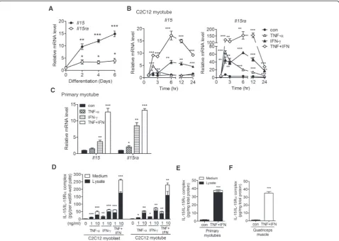
Fig. 1Skeletal muscle cells express IL-15/IL-15Rαprotein complex in response to TNF-αand IFN-γstimulation.aExpression ofIl15andIl15ra
mRNA during C2C12 myoblast differentiation. Samples were collected before (0) and 2, 4, and 6 days after differentiation induction.bExpression
ofIl15andIl15ramRNA in C2C12 myotubes treated with TNF-α(10 ng/ml), IFN-γ(10 ng/ml), TNF-α+ IFN-γ(TNF + IFN, 10 ng/ml each), or without
Co-culture of C2C12 cells with CD8+T cells
The H-2Kb (NM_001001892) coding sequence (nt. 77-1186) was cloned from the cDNA library of primary myotubes of C57BL/6J mice and then inserted into the EcoRI cloning site of lentiviral package plasmid pLKO AS3.1.EGFP3′. The full-length ovalbumin (OVA) cDNA with restriction enzyme cutting sites, 5′-NheI and 3′ -EcoRI, was amplified by PCR from pcDNA3/OVA plasmid (kindly provided by Dr. Mi-Hua Tao, Academia Sinica) and inserted into lentiviral package plasmid pLAS2-w.Ppuro. All lentiviral packaging plasmids and protocols were from the RNAi Core Facility, Academia Sinica. The EGFP-positive C2C12 myoblasts were sorted and subse-quently infected with lentivirus-carrying OVA expression cassette and selected in growth medium containing 2μg/ ml puromycin (Sigma). The expression of H-2Kband OVA was confirmed by flow cytometry and quantitative real-time PCR (qPCR), respectively. For CD8+ T cells, spleno-cytes of OT-1 mice were stimulated with OVA peptide as previously described [37] then cultured in medium contain-ing IL-15 (30 ng/ml, eBioscience). After culturcontain-ing for 8 days, more than 90 % of cells were CD8+CD44hiCD122hi, and these cells were used for experiments. C2C12 cells were treated with TNF-α and IFN-γ (1 ng/ml each) for 24 h. After washing with PBS, 24-h IL-15-deprived CD8+ T cells and 5 μg/ml brefeldin A (Sigma) were added sim-ultaneously to C2C12 cells, followed by brief centrifu-gation to make the cell-cell contact. CD8+ T cells were harvested for intracellular cytokine analysis after co-culturing for 8 h.
Flow cytometry analysis
Lymphocyte surface markers were stained with antibodies against CD19 (6D5), H57 (H57-597), CD8 (53-6.7), CD44 (IM7), CD122 (TM-b1), and NK1.1 (PK136) (eBioscience and BioLegend). C2C12 cells were stained with biotin-conjugated antibodies against IL-15 (Cat. No. 500-P173Bt, PeproTech), IL-15Rα (Cat. No. BAF551, R&D), IL-15Rβ (CD122, clone: TM-b1, eBioscience), andγc(CD132, Cat.
No. 554470, BD Biosciences) then incubated with APC-conjugated streptavidin (BD Biosciences). CD8+ T cells were stained with fixable viable dye eFluor506 (eBioscience) then intracellularly stained with anti-bodies against IFN-γ (XMG1.2, eBioscience) and gran-zyme B (NGZB, eBioscience). All data were acquired on LSRII (BD Biosciences) and analyzed by the FlowJo (Tree Star).
Induction of experimental autoimmune myositis
Preparation of mouse fast-type skeletal muscle myosin-binding protein C (C protein) fragment and induction of myositis were done as previously described with little modification [38]. C protein fragment purified from
Escherichia colilysates was washed with 60 % isopropanol
solution to remove endotoxin as previously described [39]. Female mice, 8–10 weeks old, were intradermally im-munized with 200 μg C protein emulsified in 200 μl complete Freund’s adjuvant (Sigma-Aldrich) at foot-pads, back, and tail base. Simultaneously, 0.5μg pertus-sis toxin (Calbiochem) was intraperitoneally injected. The quadriceps muscle was harvested for histology and gene expression analysis 14 days after immunization. Each muscle block was cut into four sections with in-tervals of at least 200 μm. Histopathologic scoring was based on the most severe inflammation observed in the section among four sections and graded as previously described [38].
Statistics
Results were represented as mean ± SEM. Statistical signifi-cance was determined by unpaired, two-tailed, Student’st -test using GraphPad Prism 5 (GraphPad, San Diego, CA).
pvalues less than 0.05 were considered significant.
Results
Skeletal muscle cells express IL-15/IL-15Rαcomplex
protein in response to TNF-αand IFN-γstimulation
Previous studies found up-regulation ofIl15RNA during myoblast differentiation [11] and pro-inflammatory cyto-kine stimulation [27]. Given the presence of post-transcriptional regulation of IL-15 expression and that all circulating IL-15 are in complex with IL-15Rα[9], we examined the expression of IL-15/IL-15Rαcomplex pro-tein by skeletal muscle cell under the two conditions mentioned above. During a 6-day C2C12 myoblast-to-myotube differentiation, the cells showed a greater than 10-fold and a 3-fold increase of Il15and Il15ramRNA, respectively (Fig. 1a), whereas the cell lysate and culture medium contained no detectable IL-15/IL-15Rα protein by ELISA with a sensitivity of 3.9 pg/ml. This result indi-cates that although the level of Il15 and Il15ra mRNA increased along myoblast-to-myotube differentiation, there was little production of IL-15/IL-15Rαprotein. We next examined whether pro-inflammatory cytokines in-duce IL-15 and IL-15Rα expression by skeletal muscle
cells. We found that TNF-α and IFN-γ each
up-regulated Il15 and Il15ra mRNA in C2C12 myotubes
expression of IL-15/IL-15Rα protein in the injected muscle (Fig. 1f ).
IFN-α, another cytokine up-regulated in myositis muscle [26], induced Il15and Il15ra mRNA and IL-15/ IL-15Rαcomplex protein to the level similar to those in-duced by IFN-γor TNF-α(Additional file 2: Figure S1). Other pro-inflammatory factors, such as IL-1α, IL-1β, and LPS, only transiently up-regulated Il15ra or Il15
mRNA but showed small effect on the induction of the IL-15/IL-15Rαprotein (Additional file 2: Figure S1). To-gether, these results indicate that the expression of the IL-15/IL-15Rα complex protein by skeletal muscle cells was undetectable under steady state conditions, while in-duced by TNF-α, IFN-γ, or IFN-α that associates with Th1 response in myositis.
IL-15 is present on the surface of skeletal muscle cells
As the majority of the cytokine-induced IL-15/IL-15Rα complex was in the lysates of the myoblast and myotube (Fig. 1d, e), we next examined whether the complex is present on the cell surface. We found that TNF-α and IFN-γ induced expression of IL-15 and IL-15Rα on the surface of C2C12 myoblasts as detected by flow cytome-try (Fig. 2a). For the cytokine-treated C2C12 myotubes, dissociation of IL-15 from IL-15Rα on the cell surface by washing cells with acidic glycine buffer resulted in an 80 % reduction of IL-15/IL-15Rα in the cell lysate (Fig. 2b), indicating that 80 % of IL-15 was presented by IL-15Rα on the cell surface. This reduction was not due to alteration of IL-15Rαby the acid treatment, as the level of IL-15/IL-15Rα resumed following the addition of ex-ogenous IL-15 (Fig. 2b). These results indicated that the majority of the cytokine-induced IL-15 was present on the surface of skeletal muscle cells as IL-15/IL-15Rαcomplex.
Skeletal muscle cells express a scanty amount of
IL-15Rβand only respond to a high concentration of
IL-15 hyperagonist, but not IL-IL-15
Despite of various reported IL-15 functions in the skel-etal muscle [12–16], the IL-15-induced signals in skeletal muscle cells remains unexplored. We first examined the expression of IL-15Rβ and γc on C2C12 myoblasts by
flow cytometry and detected no IL-15Rβexpression but a low level ofγcinduced by TNF-αand IFN-γtreatment
(Fig. 3a). The more sensitive qPCR also detected induc-tion of γc mRNA, but notIl15rbmRNA, in C2C12 and primary myotubes by the cytokines (Fig. 3b). Moreover, the level of Il15rb mRNA in the primary myotube was 16 times lower than that in the C2C12 myotube based on the Ct value of qPCR.
Due to the possibility of a very low level of IL-15Rβ expression, we examined whether IL-15 or an IL-15 hyperagonist induces signal transduction in C2C12 myo-tubes. The latter is a fusion protein of IL-15 and IL-15Rα-sushi domain, which possesses higher binding affinity for IL-15Rβ/γc(Kd= 780 pM) and promotes a stronger prolif-eration of IL-15Rβ/γc-bearing cells (EC50= 25 pM) than
IL-15 (Kd= 13.5 nM, EC50= 3 nM) [40]. We found that
IL-15 did not induce STAT5 phosphorylation at a concen-tration up to 400 ng/ml (26.7 nM) (Fig. 3c). Whereas the IL-15 hyperagonist induced moderate but significant STAT5 phosphorylation at concentrations of 100 ng/ml (3.4 nM) and higher (Fig. 3c), which are much higher than that required for the proliferation of IL-15Rβ/γc-bearing
cells (EC50= 25 pM) and for STAT5 phosphorylation in
pre-activated murine CD8+cells (EC50= 10 pM) [40, 41].
In addition to STAT5 phosphorylation, IL-15 and IL-15 hyperagonist did not induce phosphorylation of STAT3, AKT, and ERK in skeletal muscle cells (data not shown). The IL-15 hyperagonist-induced STAT5 phosphorylation
B
IL-15 IL-15R
% of Max
A
Unstained Control TNF+IFN
C2C12 myoblast C2C12 myotube
Fig. 2IL-15 is presented on the surface of skeletal muscle cells.aExpression of IL-15 and IL-15Rαon C2C12 myoblasts. Cells were treated with TNF-αand IFN-γ(TNF + IFN) or without cytokine (Control) for 24 h and then stained with anti-IL-15 and IL-15Rαantibodies for flow cytometry analysis.
“Unstained”indicates cells without antibody staining. Data are representative of three independent experiments with similar results.bQuantification of
A
IL-15R c
% of Max
C
p-STAT5
t-STAT5
0 50 100 200 400
IL-15 hyperagonist
D
t-STAT5
0 100 200 400 0 100 200 400
anti-IL-15R isotype
1.0 0.9 1.0 1.1 1.0 1.4 1.9 2.2
E
Fold change (p-STAT5/t-STAT5) p-STAT5
B
0 50 100 200 400
IL-15
p-STAT5
t-STAT5
0 100 200 400 0 100 200 400
IL-15 Unstained
Control TNF+IFN
ng/ml: ng/ml:
ng/ml:
C2C12 myoblast
C2C12 myotube C2C12 myotube
C2C12 myotube
0 ng/ml 50 ng/ml 100 ng/ml
IL-15
IL-15 hyper -agonist
F
C2C12 myotube 1.0 1.1 1.1 1.0 1.1 1.0 1.1 1.2Fold change (p-STAT5/t-STAT5)
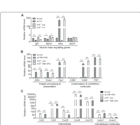
Fig. 4IL-15 deficiency does not affect the responses of primary myotube to TNF-αand IFN-γstimulation.a–cExpression profiling of genes involved in the regulation of skeletal muscle mass and immune system in wt andIl15−/−primary myotubes. Samples were collected 24 h after TNF-αand IFN-γ treatment and analyzed by qPCR. Data are mean ± SEM of triplicates. Data are representative of two independent experiments with similar results. *p< 0.05, **p< 0.01, ***p< 0.001
(See figure on previous page.)
Fig. 3High concentration of IL-15 hyperagonist, but not IL-15, induces STAT5 signaling and atrophy in skeletal muscle cells.aAnalysis of IL-15Rβ andγcexpression on C2C12 myoblasts under the same condition of Fig. 2a. Data are representative of three independent experiments with similar
results.bExpression ofIl15rbandγcmRNA in C2C12 and primary myotube. Samples were collected 24 h after cytokine treatment and analyzed by
qPCR. Data are triplicates and representative of two independent experiments with similar results.cImmunoblotting of STAT5 phosphorylation in C2C12 myotubes after treating with IL-15 or IL-15 hyperagonist for 30 min. Quantification data of four independent experiments are shown below.d
was completely blocked by the IL-15Rβ-blocking anti-body TM-b1 (Fig. 3d). However, neither 15 nor IL-15 hyperagonist induced STAT5 phosphorylation in
C2C12 myotubes pre-treated with TNF-α and IFN-γ
(Fig. 3e), which is in line with the decrease of Il15rb
mRNA after cytokine treatment (Fig. 3b). Consistent with the STAT5 phosphorylation results (Fig. 3c), 100 ng/ml of IL-15 hyperagonist, but not IL-15, induced atrophy of C2C12 myotubes (Fig. 3f ). In summary, C2C12 and primary skeletal muscle cells expressed a scanty IL-15Rβ under steady state condi-tion and TNF-α/IFN-γstimulation and only responded to a high concentration of IL-15 hyperagonist, but not IL-15. Moreover, we found thatIl15−/−mice showed nor-mal skeletal muscle mass (Additional file 3: Figure S2), myoblast differentiation (Additional file 4: Figure S3), cardiotoxin-induced muscle regeneration (Additional file 5: Figure S4), and compensatory hypertrophy of plantaris muscle (Additional file 6: Figure S5). The in vitro and in vivo results collectively suggest that the skeletal muscle cells do not use IL-15.
IL-15 deficiency does not affect the response of primary
myotube to TNF-αand IFN-γstimulation
As TNF-α and IFN-γ induced abundant expression of IL-15/IL-15Rα on skeletal muscle cells, the local con-centration of IL-15/IL-15Rα trans-presentation may be high enough to trigger signaling through the limited number of IL-15Rβ/γc on adjacent muscle cells. We
thus examined whether IL-15 affects the response of the skeletal muscle to TNF-α and IFN-γ by comparing wild type (wt) andIl15−/−primary myotubes. As TNF-α and IFN-γ induce muscle wasting under cancer cach-exia and IL-15 was shown to prevent it [16, 42, 43], we first examined genes that regulate muscle mass. Upon TNF-α and IFN-γ stimulation, wt and Il15−/− primary myotubes showed similar reduction in the
hypertrophy-related genes Igf1 and Myh4 and increase in the
atrophy-related gene iNos (Fig. 4a). We next examined the immune regulatory genes affected by IL-15 in im-mune cells [44–48]. We found that TNF-α and IFN-γ induced the expression of immune regulatory genes in wt and Il15−/− primary myotubes to similar extents, including molecules in the MHC class I antigen presentation pathway and for T-cell co-stimulation and inhibition, chemokines, and cell adhesion molecules (Fig. 4b, c). These results indicate that skeletal muscle IL-15 does not affect the expression of protein homeostasis and immune regulation genes by skeletal muscle cells in response to TNF-α and IFN-γ, which is consistent with the idea that the muscle cells do not use their own IL-15.
Skeletal muscle cells stimulated with TNF-αand IFN-γ
present antigen and provide IL-15 to memory-like CD8+
T cells
IL-15 is a well-known survival and activation factor for memory CD8+T cells. As inflammatory cytokines induce the expression of IL-15/IL-15Rα and antigen presenta-tion molecules by myoblasts and myotubes [49], we de-signed a muscle-cell-T-cell co-culture system to assess whether the muscle cells directly activate CD8+ T cells and the role of IL-15 in this process. We generated a C2C12 myoblast subline that stably expresses H-2Kb -EGFP (C2C12-Kb). As overexpression of H-2Kbimpairs myoblast differentiation in this study and [50], this co-culture system is for myoblast and T cells. We then transduced full-length OVA gene into C2C12-Kb myo-blast as an endogenous antigen and generated C2C12-Kb/OVA myoblast. TNF-α and IFN-γ greatly induced the expression of IL-15 and H-2Kb on C2C12-Kb/OVA cells (Fig. 5a). This high induction of H-2Kbmight partly result from the up-regulation ofβ2-microglobulin (B2m) by the cytokines (Fig. 4b), because B2m is essential for the stabilization of MHC class I molecule in correct con-formation to receive the peptide in the ER and to move from the ER to the cell surface [51]. The cytokine-stimulated C2C12-Kb/OVA, but not C2C12-Kb, cells in-duced production of granzyme B (grB) and IFN-γ by memory-like OT-1 cells (Fig. 5b). We then found that an IL-15Rβ-blocking antibody, but not IL-2-neutralizing antibody, suppressed grB and IFN-γ production by the memory-like OT-1 cells (Fig. 5c). As the cytokine-stimulated C2C12-Kb/OVA cells were washed before co-culturing with OT-1 cells in the presence of the exocytosis inhibitor brefeldin A, IL-15 was presumably only present on the muscle cell surface. These results indicate that myoblasts stimulated with TNF-αand IFN-γpresent anti-gen and IL-15 to memory-like CD8+ T cells to promote their effector function. Given that both myoblasts and myotubes function as antigen-presenting cells under in-flammation, what we observed in the myoblast-CD8+ T-cell co-culture is likely applicable to myotubes.
Skeletal muscle IL-15 promotes the progression of auto-immune myositis
TNF-αand IFN-γare commonly expressed in the skeletal muscle of patients suffering from inflammatory myop-athies, in which CD8+ T cells infiltrate and play a critical role in disease progression [23, 25, 52]. The enhancement of memory-like CD8+T-cell effector function by myoblast IL-15 in vitro prompted us to examine the role of skeletal muscle IL-15 in autoimmune myositis in vivo. We first generated skeletal-muscle-specificIl15−/−mice by crossing
IL-15Rα complex and NK and memory CD8+ T cells in the peripheral blood (Fig. 6b, c).
As two previously reported autoimmune myositis models, wt mice immunized with C protein [38, 53] and
Syt7−/− mice [54, 55], did not develop myositis in our hand, we immunized mice ofSyt7−/−background with C protein because impaired muscle membrane sealing due to Syt7 deficiency facilitates myositis induction [54, 56]. Similar to previously reported pathology in auto-immune myositis, we found that C-protein-immunized
Syt7−/−Il15f/f mice developed mononuclear cell infiltra-tion (Fig. 6d). The mononuclear cells predominantly infil-trated into the endomysium as well as the perimysium and perivascular region (Fig. 6d (left)). The immunized
Syt7−/−Il15f/fmice also developed focal lymphatic invasion of muscle fibers that features CD8+T-cell-mediated myo-sitis (Fig. 6d (middle)). Whereas C-protein-immunized
Syt7−/−ACTA-Il15−/−mice showed a significantly reduced mononuclear cell infiltration and histopathology score (Fig. 6d). We also found elevation of Cd4, Cd8α, and
F4/80 mRNA in the skeletal muscle of the immunized
Syt7−/−Il15f/f mice (Fig. 6e), suggesting infiltration of
CD4+ and CD8+ T cells and macrophages. The
expression of MHC class I subunit B2m;
pro-inflammatory cytokines Tnfa, Ifng, and Il1β: and ef-fector molecule Prf1 were also induced in the skeletal muscle of the immunized Syt7−/−Il15f/f mice (Fig. 6f ). Whereas all these molecules examined were not induced in the C-protein-immunized Syt7−/−ACTA-Il15−/− mice (Fig. 6e, f ). These results together demonstrate that gen-etic ablation of skeletal muscle IL-15 greatly reduced the pathogenesis of autoimmune myositis.
Discussion
In this study, we examined the expression and function of the skeletal muscle cell 15. We found that the IL-15/IL-15Rαprotein was not produced by skeletal muscle cells under steady state conditions but highly induced by TNF-αand IFN-γand presented on the cell surface. Ra-ther than being used by skeletal muscle cells, the IL-15 directly promoted the effector function of memory-like CD8+T cells in vitro and exacerbated the progression of autoimmune myositis in vivo. These results suggest that
the endogenous IL-15 of the skeletal muscle cell func-tions as an immune regulator in an inflammatory skel-etal muscle microenvironment.
IL-15 has been reported to affect skeletal muscle physiology, but IL-15 signaling in skeletal muscle cells remains unclear. Our findings shed some light on this. First, C2C12 and primary muscle cells expressed very low levels of Il15rb mRNA and undetectable levels of cell surface IL-15Rβ(Fig. 3a, b). Second, a soluble IL-15 hyperagonist, but not IL-15, induced STAT5 phosphoryl-ation in C2C12 myotube at 100 ng/ml and above (Fig. 3c). These concentrations are much higher than that required for binding to IL-15Rβ/γcand the pg/ml level of
circulating the IL-15/IL-15Rαcomplex [9], which suggests that the myotubes do not use soluble IL-15 or IL-15/IL-15Rαcomplex under steady state conditions. Consistently, our in vivo studies showed that IL-15 deficiency did not affect skeletal muscle mass, cardiotoxin-induced muscle regeneration, and compensatory hypertrophy of plantaris muscle (Additional file 3, 5, and 6: Figure S2, S4, and S5). Although trans-presentation of cell-bound IL-15/IL-15Rα among the stimulated muscle cells may reach a high enough local concentration to trigger signaling through the sparsely expressed IL-15Rβ/γc,Il15 knockout did not affect the response of the primary myotube to TNF-αand IFN-γ in vitro (Fig. 4). Collectively, our results support that the skeletal muscle cells do not use IL-15 for skeletal muscle growth, regeneration, or inflammatory responses.
Our in vitro results appear different from previous in vitro studies showing that exogenous IL-15 promotes skeletal muscle hypertrophy [12, 14]. The difference may partly result from the intrinsic differences between the C2C12 and C2 cell lines used in this and previous stud-ies, respectively. C2C12 cells have a higher differenti-ation potency and insulin-like growth factor 1 (IGF-I) level than C2 cells [57]. IGF-I is a strong stimulator for muscle hypertrophy [58]. An earlier study indicates that IL-15-induced C2 differentiation was only revealed in the absence of IGF-I signaling [13]. Therefore, it is pos-sible that the higher level of IGF-I masked IL-15-induced hypertrophy in C2C12 cells under steady state condition. However, IL-15 hyperagonist did not induce STAT5 phosphorylation in TNF-α and IFN-γ-pretreated
(See figure on previous page.)
Fig. 5Skeletal muscle cells presented antigen and provided IL-15 to promote the effector function of CD8+T cells under TNF-αand IFN-γtreatment.
myotubes (Fig. 3e), in which 90 % of endogenous IGF-I was downregulated [59]. Therefore, the scanty amount of IL-15Rβ likely limits the use of IL-15 by the muscle cells.
Although various IL-15 functions in the skeletal muscle were reported previously, some controversial re-sults exist between those in vitro and in vivo studies. In contrast to muscle hypertrophy induced by IL-15 in vitro, muscle atrophy was observed in mice carrying skeletal-muscle-specific IL-15 transgene [18] or receiving systemic infusion of exogenous IL-15 [19]. The muscle atrophy may be contributed by fatal leukemia [60] and metabolic dysregulation [61] induced by overexpression of IL-15 . In addition, recent studies show that exercise endurance increases in skeletal-muscle-specificIl15 -trans-genic mice and reduces in Il15−/− mice [18, 20], despite that their earlier ex vivo study found no difference in the fatigue index between the EDL or soleus muscle isolated fromIl15−/−mice and skeletal-muscle-specificIl15 -trans-genic mice [21]. Collectively, the inconsistency between the in vitro/ex vivo and in vivo results suggests that the change of muscle mass or exercise endurance in vivo is not caused by the direct effect of IL-15 on the skeletal muscle.
IL-15 has been reported to be involved in a number of autoimmune diseases, including rheumatoid arthritis, in-flammatory bowel disease, and multiple sclerosis, in which IL-15 promotes the effector function of cytotoxic CD8+T cells to destroy the target tissues [62]. Consider-ing that TNF-αand IFN-γinduced the expression of IL-15/IL-15Rα and T-cell interacting molecules in skeletal muscle cells, the stimulated muscle cells may directly communicate with T cells. Indeed, we clearly demon-strated in vitro that the stimulated skeletal muscle cells presented antigen and IL-15 to memory-like CD8+T cells and enhanced their effector function. Following induction of autoimmune myositis in vivo, skeletal-muscle-specific
Il15−/− mice showed reduced mononuclear cell infiltra-tion, histopathology score, and expression of inflammatory molecules. Our data together suggest a scenario in which
skeletal muscle IL-15 promotes the production of cyto-toxic molecules, such as granzyme, by memory CD8+ T cells to necrotize muscle cells, which triggers phagocyte recruitment and inflammation. The locally enriched cyto-kines, TNF-αand IFN-γ(Fig. 6f), may stimulate IL-15 ex-pression by muscle cells to form a feed-forward loop to perpetuate the inflammatory milieu, which contributes to the progression of autoimmune myositis.
Conclusions
We provide new insights into the function of skeletal muscle IL-15. Rather than being used by the muscle cell itself, the skeletal muscle IL-15 directly promotes the ef-fector function of memory-like CD8+ T cells, which fa-cilitates the formation of a pro-inflammatory skeletal muscle microenvironment during myositis progression. Given that IL-15 is not required for muscle growth and regeneration, IL-15 has the potential to be a suitable therapeutic target for autoimmune myositis.
Additional files
Additional file 1: Table S1.Primer pairs used in this study.
Additional file 2: Figure S1.IL-1α/βor LPS induced moderate expression of IL-15/IL-15Rαprotein complex in C2C12 myotubes. (A–C) C2C12 myotubes were treated with IFN-α(0 or 10 ng/ml), IL-1α/β(0 or 10 ng/ml), or LPS (0, 0.1, or 1μg/ml) and examined for the expression of
Il15andIl15ramRNA at indicated time points. Data represent mean ±
SEM of triplicates. (D–F) The level of IL-15/IL-15Rαcomplex protein in cell lysate and culture medium after 24-h treatment with IL-1α/β, LPS, or IL-1α were measured by ELISA. Data were pooled from two and three independent experiments. Data are mean ± SEM. *p< 0.05, **p< 0.01, ***p< 0.001, in comparison to“0”or“con”.
Additional file 3: Figure S2.Phenotype analysis of 12-week-old male wt,Il15−/−, andIl15ra−/−mice. (A) Body weight. (B) Absolute weight and the percentage of body weight (% BW) of heart and gonadal fat pad. (C) Absolute weight and the value normalized to tibia bone length (mg/ mm) of gastrocnemius muscle (Gas), tibialis anterior muscle (TA), soleus muscle (Sol), and EDL. One symbol represents one mouse. wt (n =14);
Il15−/−(n =12);Il15ra−/−(n =13).
Additional file 4: Figure S3.Comparative analysis of wt andIl15−/−
primary myoblast differentiation. (A) Morphology of wt andIl15−/−
primary myotubes after differentiation for 2 days. The images are representative of at least three independent experiments. (B) Fusion (See figure on previous page.)
Fig. 6Skeletal muscle IL-15 contributes to the progression of autoimmune myositis.aThe expression ofIl15mRNA in various tissues of wt (n= 3),
Il15f/f(n= 5), andACTA-Il15−/−(n= 5) mice was detected by qPCR.WATwhite adipose tissue. Data are mean ± SEM. ***p< 0.001.bThe amount of IL-15/IL-15Rαcomplex protein in the serum ofIl15f/fandACTA-Il15−/−mice (n= 9–10) was measured by ELISA. Data are mean ± SEM.cComparison of memory CD8+T cell (mCD8) and NK cell (NK) level in the peripheral blood among wt,Il15−/−,Il15f/f, andACTA-Il15−/−mice (n= 5 in each group). Fold change was calculated by normalizing the percentage of indicated cell type in mutant mice to that in wt mice. mCD8 were H57+CD19−CD8
+
CD44hiCD122hi, and NK were H57−CD19−NK1.1+. Data are mean ± SEM. ***p< 0.001.dMononuclear cell infiltration in the quadriceps muscles ofSyt7 −/−Il15f/f
andSyt7−/−ACTA-Il15−/−mice 14 days after C protein immunization (Left). Mononuclear cell infiltration was found in the endomysium (black
square), perimysium (black arrowhead), and perivascular region (white arrowhead). Focal lymphatic invasion of myofibers were observed in
C-protein-immunizedSyt7−/−ACTA-Il15−/−mice as indicated by thewhite arrowhead(Middle). The histopathology scores were compiled in theright panelwith each symbol representing one mouse (Right). Scale bar in left = 100μm; middle = 25μm. **p< 0.01.eExpression of mononuclear cell markers,Cd4,
Cd8, andF4/80, mRNA in the quadriceps muscles of C-protein-immunized mice measured by qPCR. Eachsymbolrepresenting one mouse. *p< 0.05, **p< 0.01.fExpression of immune-relevant genes mRNA in the quadriceps muscles of C-protein-immunized mice measured by qPCR. Eachsymbol
index and differentiation index of wt andIl15−/−primary myoblast. Fusion index is the percentage of nuclei in MyHC-positive cells among total nuclei. Differentiation index is the percentage of myotubes with indicated number of nuclei among total myotubes. Each index was calculated from at least 24 microscopic fields with each field containing more than 10 myotubes. Data are mean ± SEM. (C) Expression of differentiation-related genes in primary muscle cells. Samples were collected after culturing in growth medium for 24 h (GM) and after switching into differentiation medium for 1 and 2 days (day 1 and 2). Data are mean ± SEM. Data are pooled from three independent experiments.
Additional file 5: Figure S4.Cardiotoxin-induced muscle regeneration in the TA muscle of wt andIl15−/−mice. TA muscle was injected intramuscularly with cardiotoxin (50μl, 10μM, Sigma), dissected out, and fixed in formalin at days 5, 10, and 21 after injection. Fixed muscles were embedded in paraffin for histological examination. (A) Representative images of TA muscle histology after cardiotoxin injection for 5 (n =4 each genotype), 10 (n= 4 each genotype), and 21 (wtn= 2;Il15−/−n= 3) days. Muscle fibrosis was evaluated by Masson’s trichrome staining in the 21-day cardiotoxin-injected samples. (B) Expression profiling of regeneration-related genes in the TA muscle injected with cardiotoxin for 5 and 10 days using qPCR. Each group contains four mice. Scale bar = 50μm. Data are mean ± SEM. *p< 0.05, ***p< 0.001.
Additional file 6: Figure S5.Compensatory hypertrophy of plantaris muscle. Lower leg soleus and gastrocnemius muscles were removed without damaging the neurovascular supply. Fourteen days after surgery, plantaris muscles were dissected out and weighted. No body weight change was observed during the experiment. The weight of plantaris muscle of mice received surgery or sham surgery were normalized to body weight. One symbol represents one mouse. ***p< 0.001.
Abbreviations
ACTA:human alpha-skeletal actin; EDL: extensor digitorum longus muscle; Gas: gastrocnemius muscle; grB: granzyme B; 15: interleukin 15; IL-15Rα: interleukin 15 receptor alpha; MyHC: myosin heavy chain; Syt7: synaptotagmin VII; TA: tibialis anterior muscle; wt: wild type;
γc: common gamma chain.
Competing interests
The authors declare that they have no competing interests.
Authors’contributions
PLH designed the study, performed the experiments, analyzed the data, and drafted the manuscript. MSH participated in experimental design and provided technical advice. SWW generated the genetically modified mice. CLC and YHL provided technical advice and contributed to acquisition and analysis of the data. MSH, SWW, CLC, and YHL had been involved in revising the manuscript for important intellectual content. NSL substantially contributed to the study design, interpretation of the data, and revising the manuscript. All authors read and approved the final manuscript.
Acknowledgements
We thank the Taiwan Mouse Clinic (MOST 103-2325-B-001-015), funded by the National Research Program for Biopharmaceuticals at the Ministry of Science and Technology (MOST) of Taiwan, for technical support in histology experiments. We also acknowledge the assistance of FACS Facility, Transgenic Core Facility, Genomics Core Facility, Imaging Core Facility, and Scientific English Editing Core Facility at the Institute of Molecular Biology. This study was supported by MOST (NSC 98-2320-B-001-004-MY3) and Academia Sinica, Taiwan.
Received: 29 May 2015 Accepted: 8 September 2015
References
1. Budagian V, Bulanova E, Paus R, Bulfone-Paus S. IL-15/IL-15 receptor biology: a guided tour through an expanding universe. Cytokine Growth Factor Rev. 2006;17(4):259–80. doi:10.1016/j.cytogfr.2006.05.001.
2. Duitman EH, Orinska Z, Bulanova E, Paus R, Bulfone-Paus S. How a cytokine is chaperoned through the secretory pathway by complexing with its own
receptor: lessons from interleukin-15 (IL-15)/IL-15 receptor alpha. Mol Cell Biol. 2008;28(15):4851–61. doi:10.1128/mcb.02178-07.
3. Mortier E, Woo T, Advincula R, Gozalo S, Ma A. IL-15Ralpha chaperones IL-15 to stable dendritic cell membrane complexes that activate NK cells via trans presentation. J Exp Med. 2008;205(5):1213–25. doi:10.1084/jem.20071913. 4. Stonier SW, Schluns KS. Trans-presentation: a novel mechanism regulating
IL-15 delivery and responses. Immunol Lett. 2010;127(2):85–92. doi:10.1016/ j.imlet.2009.09.009.
5. Waldmann TA. The biology of interleukin-2 and interleukin-15: implications for cancer therapy and vaccine design. Nat Rev Immunol. 2006;6(8):595–601. doi:10.1038/nri1901.
6. Ellery JM, Nicholls PJ. Alternate signalling pathways from the interleukin-2 receptor. Cytokine Growth Factor Rev. 2002;13(1):27–40.
7. Marzec M, Halasa K, Kasprzycka M, Wysocka M, Liu X, Tobias JW, et al. Differential effects of interleukin-2 and interleukin-15 versus interleukin-21 on CD4+ cutaneous T-cell lymphoma cells. Cancer Res. 2008;68(4):1083–91. doi:10.1158/0008-5472.can-07-2403.
8. Fehniger TA, Caligiuri MA. Interleukin 15: biology and relevance to human disease. Blood. 2001;97(1):14–32.
9. Bergamaschi C, Bear J, Rosati M, Beach RK, Alicea C, Sowder R, et al. Circulating IL-15 exists as heterodimeric complex with soluble IL-15Ralpha in human and mouse serum. Blood. 2012;120(1):e1–8. doi:10.1182/blood-2011-10-384362.
10. Grabstein KH, Eisenman J, Shanebeck K, Rauch C, Srinivasan S, Fung V, et al. Cloning of a T cell growth factor that interacts with the beta chain of the interleukin-2 receptor. Science. 1994;264(5161):965–8.
11. Quinn LS, Strait-Bodey L, Anderson BG, Argiles JM, Havel PJ. Interleukin-15 stimulates adiponectin secretion by 3 T3-L1 adipocytes: evidence for a skeletal muscle-to-fat signaling pathway. Cell Biol Int. 2005;29(6):449–57. doi:10.1016/j.cellbi.2005.02.005.
12. Quinn LS, Anderson BG, Drivdahl RH, Alvarez B, Argiles JM. Overexpression of interleukin-15 induces skeletal muscle hypertrophy in vitro: implications for treatment of muscle wasting disorders. Exp Cell Res. 2002;280(1):55–63. 13. Quinn LS, Haugk KL, Damon SE. Interleukin-15 stimulates C2 skeletal
myoblast differentiation. Biochem Biophys Res Commun. 1997;239(1):6–10. doi:10.1006/bbrc.1997.7414.
14. Quinn LS, Haugk KL, Grabstein KH. Interleukin-15: a novel anabolic cytokine for skeletal muscle. Endocrinology. 1995;136(8):3669–72.
15. Harcourt LJ, Holmes AG, Gregorevic P, Schertzer JD, Stupka N, Plant DR, et al. Interleukin-15 administration improves diaphragm muscle pathology and function in dystrophic mdx mice. Am J Pathol. 2005;166(4):1131–41. doi:10.1016/s0002-9440(10)62333-4.
16. Carbo N, Lopez-Soriano J, Costelli P, Busquets S, Alvarez B, Baccino FM, et al. Interleukin-15 antagonizes muscle protein waste in tumour-bearing rats. Br J Cancer. 2000;83(4):526–31. doi:10.1054/bjoc.2000.1299.
17. Quinn LS, Anderson BG, Strait-Bodey L, Stroud AM, Argiles JM. Oversecretion of interleukin-15 from skeletal muscle reduces adiposity. Am J Physiol Endocrinol Metab. 2009;296(1):E191–202. doi:10.1152/ajpendo.90506.2008. 18. Quinn LS, Anderson BG, Conner JD, Wolden-Hanson T. IL-15 overexpression
promotes endurance, oxidative energy metabolism, and muscle PPARdelta, SIRT1, PGC-1alpha, and PGC-1beta expression in male mice. Endocrinology. 2013;154(1):232–45. doi:10.1210/en.2012-1773.
19. Pistilli EE, Alway SE. Systemic elevation of interleukin-15 in vivo promotes apoptosis in skeletal muscles of young adult and aged rats. Biochem Biophys Res Commun. 2008;373(1):20–4. doi:10.1016/ j.bbrc.2008.05.188.
20. Quinn LS, Anderson BG, Conner JD, Wolden-Hanson T, Marcell TJ. IL-15 is required for postexercise induction of the pro-oxidative mediators PPARdelta and SIRT1 in male mice. Endocrinology. 2014;155(1):143–55. doi:10.1210/en.2013-1645.
21. Pistilli EE, Bogdanovich S, Garton F, Yang N, Gulbin JP, Conner JD, et al. Loss of IL-15 receptor alpha alters the endurance, fatigability, and metabolic characteristics of mouse fast skeletal muscles. J Clin Invest.
2011;121(8):3120–32. doi:10.1172/jci44945.
22. Dalakas MC. Immunotherapy of myositis: issues, concerns and future prospects. Nat Rev Rheumatol. 2010;6(3):129–37. doi:10.1038/ nrrheum.2010.2.
23. Dalakas MC. Pathogenesis and therapies of immune-mediated myopathies. Autoimmun Rev. 2012;11(3):203–6. doi:10.1016/j.autrev.2011.05.013. 24. Malmstrom V, Venalis P, Albrecht I. T cells in myositis. Arthritis Res Ther.
25. Zong M, Lundberg IE. Pathogenesis, classification and treatment of inflammatory myopathies. Nat Rev Rheumatol. 2011;7(5):297–306. doi:10.1038/nrrheum.2011.39.
26. De Paepe B, Creus KK, De Bleecker JL. Role of cytokines and chemokines in idiopathic inflammatory myopathies. Curr Opin Rheumatol. 2009;21(6):610–6. doi:10.1097/BOR.0b013e3283317b31.
27. Sugiura T, Harigai M, Kawaguchi Y, Takagi K, Fukasawa C, Ohsako-Higami S, et al. Increased IL-15 production of muscle cells in polymyositis and dermatomyositis. Int Immunol. 2002;14(8):917–24.
28. Sugiura T, Kawaguchi Y, Harigai M, Takagi K, Ohta S, Fukasawa C, et al. Increased CD40 expression on muscle cells of polymyositis and dermatomyositis: role of CD40-CD40 ligand interaction in IL-6, IL-8, IL-15, and monocyte chemoattractant protein-1 production. J Immunol. 2000;164(12):6593–600.
29. Abadie V, Jabri B. IL-15: a central regulator of celiac disease
immunopathology. Immunol Rev. 2014;260(1):221–34. doi:10.1111/imr.12191. 30. McInnes IB, Schett G. Cytokines in the pathogenesis of rheumatoid arthritis.
Nat Rev Immunol. 2007;7(6):429–42. doi:10.1038/nri2094.
31. Waldmann TA, Tagaya Y. The multifaceted regulation of interleukin-15 expression and the role of this cytokine in NK cell differentiation and host response to intracellular pathogens. Annu Rev Immunol. 1999;17:19–49. doi:10.1146/annurev.immunol.17.1.19.
32. Chang CL, Lai YG, Hou MS, Huang PL, Liao NS. IL-15Ralpha of radiation-resistant cells is necessary and sufficient for thymic invariant NKT cell survival and functional maturation. J Immunol. 2011;187(3):1235–42. doi:10.4049/jimmunol.1100270.
33. Liou YH, Wang SW, Chang CL, Huang PL, Hou MS, Lai YG, et al. Adipocyte IL-15 regulates local and systemic NK cell development. J Immunol. 2014;193(4):1747–58. doi:10.4049/jimmunol.1400868.
34. Blanco-Bose WE, Yao CC, Kramer RH, Blau HM. Purification of mouse primary myoblasts based on alpha 7 integrin expression. Exp Cell Res.
2001;265(2):212–20. doi:10.1006/excr.2001.5191.
35. Dubois S, Mariner J, Waldmann TA, Tagaya Y. IL-15Ralpha recycles and presents IL-15 in trans to neighboring cells. Immunity. 2002;17(5):537–47. 36. Spandidos A, Wang X, Wang H, Seed B. PrimerBank: a resource of human
and mouse PCR primer pairs for gene expression detection and quantification. Nucleic Acids Res. 2010;38(Database issue):D792–9. doi:10.1093/nar/gkp1005.
37. Pulle G, Vidric M, Watts TH. IL-15-dependent induction of 4-1BB promotes antigen-independent CD8 memory T cell survival. J Immunol.
2006;17(5):2739–48.
38. Sugihara T, Sekine C, Nakae T, Kohyama K, Harigai M, Iwakura Y, et al. A new murine model to define the critical pathologic and therapeutic mediators of polymyositis. Arthritis Rheum. 2007;56(4):1304–14. doi:10.1002/art.22521. 39. Linares D, Echevarria I, Mana P. Single-step purification and refolding of
recombinant mouse and human myelin oligodendrocyte glycoprotein and induction of EAE in mice. Protein Expr Purif. 2004;34(2):249–56. doi:10.1016/ j.pep.2003.11.016.
40. Mortier E, Quemener A, Vusio P, Lorenzen I, Boublik Y, Grotzinger J, et al. Soluble interleukin-15 receptor alpha (IL-15R alpha)-sushi as a selective and potent agonist of 15 action through 15R beta/gamma. Hyperagonist IL-15 x IL-IL-15R alpha fusion proteins. J Biol Chem. 2006;281(3):1612–9. doi:10.1074/jbc.M508624200.
41. Ring AM, Lin JX, Feng D, Mitra S, Rickert M, Bowman GR, et al. Mechanistic and structural insight into the functional dichotomy between IL-2 and IL-15. Nat Immunol. 2012;13(12):1187–95. doi:10.1038/ni.2449.
42. Acharyya S, Ladner KJ, Nelsen LL, Damrauer J, Reiser PJ, Swoap S, et al. Cancer cachexia is regulated by selective targeting of skeletal muscle gene products. J Clin Invest. 2004;114(3):370–8. doi:10.1172/jci20174.
43. Guttridge DC, Mayo MW, Madrid LV, Wang CY, Baldwin Jr AS. NF-kappaB-induced loss of MyoD messenger RNA: possible role in muscle decay and cachexia. Science. 2000;289(5488):2363–6.
44. Agostini C, Zambello R, Facco M, Perin A, Piazza F, Siviero M, et al. CD8 T-cell infiltration in extravascular tissues of patients with human
immunodeficiency virus infection. Interleukin-15 upmodulates costimulatory pathways involved in the antigen-presenting cells-T-cell interaction. Blood. 1999;93(4):1277–86.
45. Gil M, Park SJ, Chung YS, Park CS. Interleukin-15 enhances proliferation and chemokine secretion of human follicular dendritic cells. Immunology. 2010;130(4):536–44. doi:10.1111/j.1365-2567.2010.03252.x.
46. Perera LP, Goldman CK, Waldmann TA. IL-15 induces the expression of chemokines and their receptors in T lymphocytes. J Immunol. 1999;162(5):2606–12.
47. Tourkova IL, Shurin GV, Chatta GS, Perez L, Finke J, Whiteside TL, et al. Restoration by IL-15 of MHC class I antigen-processing machinery in human dendritic cells inhibited by tumor-derived gangliosides. J Immunol. 2005;175(5):3045–52.
48. Tourkova IL, Yurkovetsky ZR, Gambotto A, Makarenkova VP, Perez L, Balkir L, et al. Increased function and survival of IL-15-transduced human dendritic cells are mediated by up-regulation of IL-15Ralpha and Bcl-2. J Leukoc Biol. 2002;72(5):1037–45.
49. Marino M, Scuderi F, Provenzano C, Bartoccioni E. Skeletal muscle cells: from local inflammatory response to active immunity. Gene Ther. 2011;18(2):109–16. doi:10.1038/gt.2010.124.
50. Pavlath GK. Regulation of class I MHC expression in skeletal muscle: deleterious effect of aberrant expression on myogenesis. J Neuroimmunol. 2002;125(1–2):42–50.
51. Williams DB, Barber BH, Flavell RA, Allen H. Role of beta 2-microglobulin in the intracellular transport and surface expression of murine class I histocompatibility molecules. J Immunol. 1989;142(8):2796–806.
52. Figarella-Branger D, Civatte M, Bartoli C, Pellissier JF. Cytokines, chemokines, and cell adhesion molecules in inflammatory myopathies. Muscle Nerve. 2003;28(6):659–82. doi:10.1002/mus.10462.
53. Sugihara T, Okiyama N, Suzuki M, Kohyama K, Matsumoto Y, Miyasaka N, et al. Definitive engagement of cytotoxic CD8 T cells in C protein-induced myositis, a murine model of polymyositis. Arthritis Rheum.
2010;62(10):3088–92. doi:10.1002/art.27625.
54. Chakrabarti S, Kobayashi KS, Flavell RA, Marks CB, Miyake K, Liston DR, et al. Impaired membrane resealing and autoimmune myositis in synaptotagmin VII-deficient mice. J Cell Biol. 2003;162(4):543–9. doi:10.1083/jcb.200305131. 55. Young NA, Sharma R, Friedman AK, Kaffenberger BH, Bolon B, Jarjour WN.
Aberrant muscle antigen exposure in mice is sufficient to cause myositis in a Treg cell-deficient milieu. Arthritis Rheum. 2013;65(12):3259–70. doi:10.1002/art.38184.
56. Han R. Muscle membrane repair and inflammatory attack in dysferlinopathy. Skeletal Muscle. 2011;1(1):10. doi:10.1186/2044-5040-1-10.
57. Sharples AP, Al-Shanti N, Stewart CE. C2 and C2C12 murine skeletal myoblast models of atrophic and hypertrophic potential: relevance to disease and ageing? J Cell Physiol. 2010;225(1):240–50. doi:10.1002/ jcp.22252.
58. Rommel C, Bodine SC, Clarke BA, Rossman R, Nunez L, Stitt TN, et al. Mediation of IGF-1-induced skeletal myotube hypertrophy by PI(3)K/Akt/ mTOR and PI(3)K/Akt/GSK3 pathways. Nat Cell Biol. 2001;3(11):1009–13. doi:10.1038/ncb1101-1009.
59. Fernandez-Celemin L, Pasko N, Blomart V, Thissen JP. Inhibition of muscle insulin-like growth factor I expression by tumor necrosis factor-alpha. Am J Physiol Endocrinol Metab. 2002;283(6):E1279–90. doi:10.1152/
ajpendo.00054.2002.
60. Fehniger TA, Suzuki K, Ponnappan A, VanDeusen JB, Cooper MA, Florea SM, et al. Fatal leukemia in interleukin 15 transgenic mice follows early expansions in natural killer and memory phenotype CD8+T cells. J Exp
Med. 2001;193(2):219–31.
61. Barra NG, Chew MV, Reid S, Ashkar AA. Interleukin-15 treatment induces weight loss independent of lymphocytes. PLoS One. 2012;7(6):e39553. doi:10.1371/journal.pone.0039553.