R E S E A R C H A R T I C L E
Open Access
Turning T cells on: epigenetically enhanced
expression of effector T-cell costimulatory
molecules on irradiated human tumor cells
Anita Kumari, Ercan Cacan, Susanna F Greer and Charlie Garnett-Benson
*Abstract
Background:Sub-lethal doses of radiation can alter the phenotype of target tissue by modulating gene expression and making tumor cells more susceptible to T-cell-mediated immune attack. We have previously shown that sub-lethal tumor cell irradiation enhances killing of colorectal carcinoma cells by tumor-specific cytotoxic T cells by unknown mechanisms. Recent data from our lab indicates that irradiation of tumor cells results in the upregulation of OX40L and 41BBL, and that T cells incubated with irradiated tumor cells displayed improved CTL survival, activation and effector activity. The objective of this current study was to determine the mechanism of enhanced OX40L and 41BBL expression in human colorectal tumor cells.
Methods:Two colorectal carcinoma cell lines, HCT116 and SW620, were examined for changes in the expression of 41BBL and OX40L in response to inhibition of histone deacetylases (using TSA) and DNA methyltransferases (using 5-Aza-2′-deoxycytidine) to evaluate if epigenetic mechanisms of gene expression can modulate these genes. Tumor cells were treated with radiation, TSA, or 5-Aza-dC, and subsequently evaluated for changes in gene expression using RT-qPCR and flow cytometry. Moreover, we assessed levels of histone acetylation at the 41BBL promoter using chromatin immunoprecipitation assays in irradiated HCT116 cells.
Results:Our data indicate that expression of 41BBL and OX40L can indeed be epigenetically regulated, as
inhibition of histone deacetylases and of DNA methyltransferases results in increased OX40L and 41BBL mRNA and protein expression. Treatment of tumor cells with TSA enhanced the expression of these genes more than
treatment with 5-Aza-dC, and co-incubation of T cells with TSA-treated tumor cells enhanced T-cell survival and activation, similar to radiation. Furthermore, chromatin immunoprecipitation experiments revealed significantly increased histone H3 acetylation of 41BBL promoters specifically following irradiation.
Conclusions:Full understanding of specific mechanisms of immunogenic modulation (altered expression of immune relevant genes) of irradiated tumor cells will be required to determine how to best utilize radiation as a tool to enhance cancer immunotherapy approaches. Overall, our results suggest that radiation can be used to make human tumors more immunogenic through epigenetic modulation of genes stimulatory to effector T-cells.
Keywords:External beam radiation, Immunogenic modulation, CTLs, Epigenetic, Effector co-stimulation
* Correspondence:cgarnettbenson@gsu.edu
Department of Biology, Center for Inflammation, Infection and Immunity, Georgia State University, 161 Jesse Hill Jr. Dr, Atlanta, GA, USA
Background
Previous reports by us and others demonstrate that sub-lethal doses of radiation alter the expression of genes within tumor cells [1-3]. Furthermore, it has been dir-ectly demonstrated that tumor irradiation, as well as treatment with some chemotherapy drugs, results in in-creased susceptibility to killing of tumor cells by cyto-toxic T cells (CTLs) [1,4,5]. Notably, many genes that are important for T-cell anti-tumor effector activity are up-regulated following treatment with sub-lethal doses of radiation [2,4,6]. However, the mechanisms of radiation-mediated changes in the expression of such immune stimulatory genes are poorly understood.
It is clear that human cells respond to DNA-damage from ionizing radiation (IR) by inducing the expression of a number of genes at the transcriptional level [4,7,8]. Induction of altered gene expression can be due to direct cellular radiation effects or to radiation-induced changes in cellular milieu. Direct cellular effects appear to be regulated through parallel signaling pathways that ori-ginate from the nucleus following DNA damage, as well as signaling pathways that originate in the cytoplasm via reactive oxygen species production [7,9]. These pathways induce NF-kB activation and nuclear translocation [10,11]. As would be expected, DNA damage by IR can induce cellular stress responses, which result in activa-tion of DNA damage repair pathways and apoptotic pathways [6,12]. Interestingly, regulation of the expres-sion of a variety of genes, not related to known or typical DNA repair or apoptotic pathways, also occurs [2,13,14]. Indeed, we previously examined 23 human carcinoma cell lines for their phenotypic response to sub-lethal doses of IR [4], and found that RT increased the expres-sion of several genes commonly down-regulated by tu-mors to escape immune recognition and elimination [15-20], including Fas (CD95), Intercellular adhesion molecule-1 (ICAM-1/CD54), tumor associated antigens (TAA) and major histocompatibility (MHC)-Class I. Most recently we found that radiation enhances the ex-pression of OX40 ligand (OX40L/TNFSF4/CD134L/ CD252) and 41BB ligand (41BBL/TNFSF9/CD137L), im-portant co-stimulators of effector CTLs on tumor cells (submitted manuscript).
To elicit an effective immune response against tumors, T cells need to recognize tumor antigens presented by MHC in conjunction with appropriate co-stimulation [21,22]. In the absence of proper co-stimulation, these anti-tumor T cells become anergic. Proteins such as 41BBL and OX40L represent important co-stimulators of effector CTL activity [23-26], and we have seen sub-lethal doses of radiation increase their expression in human tumor cells; however, the mechanisms regulating radiation-enhanced modulation of the expression of these two genes remain unclear. OX40 (TNFRSF4/
CD134) was originally characterized as a transiently expressed co-stimulatory molecule regulating CD4 and CD8 immunity [27], and signaling through OX40 promotes T-cell survival and expansion [28]. 41BBL co-stimulation of 41BB (TNFRSF9/CD137) on tumor-specific T cells is important for T-cell proliferation [29,30], cytokine production, and activation [31]. En-gagement of OX40 and 41BB by agonist antibodies increases immunity against tumors, resulting in long-term survival [32] in a number of murine tumor models [33,34]. Recent evidence indicates that expression of 41BBL is transcriptionally activated by HDAC inhibitors in leukemia cell lines [35], and that HDAC11 plays an essential role in regulating OX40L expression [36]. Inter-estingly, radiation has been shown to inhibit the expres-sion of HDAC1 and HDAC2 [37], and we have seen enhanced cytolysis by T-cells following tumor irradi-ation. Thus, epigenetic mechanisms may be at work dur-ing radiation-enhanced susceptibility to T-cell killdur-ing.
Epigenetic changes such as histone modifications and DNA methylation play important roles in regulating gene expression. DNA methyltransferase enzyme (DNM T1) adds methyl group to cytosine residues [38]. DNA hypermethylation of CpG dinucleotides accumulates in promoter regions of genes and contributes to their loss through epigenetic silencing. Promoter hypermethylation and genome-wide hypomethylation alters genes expres-sion in colorectal cancer [39]. It has been found that genes having hypermethylation also exhibit altered acetylation and methylation of histones [40]. Histone acetylation via histone acetyltransferases (HATs) is an-other major epigenetic mechanism controlling gene ex-pression [41-43]. Gains in histone acetylation neutralize the positive charge on lysine residues and contribute to disrupted nucleosome structure, allowing unfolding of DNA, increased transcription factor access and en-hanced gene expression [44-46]. HDACs remove acetyl groups from histones and return DNA to a less access-ible conformation, thereby decreasing transcription [47-49]. Alterations in HAT and HDAC activity have been identified in many human cancers [50,51]. HDAC inhibitors (HDACi) therefore promote hyperacetylation of histones, which in turn leads to chromatin relaxation and selective expression of genes.
study to report thata) OX40L and 41BBL expression in-creases in CRC cells when DNMTs are inhibited, b) ex-pression of OX40L and 41BBL increases in human CRC cells when HDACs are inhibited,c) HDAC inhibition in CRC cells can increase the activation and survival of T cells, andd) radiation treatment of tumor cells results in epigenetic modification of the histones in the promoter of the costimulatory gene 41BBL. The use of ionizing ra-diation to specifically enhance cancer immunotherapy (CIT) strategies through epigenetic modulation of genes stimulatory to CTLs will have a broad impact on cancer therapy approaches and will extend the use of radiation into new directions.
Methods
Cell lines
Human colorectal carcinoma cell lines HCT116 cells were obtained from the laboratory of tumor immun-ology and biimmun-ology, NCI, NIH. The cell line SW620 was kindly provided by Zhi-Ren Liu [52] from Georgia State University, Department of Biology. All cells were cul-tured as recommended by ATCC and tested periodically to ensure absence of Mycoplasma. Cells were incubated at 37°C incubator with 5% CO2.
Reagents
5-Aza-2′-deoxycytidine (5-Aza-dC) and Trichostatin A (TSA) were purchased from Sigma-Aldrich (St. Louis, MO). Antibodies recognizing histone H3 and acetylated histone H3 were from Millipore (Lake Placid, NY). Cell viability following treatment was determined using Try-pan blue dye exclusion on a Countess automated cell counter (Life Technologies).
Irradiation
A RS-2000 biological X-ray irradiator (Rad source tech-nology, Suwanee, GA) was used to irradiate tumor cells. Cells were irradiated at a dose rate of 2Gy/min by set-ting irradiator voltage and current at 160 kV and 25 mA, respectively. Cells were maintained in suspension and kept on ice during irradiation. Immediately after irradi-ation, the culture media was replaced with the fresh media.
Quantitative real time PCR
Cells were plated and treated with 5AZA-dC (20 uM), TSA or 10Gy radiation. Untreated control cells were cul-tured with the equivalent amount of DMSO present in drug treated samples. Adherent and viable cells were collected and RNA was extracted from tumor cells using RNeasy mini kit (Qiagen Inc. Valencia, CA) according to manufacturer’s instructions. Purified RNA was DNase-treated by Rnase-free DNase (Qiagen Inc. Valencia, CA) following manufacturer’s instructions. Expression of
OX40L and 41BBL mRNA was determined using real time RT-PCR. cDNA was synthesized using 500 ng of mRNA. Amplification of cDNA was done using Dynamo cDNA synthesis kit (Finnzymes. Vantaa, Finland). Quantitative RT-PCR was conducted using TaqMan gene expression assay (Applied Biosystems; OX40L; Hs00967195, 41BBL; Hs00169409, and HPRT; Hs99999909) according to man-ufacturer’s protocol. PCR thermal cycling condition was 50°C for 2 min, 95°C for 10 min, 40 cycles of 95°C for 15 sec and 60°C for 1 min in a total volume of 20μl/reaction. Data were collected using a 7500 Real Time PCR System. All samples were run in duplicate. Hypoxanthine phosphoribosyltransferase (HPRT) was used as an en-dogenous house-keeping control gene and samples were normalized to expression of this gene, which was un-changed by treatment. Data were analyzed using the com-parative Ct method [53].
Flow cytometry
Cells were stained with primary labeled mAb CD137L (41BBL)-PE, and CD252 (OX40L)-PE purchased from BioLegend (San Diego, CA). Surface staining was done in cell staining buffer for 30 min on ice. Flow cytometry data were acquired on BD Fortessa and analyzed with FlowJo software (TreeStar, version 9.6). Isotype control was kept less than 5% in all the samples. Expression was considered increased if the absolute percent positive population increased by 10% or greater.
Chromatin Immunoprecipitation (ChIP) Assay
DOC, 1 mM EDTA, 10 mM Tris pH 8.0), and 1xTE buf-fer. DNA was eluted with SDS elution buffer (1% SDS, 0.1 M NaHCO3) and then cross-links were reversed overnight with 5 M NaCl at 65°C and immunoprecipitated DNA was isolated using phenol:chloroform:isopropanol mix (Invitrogen) as per the manufacturer’s instructions. Iso-lated DNA was quantified by real time PCR on an ABI prism 7900 (Applied Biosystems, Foster City, CA) using the following primers and probe for 4-1BBL: forward, 5’ -GCA CGC ATA GAC ATA AAT TGG C-3’, reverse, 5’ -TCT GTG -TCT CCC CGT TAA C -3’and probe, 5’-TCC
ACC CAC TGC AGA GGC AAT CAA-3’; for GAPDH:
forward, 5’-AAT GAA TGG GCA GCC GTT A-3’, reverse, 5’-TAG CCT CGC TCC ACC TGA CT-3’and probe, 5’
-CCT GCC GGT GAC TAA CCC TGC GCT -CCT-3’; and
for CIITA: forward, 5’-CAG TTG GGA TGC CAC TTC TGA-3’, reverse, 5’- TGG AGC AAC CAA GCA CCT ACT-3’ and probe, 5’-AAG CAC GTG GTG GC-3’. Values generated from real time PCR reactions were cal-culated based on standard curves generated, were run in triplicate reactions and were analyzed using the SDS 2.0 program.
Generation TAA-specific cytotoxic T-lymphocytes
PBMCs from HLA-A2+ donors were purchased from Hemacare (Van Nuys, CA) for the generation of antigen specific CTLs as described elsewhere [4,55,56]. These leukapheresis samples, derived from HLA-A2+ patients, were obtained from Hemacare with appropriate in-formed consent. The use of these de-identified and commercially purchased tissues is under a human investi-gation protocol approved by the GSU IRB (exempt ap-proval #H13305). Briefly, PMBCs were allowed to adhere to T150 flask for 2 hr in AIM-V media. After 2 hr, non-adherent cells were removed for lymphocyte isolation. Ad-herent cells were cultured for seven days in the presence of 100 ng/ml of human granulocyte colony stimulating factor (GM-CSF) and 20 ng/ml of IL-4 (Miltenyi Biotec, Inc. Auburn, CA) in AIM-V media and 500 ng/ml CD40L (Millipore corporation, Temecula, CA) was added on day five to mature the DCs. On day seven DCs were collected and pulsed with 40μg/mL of HLA-A2 binding CEA pep-tide (YLSGANLNL (CAP-1; [56],) peppep-tide for 4 hr in a 37°C 5% CO2 incubator. Unused DCs were frozen and stored in liquid nitrogen for subsequent restimulations. DCs loaded with peptide were subsequently irradiated with 50Gy. Immunomagnetic beads (Miltenyi Biotec Inc. Auburn, CA) were used to isolate CD8+ T cells from the non-adherent cells, following manufacturer instructions. Subsequently, isolated CD8+ T cells were co-cultured with peptide pulsed DCs. IL-7 (Millipore, Temecula, CA) at 10 ng/ml and IL2 (Millipore, Temecula, CA) at 30U/ml were added to each well on the first and third day, respect-ively. T-cells were restimulated in this manner weekly
using mature autologous DCs. Restimulated T cells were isolated over ficoll on day seven of culture, and used in a T-cell activation and survival assays.
T-cell activation and survival assay
1 × 10³colorectal tumor cells were irradiated or treated with TSA and plated in 96-well plate for 48 hr. 1 × 10⁴ human CEA specific CD8+ T cells were subsequently added and co-cultured with the colorectal tumor cells for 48 hr. The percent of CD8+ T cells expressing CD69 or CD25 was measured by flow-cytometry. In parallel experiments, 7AAD was used to measure T-cell death. Flow cytometry data were acquired on BD Fortessa and analyzed with FlowJo software (TreeStar, version 9.6). The live cells population was gated on the FSC and SSC scatter plots for analysis of surface proteins. No live cells gate was used for cell death analysis samples. T cell stimulation for 24 h using a (1×) cocktail of PMA and ionomycin (eBioscience) was used as a positive control for activation of TAA-specific T-cells.
Statistical analysis
Statistical differences between groups were calculated using un-paired two-tailed student T-test and calculated at 95% confidence using Graphpad by Prism. P-values less that 0.05 were considered statistically significant.
Results
OX40L and 41BBL transcripts increase when DNMTs and HDACs are inhibited
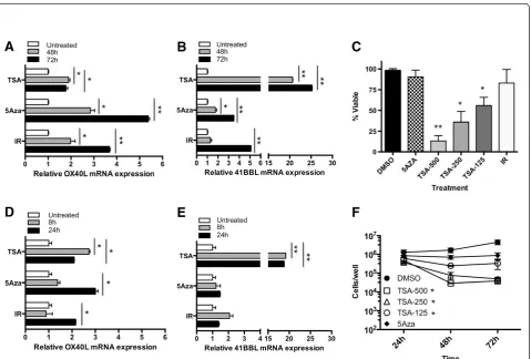
HCT116 was treated with 5-Aza-dC for 48 or 72 hr, and OX40L and 41BBL mRNA was quantified. OX40L mRNA increased 1.4-fold (Figure 1A) and 41BBL mRNA in-creased approximately 2-fold (Figure 1B) at both 48 and 72 hr post-treatment with 5-Aza-dC. OX40L mRNA increased over time in tumor cells treated with radiation, as there was a 2.3-fold increase at 48 hr and a 3.6-fold in-crease at 72 hr (Figure 1A). Radiation induced a similar increase in 41BBL transcript levels. Interestingly, this tem-poral increase was not observed in tumor cells treated with 5-Aza-dC as relatively equal levels of both OX40L and 41BBL mRNA were detected after 48 hr (gray bar) and 72 hr (black bar) drug treatment. Moreover, the level of OX40L mRNA in cells treated 5-Aza-dC never exceeded those observed 72 h post-IR.
HDACs enzymes remove acetyl groups from histones and suppress gene transcription. Recent studies have shown that HDAC inhibitors also have immune-modulatory properties, such as increasing expression of HLA-DR, ICAM-1 and B7-2 in acute myeloid leukemia cell lines [60]. We next asked if inhibition of HDACs would result in increased expression of OX40L and 41BBL similar to the increase seen in radiation-treated cells. For these experiments we used Trichostatin A (TSA), an inhibitor of the class I and class II family of HDAC enzymes, and evaluated OX40L and 41BBL mRNA expression. HCT116 cells treated with TSA for 48 hr (gray bar) contained more OX40L (Figure 1A) and 41BBL mRNA (Figure 1B) as compared to cells treated with 5-Aza-dC for 48 or 72 hr. Messenger RNA levels
decreased after 72 hr (gray bar) of TSA treatment; we note that these cells were sensitive to TSA toxicity and began dying after 48 hr TSA treatment though this loss of viability did not reach significance (Figure 1C). It is likely that mRNA expression at 48 and 72 h is not repre-sentative of early radiation events. As changes in pro-moter activation are often an early event we next evaluated cells at 8 and 24 h post-treatment. We found no significant increase in OX40L mRNA. Surprisingly, while radiation did not induce a significant increase in 41BBL RNA at 8 or 24 h, TSA did at both time points (Figure 1). Indeed the increase in 41BBL mRNA at 24 h (4-fold) exceeded levels observed after 48 h treatment (Figure 1B). 5-Aza-dC began to increase 41BBL as early as 24 h after treatment by slightly greater that 2-fold
(Figure 1E) and this increase was maintained during 48 and 72 h treatment (Figure 1B). However, both radiation and TSA induced more 41BBL mRNA than 5-Aza-dC at their respective times of maximum induction. Overall, inhibition of both HDACs and DNMTs increased the levels of OX40L and 41BBL mRNA in HCT116 cells.
To determine if epigenetic regulation of these genes was a common mechanism observable in carcinoma cells, we evaluated a second human CRC cell line, SW620. Again, SW620 cells were treated with 5-Aza-dC and TSA for 48 or 72 hr and mRNA expression was measured by qRT-PCR. Overall, SW620 cells were more responsive to these treatments than HCT116 cells. 5-Aza-dC upregulated the expression of OX40L by 5.3 fold (Figure 2A) and 41BBL by 3.5 fold (Figure 2B) in SW620
cells treated for 72 hr (gray bar). HDAC inhibition by TSA robustly altered the expression of 41BBL mRNA resulting in a 25-fold increase (Figure 2B), and again resulted in a more modest upregulation of OX40L by 1.8-fold in SW620 cells treated for 72 hr (Figure 2A). Interestingly, these cells were more sensitive to TSA tox-icity (Figure 2C) and displayed significantly reduced cell numbers following 48 and 72 h treatment with TSA concentrations ranging from 500 nM to 125 nM (Figure 2F). Viable cell numbers decreased with TSA treatment time and dose (Figure 2C), however, RNA was isolated and analyzed from the adherent and viable cells remaining in the culture (Figure 2F) for our experiments (Figure 2A & B). Moreover, we observed similar cell numbers remaining between the treatment groups after 24 h treatment with TSA and next evaluated changes in gene expression after 8 and 24 h treatment. Increased message for OX40L could be detected as early as 24 h in cells treated with radiation and 5-Aza-dC (Figure 2D) and was further increased after 48 and 72 h (Figure 2A). The largest increase in OX40L in response to TSA treat-ment in SW620 cells was detected following treattreat-ment for 8 h (2.7-fold) and was reduced slightly thereafter (2.1-fold). We also evaluated 41BBL expression after 8 and 24 h treatment. No significant change in 41BBL mRNA was observed at either of the earlier time points in cells treated with 5-Aza-dC or radiation. In contrast, a significant and robust increase in 41BBL expression could be detected after both 8 and 24 hr TSA treatment (20-fold) (Figure 2E) that was further increased after 72 hr treatment (Figure 2B). We noted that the relative level of 41BBL mRNA in untreated control cells appeared to be higher than OX40L mRNA levels in both cell lines evaluated. Overall, the largest increases in mRNA were detected for 41BBL mRNA following treat-ment of CRC cells with TSA. We also found that TSA induced robust mRNA changes at earlier times of treat-ment (8 h and 24 h) while radiation-induced changes took longer and were greatest at later times of treatment (48 h and 72 h).
Following tumor cell irradiation only adherent and proliferating cells were harvested for analysis. We have previously demonstrated that irradiated tumor cells con-tinue to proliferate and remain viable using this method [6] (Figures 1C & 2C). HCT116 cells appear to be less sensitive to TSA than SW620 cells as significantly re-duced proliferation of treated HCT116 cells was detected only when the highest dose of TSA (500 nM) was used (Figure 1F). In contrast to TSA, there was very little impact of 5-Aza-dC on viability of tumor cells 48 h after treatment in either cell line (Figures 1C & 2C). Though cell numbers were slightly reduced following 5-Aza-dC treatment of SW620 cells this was not signifi-cant (Figure 2F).
Surface expression of OX40L and 41BBL protein increases when DNMTs and HDACs are inhibited.
The largest increase in mRNA was detected in SW620 cells treated with 5-Aza-dC (OX40L, Fig-ure 2A) or TSA (41BBL, FigFig-ure 2B), and we wanted to determine if increased protein expression also oc-curred. There was no significant difference in the total cell number (Figure 2F) or the viability (data not shown) of SW620 cells following 24 h hour
treatment with 125 nM TSA. As such,
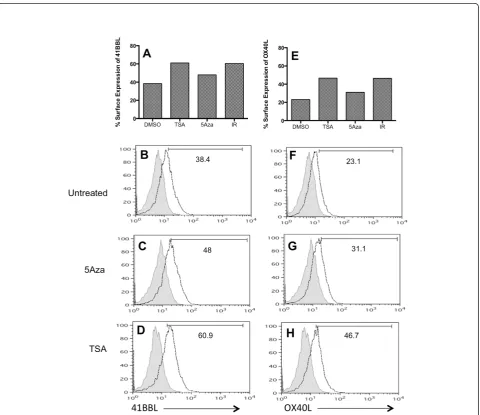
we evaluated surface expression of 41BBL protein by flow cytometry after 24 hr treatment with either TSA (125 nM) or 5-Aza-dC. Untreated SW620 cells expressed modest amounts of 41BBL on the surface (38.4%), and as expected radiation increased the fre-quency to 60.4% (Figure 3A). Treatment with 5-Aza -dC had less of an impact on protein expression and 48% of cells expressed 41BBL after treatment with the drug (Figure 3C). In contrast, TSA had a much larger impact on protein expression and, similar to radiation-induced expression, 61% of TSA-treated SW620 cells expressed 41BBL (Figure 3D) (66% in cells treated with 500 nM). Thus, relative changes in 41BBL protein expression (Figure 3A) and 41BBL mRNA quantities (Figure 2B) were similar in this cell line.
We next evaluated OX40L protein expression. SW620 tumor cells increased surface OX40L following exposure to 10Gy of radiation (IR; 46.4%), as compared to un-treated cells (DMSO; 23.1%) (Figure 3E). TSA increased protein expression of OX40L to a similar magnitude (46.7%) as irradiated cells. Again, as seen with 41BBL, there was a smaller increase in surface OX40L detected (31.1%) following treatment with 5-Aza-dC. This was surprisingly low given the 3- to 5-fold increase in OX40L mRNA seen in these cells upon 5-Aza-dC treat-ment (Figures 2A & 2D). Thus, mRNA modulation of the two genes (Figure 2) was similar to protein changes by TSA and radiation (Figure 3), but not 5-Aza-dC. Fur-thermore, the modulation of OX40L protein was less ro-bust than that observed for 41BBL protein in SW620 cells (Figure 3B-3D & 3F-3H).
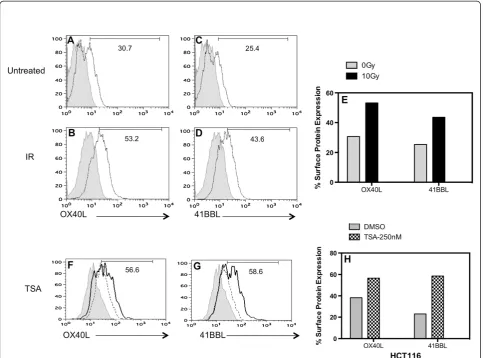
Expression of 41BBL was also enhanced too much greater levels following treatment with both IR (43.6% 10Gy) (Figure 4C-D & 4E) and TSA (58.6%-250 nM TSA versus 23%-untreated) at 48 hr (Figure 4G). The relative change in 41BBL surface expression compared to untreated cells was larger that the change in OX40L following TSA treatment in HCT116 cells (Figure 4H). Ele-vated levels of these co-stimulatory proteins could still be detected after 3- to 4-days of TSA treatment and radiation-induced changes where greater after 72 h (data not shown). Overall, both HCT116 and SW620 cells showed a more robust increase in expression of 41BBL as compared to OX40L protein expression upon TSA treatment.
Radiation increases histone H3 acetylation at the 41BBL promoter
Our data indicates that 41BBL and OX40L are epigeneti-cally regulated and radiation increases expression of these genes in CRC cell lines. Histone acetylation facili-tates transcription initiation by loosening interactions between the histones and DNA. Whereas, HDACs re-move these acetyl groups from histones which reduces transcription. We observed that inhibition of HDACs by TSA increased 41BBL mRNA expression and surface protein levels in tumor cells. We observed that radiation increased 41BBL gene expression in a similar manner but was more robust at later times during treatment. As
Figure 3TSA and ionizing radiation increase surface expression of co-stimulatory molecules in SW620 cells more than 5AZA. (A-D)
radiation has been reported to inhibit HDACs [37], we next wanted to determine if radiation could be increas-ing 41BBL expression by promotincreas-ing increased promoter histone acetylation. To explore whether histone modifi-cations are regulated in part by radiation, we assessed levels of histone acetylation at the 41BBL promoters using chromatin immunoprecipitation (ChIP) assays in both non-radiated and irradiated HCT116 cells. We evaluated promoter acetylation at 48 h post-IR when radiation-induced changes in mRNA levels were robust (Figure 1). TSA-treated HCT116 cells were used as a positive control for 41BBL promoter acetylation. As TSA inhibits HDAC activity, we expect to see robust in-creases in histone acetylation status following TSA treatment. As expected, Figure 5A shows increased
acetylation at the 41BBL promoter following TSA treat-ment (gray bar) as compared to untreated control cells (white bar). Surprisingly, acetylated H3 histone levels were significantly higher at 41BBL promoters in irradi-ated cells (black bar). In contrast, similar levels of acety-lated histone H3 were associated with the GAPDH promoter in both untreated and irradiated HCT116 cells (Figure 5B). Moreover, total levels of histone H3 were similar at 41BBL and GAPDH promoters revealing that there was no global change in overall histone levels (data not shown). These data indicate that radiation increases 41BBL expression by increasing histone acetylation. To determine if radiation non-specifically increases histone acetylation levels at other genes, his-tone H3 ChIP assays were performed on the Class II
Transactivator (CIITA) promoter IV. Histone H3 acetylation levels were similar for non-irradiated, irradiated and TSA treated cells at CIITA promoter IV (Figure 5C), which suggests gene-specificity for radiation-induced 41BBL promoter acetylation, likely via HDAC inhibition.
Treatment of CRC cells with TSA enhances T-cell survival and activation similar to co-incubation with irradiated tumor cells
To investigate the impact of HDAC inhibition in tumor cells on T-cell survival, we measured T-cell death by 7AAD staining after 48 hr co-incubation with tumor cells. 7AAD + staining determined cell death of 8.96% of CD8+ T cells incubated alone (Figure 6A). The frequency of dead CD8+ T cells increased to 24.8% following co-incubation with untreated SW620 cells (Figure 6B). Death of T-cells following interaction with tumor cells has been reported
by others, and is thought to be caused by tumor expressed PDL1, FasL and/or activation induced cell death (AICD) [61-63]. Incubation of T-cells with SW620 cells, which had been treated with TSA for 48 hr, reduced the percentage of dead T cells to 17.6% (Figure 6D) similar to incubation with irradiated tumor cells (16.6%). A reduc-tion in T-cell death (18%) was also observed when T-cells were co-incubated with TSA-treated HCT116 cells as compared to untreated tumor cells (26%) (Figure 6E). These data indicate that HDAC inhibition by TSA treat-ment of tumor cells increases the survival of CD8+T cells following co-incubation with tumor cells.
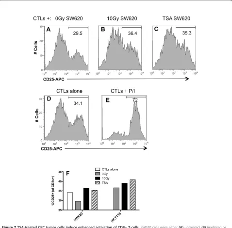
CD25 and CD69 are surface markers expressed on activated T cells [64]. Data from our lab supports the hypothesis that changes in the expression of tumor-expressed 41BBL and OX40L contribute to increased kill-ing of irradiated tumor cells by CTLs (submitted manu-script). We have also observed increased expression of
CD25 and CD69 on T cells following co-incubation with ir-radiated tumor cells compared to non-irir-radiated tumor cells. Lastly, we have observed increased viability of T cells cultured with irradiated tumor cells. We next determined if tumor cells treated with HDACi induced similar changes in T cell activation. Non-treated, irradiated or TSA treated tumor cells were co-cultured with CD8+ T cells, and after 48 hr the expression of CD25 on T cells was measured by flow cytometry. We found that 29.5% of CD8+ T cells incu-bated with untreated tumor cells expressed CD25 (Figure 7A), and this frequency was reduced compared to activation of T cells incubated alone (34.1%) (Figure 7D). This reduction is not surprising as reduced activity and ac-tivation of T-cells following interaction with tumor cells has been described by others [61-63]. The frequency of CD25+ within the CD8+T cell population increased following co-incubation with either radiation-treated (Fig-ure 7B) or TSA-treated tumor cells to 35.3% (Fig(Fig-ure 7C). In fact, the frequency of activated T cells following co-incubation with TSA-treated cells was equal to T cells not co-incubated with tumor cells (34.1%). CD25 expression in T-cells activated with PMA and ionomycin are shown as a positive control (Figure 7E). We evaluated a second CRC cell line and found that TSA-treated HCT116 cells also in-creased the frequency of CD8+CD25+cells to 41%, as com-pared to the frequency activated in the presence of
untreated HCT116 cells (36.6%) (Figure 7F). Irradiated tumor cells also increased CD25+ expression to 36.4% and the dynamics of T-cell activated were similar in repeat ex-periments. We observed a similar increase in the frequency of CD69+ T cells following co-incubated with TSA-treated or irradiated tumor cells (data not shown). These data sug-gest that T cells exposed to TSA treated tumor cells have improved activation. As a component of the IL-2 receptor, CD25 it has been linked to increased survival in studies by others and thus could be a contributor to the increased sur-vival we observe following TSA treatment (Figure 6).
Discussion
Modulation of costimulatory molecules such as OX40L and 41BBL appear to be particularly important for maintaining effective immune responses against self-antigens presented by tumor cells. Here, we report that costimulatory molecule promoter histones can be acety-lated in colorectal tumors in response to sub-lethal radi-ation (Figure 5A). Most studies of radiradi-ation-induced gene expression have used large cytotoxic doses of radi-ation, and mechanisms of altered gene expression are much less explored in cells receiving low or sub-lethal doses of radiation. Results of this study suggest that radi-ation therapy may be useful to specifically modulate gene expression within tumor targets. This mechanism
would be useful against radioresistant cancer cells, and could occur even in the absence of immunogenic cell death (cell death that invokes enhanced antigen process-ing and presentation) [65]. Full understandprocess-ing of specific mechanisms of immunogenic modulation (altered ex-pression of immune relevant genes) [66] of irradiated tumor cells will be required to determine how to best utilize radiation as a“tool”to enhance cancer immuno-therapy approaches.
Dramatic changes in DNA methylation are common in cancer, and manifest primarily as global DNA hypomethylation, paralleled by local hypermethylation at gene promoters resulting in loss of gene expression [67,68]. Tumor cells down-regulate the expression of many genes needed for induction of effective anti-tumor immune activity [15,16,18,19], and DNA methylation may be one mechanism employed to accomplish this. Our studies reveal that inhibition of DNMT in tumor
cells using 5-Aza-dC could induce mRNA expression of both OX40L and 41BBL on two different CRC cell lines (Figure 1 & 2). Although a greater than 5-fold induction of mRNA was detected in SW620 cells treated with 5-Aza-dC, we did not observe a robust increase in protein expression upon 5-Aza-dC treatment of these cells (Figure 3). These discordant results could simply be a re-sult of the time of evaluation post-treatment. 41BBL mRNA was maximally increased 72 hr post-treatment with 5-Aza-dC, while protein expression was evaluated after 24 h of treatment to keep cell death low at time of evaluation. Current studies are underway to determine if 5-Aza-dC can indeed upregulate protein expression at later times post-treatment.
HDAC inhibition has been shown to be involved in modulating the expression of TNF family members [69,70]. In this study we extended analysis to other TNF family members and found that both 41BBL and OX40L expression could also be modulated by inhibition of HDACs. We found that the expression of both OX40L and 41BBL was increased on the surface of tumor cells treated with TSA for 24 hr (Figure 3) or 48 hr (Figure 4). Interestingly, the impact of HDAC inhibition by TSA on 41BBL protein expression was much more robust than changes observed in the expression of OX40L protein following TSA treatment. Studies are currently under-way to evaluate changes in histone acetylation at the OX40L promoter to determine how acetylation is im-pacted by TSA inhibition of HDACs. We also observed increased expression of co-stimulatory proteins as long as four days after TSA-treatment and irradiation. While many of the cellular stress response genes are acute re-sponse genes whose expression is altered transiently, other genes remain altered for prolonged periods of time [71-73]. As such, altered gene expression following radi-ation treatment that is sustained is not unexpected.
The TNF family includes numerous costimulatory molecules known to play an important role in CD8+ T cell activation and survival. We found that inhibition of HDACs in tumor cells resulted in enhanced T-cell sur-vival (Figure 6) and activation (Figure 7). To our know-ledge this is the first study to explore the impact of radiation-induced epigenetic changes in tumor cells on the quality of anti-tumor CTLs. We are currently inves-tigating if, by promoting T-cell survival and activation, the altered expression of these specific genes by HDACi enhances the tumor cells’ susceptibility to T-cell-medi-ated immune attack in a manner similar to observations in irradiated tumor cells (submitted manuscript). Future studies seek to more fully investigate if increased signal-ing through CD25 is directly responsible for the in-creased survival of T-cells by evaluating T cells after shorter periods of co-incubation as well as investing intracellular regulators of T-cell apoptosis.
HDACs enzymes reverse the activity of HATs by re-moving acetyl group and thus suppressing gene tran-scription. In several tumors, the expression of HATs is down-regulated, whereas HDACs is upregulated [74,75]. As previously mentioned, alteration of HAT and HDAC activity has been observed in tumor cell lines. HDACi induce a potent anticancer response by inhibiting HDACs [76,77]. HDACi have various biological effects, such as inhibition of cell cycle at G1/G2 phase, induc-tion of differentiainduc-tion and apoptosis of tumor cells [78-80]. Our results reveal that radiation treatment changes the epigenetic landscape of the 41BBL gene via an increase in histone acetylation, displaying a marked increase in H3 acetylation at this specific promoter, as compared to our positive control of cells treated with the HDACi, TSA. We also observed that TSA induced robust 41BBL mRNA changes at earlier times of treat-ment (8 h and 24 h) while radiation-induced changes took longer and were greatest at later times of treatment (48 h and 72 h). These data, in combination with increase promoter acetylation, suggest that radiation me-diated effects take longer to modulate histone acetyl-ation events than direct modulators such as TSA. This could be related to differences in modulation of HATs versus HDAC inhibitors. Current lab efforts are pursuing the mechanism for these epigenetic changes in primary carcinoma cells; specifically, does IR treatment change the activity of HATs, HDACs or both? If HDACs are involved, specific HDAC inhibitors will be utilized to identify which HDACs suppression(s) are vital for the upregulation of 41BBL expression. Also, how long can these epigenetic changes be maintained to promote increased effector T-cell function? Finally, we note that expression of OX40L and 41BBL varied with different concentrations of drug exposure. Our focus here is to describe a novel gene regulatory mechanism by epigen-etic modification in response to irradiation. However, the application of clinically relevant doses of TSA and 5-Aza-dC, which might be combined with radiation, will also require a further investigation in a broad range of tumor cells.
Conclusions
ways of triggering these signal pathways would be widely applicable in current CIT approaches. Furthermore, if ra-diation is shown to have a profound and consistent effect on immune stimulatory gene expression, this would provide support for using IR in conjunction with CIT strategies to specifically enhance such signals to T-cells arriving at tumor sites and optimize anti-tumor CTL responses.
Abbreviations
5-Aza-dC:5-Aza-2′-deoxycytidine; CEA: Carcinoembryonic antigen; CIT: Cancer immunotherapy; ChIP: Chromatin immunoprecipitation; CRC: Colorectal carcinoma; CTL: Cytotoxic T cells; DMSO: Dimethyl sulfoxide; DNMTs: DNA methyltransferases; HDACi: HDAC inhibitors; HDACs: Histone deacetylases; IR: Ionizing radiation; RT: Radiation therapy; TSA: Trichostatin A.
Competing interests
The authors declare that they have no competing interests.
Authors’contributions
Conceived and designed the experiments: CGB and SFG. Performed the experiments: AK, EC and CGB. Analyzed the data: AK, EC and CGB.
Contributed reagents/materials/analysis tools: CGB and SFG. Wrote the paper: CGB, AK, EC and SFG. All authors read and approved the final manuscript.
Authors’information
C. Garnett-Benson previously published under Garnett, C.T.
Acknowledgement
The authors would like to thank Melissa Heffner and Dr. Alex Spring for help in editing the manuscript. This research was supported by a Mentored Award Grant from Georgia State University.
Received: 23 April 2013 Accepted: 12 September 2013 Published: 23 September 2013
References
1. Chakraborty M, Abrams SI, Camphausen K, Liu K, Scott T, Coleman CN, Hodge JW:Irradiation of tumor cells up-regulates Fas and enhances CTL lytic activity and CTL adoptive immunotherapy.J Immunol2003, 170:6338–6347.
2. Friedman EJ:Immune modulation by ionizing radiation and its implications for cancer immunotherapy.Curr Pharm Des2002, 8:1765–1780.
3. Demaria S, Kawashima N, Yang AM, Devitt ML, Babb JS, Allison JP, Formenti SC:Immune-mediated inhibition of metastases after treatment with local radiation and CTLA-4 blockade in a mouse model of breast cancer. Clin Cancer Res2005,11:728–734.
4. Garnett CT, Palena C, Chakraborty M, Tsang KY, Schlom J, Hodge JW: Sublethal irradiation of human tumor cells modulates phenotype resulting in enhanced killing by cytotoxic T lymphocytes.Cancer Res 2004,64:7985–7994.
5. Gelbard A, Garnett CT, Abrams SI, Patel V, Gutkind JS, Palena C, Tsang KY, Schlom J, Hodge JW:Combination chemotherapy and radiation of human squamous cell carcinoma of the head and neck augments CTL-mediated lysis.Clin Cancer Res2006,12:1897–1905.
6. Ifeadi V, Garnett-Benson C:Sub-lethal irradiation of human colorectal tumor cells imparts enhanced and sustained susceptibility to multiple death receptor signaling pathways.PLoS One2012,7:e31762. 7. Brzoska K, Szumiel I:Signalling loops and linear pathways: NF-kappaB
activation in response to genotoxic stress.Mutagenesis2009,24:1–8. 8. Makinde AY, John-Aryankalayil M, Palayoor ST, Cerna D, Coleman CN:
Radiation survivors: understanding and exploiting the phenotype following fractionated radiation therapy.Mol Cancer Res2013,11:5–12. 9. Janssens S, Tschopp J:Signals from within: the DNA-damage-induced
NF-kappaB response.Cell Death Differ2006,13:773–784. 10. Li N, Karin M:Ionizing radiation and short wavelength UV activate
NF-kappaB through two distinct mechanisms.Proc Natl Acad Sci USA 1998,95:13012–13017.
11. Schreck R, Albermann K, Baeuerle PA:Nuclear factor kappa B: an oxidative stress-responsive transcription factor of eukaryotic cells (a review). Free Radic Res Commun1992,17:221–237.
12. Amundson SA, Do KT, Fornace AJ Jr:Induction of stress genes by low doses of gamma rays.Radiat Res1999,152:225–231.
13. Gasser S, Raulet DH:The DNA damage response arouses the immune system.Cancer Res2006,66:3959–3962.
14. Sreekumar A, Nyati MK, Varambally S, Barrette TR, Ghosh D, Lawrence TS, Chinnaiyan AM:Profiling of cancer cells using protein microarrays: discovery of novel radiation-regulated proteins.Cancer Res2001, 61:7585–7593.
15. Bubenik J:Tumour MHC class I downregulation and immunotherapy (Review).Oncol Rep2003,10:2005–2008.
16. French LE, Tschopp J:Defective death receptor signaling as a cause of tumor immune escape.Semin Cancer Biol2002,12:51–55.
17. Kojima H, Shinohara N, Hanaoka S, Someya-Shirota Y, Takagaki Y, Ohno H, Saito T, Katayama T, Yagita H, Okumura K,et al:Two distinct pathways of specific killing revealed by perforin mutant cytotoxic T lymphocytes. Immunity1994,1:357–364.
18. Zamai L, Rana R, Mazzotti G, Centurione L, Di Pietro R, Vitale M: Lymphocyte binding to K562 cells: effect of target cell irradiation and correlation with ICAM-1 and LFA-3 expression.Eur J Histochem1994, 38(Suppl 1):53–60.
19. Slavin-Chiorini DC, Catalfamo M, Kudo-Saito C, Hodge JW, Schlom J, Sabzevari H:Amplification of the lytic potential of effector/memory CD8+ cells by vector-based enhancement of ICAM-1 (CD54) in target cells: implications for intratumoral vaccine therapy.Cancer Gene Ther2004, 11:665–680.
20. Modrak DE, Gold DV, Goldenberg DM, Blumenthal RD:Colonic tumor CEA, CSAp and MUC-1 expression following radioimmunotherapy or chemotherapy.Tumour Biol2003,24:32–39.
21. Jensen SM, Maston LD, Gough MJ, Ruby CE, Redmond WL, Crittenden M, Li Y, Puri S, Poehlein CH, Morris N,et al:Signaling through OX40 enhances antitumor immunity.Semin Oncol2010,37:524–532.
22. Kroczek RA, Mages HW, Hutloff A:Emerging paradigms of T-cell co-stimulation.Curr Opin Immunol2004,16:321–327.
23. Watts TH:TNF/TNFR family members in costimulation of T cell responses. Annu Rev Immunol2005,23:23–68.
24. Kober J, Leitner J, Klauser C, Woitek R, Majdic O, Stockl J, Herndler-Brandstetter D, Grubeck-Loebenstein B, Reipert BM, Pickl WF,et al:The capacity of the TNF family members 4-1BBL, OX40L, CD70, GITRL, CD30L and LIGHT to costimulate human T cells.Eur J Immunol2008,38:2678–2688.
25. Curtsinger JM, Lins DC, Mescher MF:Signal 3 determines tolerance versus full activation of naive CD8 T cells: dissociating proliferation and development of effector function.J Exp Med2003,197:1141–1151. 26. Mescher MF, Curtsinger JM, Agarwal P, Casey KA, Gerner M, Hammerbeck
CD, Popescu F, Xiao Z:Signals required for programming effector and memory development by CD8+ T cells.Immunol Rev2006,211:81–92. 27. al-Shamkhani A, Birkeland ML, Puklavec M, Brown MH, James W, Barclay AN:
OX40 is differentially expressed on activated rat and mouse T cells and is the sole receptor for the OX40 ligand.Eur J Immunol1996,26:1695–1699. 28. Garber K:Beyond ipilimumab: new approaches target the immunological
synapse.J Natl Cancer Inst2011,103:1079–1082.
29. Waller EC, McKinney N, Hicks R, Carmichael AJ, Sissons JG, Wills MR: Differential costimulation through CD137 (4-1BB) restores proliferation of human virus-specific "effector memory" (CD28(-) CD45RA(HI)) CD8(+) T cells.Blood2007,110:4360–4366.
30. Habib-Agahi M, Jaberipour M, Searle PF:4-1BBL costimulation retrieves CD28 expression in activated T cells.Cell Immunol2009,256:39–46. 31. Curran MA, Kim M, Montalvo W, Al-Shamkhani A, Allison JP:Combination
CTLA-4 blockade and 4-1BB activation enhances tumor rejection by increasing T-cell infiltration, proliferation, and cytokine production. PLoS One2011,6:e19499.
32. Pan PY, Zang Y, Weber K, Meseck ML, Chen SH:OX40 ligation enhances primary and memory cytotoxic T lymphocyte responses in an immunotherapy for hepatic colon metastases.Mol Ther2002, 6:528–536.
34. Murata S, Ladle BH, Kim PS, Lutz ER, Wolpoe ME, Ivie SE, Smith HM, Armstrong TD, Emens LA, Jaffee EM, Reilly RT:OX40 costimulation synergizes with GM-CSF whole-cell vaccination to overcome established CD8+ T cell tolerance to an endogenous tumor antigen.J Immunol2006, 176:974–983.
35. Vire B, de Walque S, Restouin A, Olive D, Van Lint C, Collette Y: Anti-leukemia activity of MS-275 histone deacetylase inhibitor implicates 4-1BBL/4-1BB immunomodulatory functions.PLoS One2009,4:e7085. 36. Buglio D, Khaskhely NM, Voo KS, Martinez-Valdez H, Liu YJ, Younes A:
HDAC11 plays an essential role in regulating OX40 ligand expression in Hodgkin lymphoma.Blood2011,117:2910–2917.
37. Han Y, Wang Y, Xu HT, Yang LH, Wei Q, Liu Y, Zhang Y, Zhao Y, Dai SD, Miao Y, et al:X-radiation induces non-small-cell lung cancer apoptosis by upregulation of Axin expression.Int J Radiat Oncol Biol Phys2009,75:518–526. 38. Gal-Yam EN, Saito Y, Egger G, Jones PA:Cancer epigenetics: modifications,
screening, and therapy.Annu Rev Med2008,59:267–280.
39. Cheng YW, Pincas H, Bacolod MD, Schemmann G, Giardina SF, Huang J, Barral S, Idrees K, Khan SA, Zeng Z,et al:CpG island methylator phenotype associates with low-degree chromosomal abnormalities in colorectal cancer.Clin Cancer Res2008,14:6005–6013.
40. Kouzarides T:Chromatin modifications and their function.Cell2007, 128:693–705.
41. Roth SY, Denu JM, Allis CD:Histone acetyltransferases.Annu Rev Biochem 2001,70:81–120.
42. Seo SB, McNamara P, Heo S, Turner A, Lane WS, Chakravarti D:Regulation of histone acetylation and transcription by INHAT, a human cellular complex containing the set oncoprotein.Cell2001,104:119–130. 43. Eberharter A, Becker PB:Histone acetylation: a switch between repressive
and permissive chromatin. Second in review series on chromatin dynamics.EMBO Rep2002,3:224–229.
44. Richon VM, Sandhoff TW, Rifkind RA, Marks PA:Histone deacetylase inhibitor selectively induces p21WAF1 expression and gene-associated histone acetylation.Proc Natl Acad Sci USA2000,97:10014–10019. 45. Dion MF, Altschuler SJ, Wu LF, Rando OJ:Genomic characterization reveals
a simple histone H4 acetylation code.Proc Natl Acad Sci USA2005, 102:5501–5506.
46. Choudhary C, Kumar C, Gnad F, Nielsen ML, Rehman M, Walther TC, Olsen JV, Mann M:Lysine acetylation targets protein complexes and co-regulates major cellular functions.Science2009,325:834–840. 47. Kadosh D, Struhl K:Repression by Ume6 involves recruitment of a
complex containing Sin3 corepressor and Rpd3 histone deacetylase to target promoters.Cell1997,89:365–371.
48. Wang Z, Zang C, Cui K, Schones DE, Barski A, Peng W, Zhao K: Genome-wide mapping of HATs and HDACs reveals distinct functions in active and inactive genes.Cell2009,138:1019–1031.
49. Glozak MA, Seto E:Histone deacetylases and cancer.Oncogene2007, 26:5420–5432.
50. Barneda-Zahonero B, Parra M:Histone deacetylases and cancer. Mol Oncol2012,6:579–589.
51. Marks P, Rifkind RA, Richon VM, Breslow R, Miller T, Kelly WK:Histone deacetylases and cancer: causes and therapies.Nat Rev Cancer2001, 1:194–202.
52. Wang H, Gao X, Yang JJ, Liu ZR:Interaction between p68 RNA helicase and Ca2+-calmodulin promotes cell migration and metastasis. Nat Commun2013,4:1354.
53. Livak KJ, Schmittgen TD:Analysis of relative gene expression data using real-time quantitative PCR and the 2(-Delta Delta C(T)) Method. Methods2001,25:402–408.
54. Ali MW, Cacan E, Liu Y, Pierce JY, Creasman WT, Murph MM, Govindarajan R, Eblen ST, Greer SF, Hooks SB:Transcriptional suppression, DNA methylation, and histone deacetylation of the regulator of G-protein signaling 10 (RGS10) gene in ovarian cancer cells.PLoS One2013,8:e60185. 55. Tsang KY, Zaremba S, Nieroda CA, Zhu MZ, Hamilton JM, Schlom J:
Generation of human cytotoxic T cells specific for human carcinoembryonic antigen epitopes from patients immunized with recombinant vaccinia-CEA vaccine.J Natl Cancer Inst1995, 87:982–990.
56. Tsang KY, Zhu M, Nieroda CA, Correale P, Zaremba S, Hamilton JM, Cole D, Lam C, Schlom J:Phenotypic stability of a cytotoxic T-cell line directed against an immunodominant epitope of human carcinoembryonic antigen.Clin Cancer Res1997,3:2439–2449.
57. Liang G, Gonzales FA, Jones PA, Orntoft TF, Thykjaer T:Analysis of gene induction in human fibroblasts and bladder cancer cells exposed to the methylation inhibitor 5-aza-2'-deoxycytidine.Cancer Res2002, 62:961–966.
58. Dubovsky JA, McNeel DG, Powers JJ, Gordon J, Sotomayor EM, Pinilla-Ibarz JA:Treatment of chronic lymphocytic leukemia with a hypomethylating agent induces expression of NXF2, an immunogenic cancer testis antigen.Clin Cancer Res2009,15:3406–3415.
59. Kane MF, Loda M, Gaida GM, Lipman J, Mishra R, Goldman H, Jessup JM, Kolodner R:Methylation of the hMLH1 promoter correlates with lack of expression of hMLH1 in sporadic colon tumors and mismatch repair-defective human tumor cell lines.Cancer Res1997,57:808–811. 60. Maeda T, Towatari M, Kosugi H, Saito H:Up-regulation of costimulatory/
adhesion molecules by histone deacetylase inhibitors in acute myeloid leukemia cells.Blood2000,96:3847–3856.
61. Zaks TZ, Chappell DB, Rosenberg SA, Restifo NP:Fas-mediated suicide of tumor-reactive T cells following activation by specific tumor: selective rescue by caspase inhibition.J Immunol1999,162:3273–3279. 62. Prado-Garcia H, Romero-Garcia S, Morales-Fuentes J, Aguilar-Cazares D,
Lopez-Gonzalez JS:Activation-induced cell death of memory CD8+ T cells from pleural effusion of lung cancer patients is mediated by the type II Fas-induced apoptotic pathway.Cancer Immunol Immunother2012, 61:1065–1080.
63. Chiou SH, Sheu BC, Chang WC, Huang SC, Hong-Nerng H:Current concepts of tumor-infiltrating lymphocytes in human malignancies. J Reprod Immunol2005,67:35–50.
64. Zola H:Markers of cell lineage, differentiation and activation.J Biol Regul Homeost Agents2000,14:218–219.
65. Tesniere A, Panaretakis T, Kepp O, Apetoh L, Ghiringhelli F, Zitvogel L, Kroemer G:Molecular characteristics of immunogenic cancer cell death. Cell Death Differ2008,15:3–12.
66. Hodge JW, Garnett CT, Farsaci B, Palena C, Tsang KY, Ferrone S, Gameiro SR: Chemotherapy-induced immunogenic modulation of tumor cells enhances killing by cytotoxic T lymphocytes and is distinct from immunogenic cell death.Int J Cancer2013,133(3):624–636. 10.1002/ijc.28070.
67. Feinberg AP, Vogelstein B:Hypomethylation distinguishes genes of some human cancers from their normal counterparts.Nature1983,301:89–92. 68. Rhee I, Bachman KE, Park BH, Jair KW, Yen RW, Schuebel KE, Cui H, Feinberg
AP, Lengauer C, Kinzler KW,et al:DNMT1 and DNMT3b cooperate to silence genes in human cancer cells.Nature2002,416:552–556. 69. Sutheesophon K, Nishimura N, Kobayashi Y, Furukawa Y, Kawano M, Itoh K,
Kano Y, Ishii H:Involvement of the tumor necrosis factor (TNF)/TNF receptor system in leukemic cell apoptosis induced by histone deacetylase inhibitor depsipeptide (FK228).J Cell Physiol2005,203:387–397.
70. Earel JK Jr, VanOosten RL, Griffith TS:Histone deacetylase inhibitors modulate the sensitivity of tumor necrosis factor-related apoptosis-inducing ligand-resistant bladder tumor cells.Cancer Res2006,66:499–507.
71. Fornace AJ Jr, Amundson SA, Do KT, Meltzer P, Trent J, Bittner M: Stress-gene induction by low-dose gamma irradiation.Mil Med2002,167:13–15. 72. Woloschak GE, Paunesku T:Mechanisms of radiation-induced gene
responses.Stem Cells1997,15(Suppl 2):15–25.
73. Chen M, Quintans J, Fuks Z, Thompson C, Kufe DW, Weichselbaum RR: Suppression of Bcl-2 messenger RNA production may mediate apoptosis after ionizing radiation, tumor necrosis factor alpha, and ceramide. Cancer Res1995,55:991–994.
74. Ropero S, Esteller M:The role of histone deacetylases (HDACs) in human cancer.Mol Oncol2007,1:19–25.
75. Sharma NL, Groselj B, Hamdy FC, Kiltie AE:The emerging role of histone deacetylase (HDAC) inhibitors in urological cancers.BJU Int2013, 111:537–542.
76. Redner RL, Wang J, Liu JM:Chromatin remodeling and leukemia: new therapeutic paradigms.Blood1999,94:417–428.
77. Saunders N, Dicker A, Popa C, Jones S, Dahler A:Histone deacetylase inhibitors as potential anti-skin cancer agents.Cancer Res1999, 59:399–404.
78. Yoshida M, Horinouchi S:Trichostatin and leptomycin. Inhibition of histone deacetylation and signal-dependent nuclear export.Ann N Y Acad Sci1999,886:23–36.
leukemia: a new approach to anti-leukemia therapy.Leukemia1999, 13:1316–1324.
80. Rajgolikar G, Chan KK, Wang HC:Effects of a novel antitumor
depsipeptide, FR901228, on human breast cancer cells.Breast Cancer Res Treat1998,51:29–38.
81. Weinberg AD, Morris NP, Kovacsovics-Bankowski M, Urba WJ, Curti BD: Science gone translational: the OX40 agonist story.Immunol Rev2011, 244:218–231.
doi:10.1186/2051-1426-1-17
Cite this article as:Kumariet al.:Turning T cells on: epigenetically enhanced expression of effector T-cell costimulatory molecules on irradiated human tumor cells.Journal for ImmunoTherapy of Cancer 20131:17.
Submit your next manuscript to BioMed Central and take full advantage of:
• Convenient online submission
• Thorough peer review
• No space constraints or color figure charges
• Immediate publication on acceptance
• Inclusion in PubMed, CAS, Scopus and Google Scholar
• Research which is freely available for redistribution