ULTRA PERFORMANCE LIQUID CHROMATOGRAPHY (UPLC) METHOD
DEVELOPMENT AND VALIDATION FOR THE ESTIMATION OF
L-ALANYL-L-GLUTAMINE IN PHARMACEUTICAL DOSAGE FORM
(INFUSIONS).
DEVANG. N. WADIA AND HEMANT. T. DESAI *
Formulation & Development Department, Nirlife Healthcare Division, Nirma Ltd, Sachana, Ahmedabad-382150. Gujarat. India.
ABSTRACT
A simple, precise, accurate and validated reverse phase UPLC method has been developed for the estimation of L-Alanyl L-Glutamine (20% w/v) in infusion. The quantification was carried out using amino-bonded silica gel column, packed with octadecylsilane (2.1 mm × 100 mm, 1.7 μm) and the mobile phase used was a mixture of acetonitrile and 0.05 M phosphate buffer, pH 4, (70:30) at a flow rate of 0.25 ml / min. The detection wavelength was 215 nm and column temperature was 400C. The retention time was found to be 0.77 min. The results obtained showed a good agreement with the declared content. Recovery values for L-Alanyl -L-Glutamine were 99.19 - 100.82 %. The proposed method is reliable, rapid, precise, selective and may be used for the quantitative analysis of L-Alanyl -L-Glutamine in infusions.
Key words:
L-Alanyl L-Glutamine, UPLC, method development, validation.
1. INTRODUCTION
Glutamine is the most abundant free amino acid in the extracellular and intracellular compartments, contributing to more than 50% of the body’s free amino acid pool(Young VR et al.2001). Glutamine is involved in a wide variety of metabolic and synthetic biochemical processes and supports rapidly proliferating cells, such as lymphocytes and enterocytes (Souba WW.1997). and acts as nitrogen and ammonium carrier to the liver and kidney. In conditions of excessive organ/tissue demand of Glutamine during episodes of sepsis following trauma, major surgery, and other catabolic stress situations, endogenous Glutamine
intestinal proinflammatory cytokine production(Ameho CK et al. 1997). In humans, Glutamine supplementation reduced clinical infections after bone-marrow transplantation(Ziegler TR et al. 1992) and maintained gut integrity during total parenteral nutrition (TPN)(van der Hulst RR et al. 1993).
UPLC refers to Ultra Performance Liquid Chromatography. It improve in three areas: chromatographic resolution, speed and sensitivity analysis. It uses fine particles and saves time and reduces solvent consumption.(Jerkovich AD et al. 2003; Wu N et al. 2001; Unger KK et al. 2000; Swartz ME 2004).
UPLC comes from HPLC. HPLC has been the evolution of the packing materials used to effect the separation. An underlying principle of HPLC dictates that as column packing particle size decreases, efficiency and thus resolution also increases. As particle size decreases to less than 2.5μm, there is a significant gain in efficiency and it’s doesn’t diminish at increased linear velocities or flow rates according to the common Van Deemter equation(Van Deemter et al. 1956) . By using smaller particles, speed and peak capacity (number of peaks resolved per unit time) can be extended to new limits which is known as Ultra Performance.
The classic separation method is of HPLC (High Performance Liquid Chromatography) with many advantages like robustness, ease of use, good selectivity and adjustable sensitivity. It’s main limitation is the lack of efficiency compared to gas chromatography or the capillary electrophoresis(Zhang YH et al. 2000; Zhou C et al. 2005) due to low diffusion coefficients in liquid phase, involving slow diffusion of analytes in the stationary phase. The Van Deemter equation shows that efficiency increases with the use of smaller size particles but this leads to a rapid increase in back pressure, while most of the HPLC system can operate only up to 400 bar. That is why short columns filled with particles of about 2μm are used with these systems, to accelerate the analysis
without loss of efficiency, while maintaining an acceptable loss of load.
To improve the efficiency of HPLC separations, the following can be done :-
a. work at higher temperatures b. use of monolithic columns
1.1 Use of the UPLC system
Elevated-temperature chromatography also allows for high flow rates by lowering the viscosity of the mobile phase, which significantly reduces the column backpressure(Zhu J et al. 2005; Greibrokk T et al. 2003). Monolithic columns contain a polymerized porous support structure that provide lower flow resistances than conventional particle-packed columns(Gerber F et al. 2004; Tanaka N et al. 2001; Wu N et al. 2004).
No methods have been reported in the literature for the determination of drug on UPLC.
The goal of this study was to develop a rapid, more accurate, precisely reliable , less expensive and least time UPLC method for the analysis of Alanyl L-Glutamine in infusions.
2. MATERIALS AND METHODS
All the chemicals and reagents used were of AR/HPLC grade were from Merck . Pure standard and samples of L-Alanyl L-Glutamine were obtained from Kyuwa Hakko Bio Ltd, Singapore. The purities of this standard was 99.92 %.
A reverse phase UPLC system (WATERS, ACQUITY UPLC), consisting of Binary Solvent Manager along with TUV detector and Sample Manager, having Empower2 software was used for analysis. UPLC Amino Acid Column, AccQ-Tag Ultra consisted of Amino-Bonded silica gel, packed with octadecylsilane (2.1 mm X 100 mm), 1.7 µm was used for analysis.
2.2 Preparation of Mobile Phase
adjusted to 4.0 with o-phosphoric acid/tri ethyl amine. The mixture was filtered through 0.22 µ membrane filters and was degassed.
2.3 Standard Preparation
A 40.0 mg of L-Alanyl L-Glutamine working Standard was weighed and transferred accurately into a 100 ml clean and dry volumetric flasks. It was further diluted to volume with HPLC grade water. Resulting into final concentration of 400 ppm .
2.4 Sample Preparation
A 1 ml of sample solution from Alanyl L-Glutamine infusion (20% w/v) was taken and transferred accurately to 50 ml clean and dry volumetric flask. It was diluted to volume with HPLC grade water. From this further dilution was prepared to get the final concentration of 400 ppm. The solution was filtered through 0.22 µ membrane filters and it was degassed.
2.5 Chromatographic Conditions
Freshly prepared Buffer and Acetonitrile 30:70 (v/v) mobile phase and adjust pH to 4, were filtered through 0.22 µ membrane filter and sonicated before use.
Flow rate of Mobile phase was maintained at 0.25 ml/min. The column temperature was maintained at 40 0C ± 0.5 0C. The detection was carried out at 215 nm, Injection volume was 0.4 µl and total run time was 3 min. Column used was AccQ-Tag Ultra consisted of Amino-Bonded silica gel, packed with octadecylsilane (2.1 mm X 100 mm), 1.7 µm.
2.6 Assay Procedure
A 0.4 µl of placebo, standard preparation (6 times) and sample preparation (3 times) were separately injected into the chromatographic systems. Then the chromatograms and the peak responses were measured. The placebo chromatogram was examined for any extraneous peaks that were observed in the chromatograms of sample and standard preparations. Chromatogram of the standard preparation was recorded and the peak responses were measured. The tailing factor for the principal peak should not be more than 4.0 and the
number of the theoretical plates should not be less than 5000. The % RSD (Relative Standard Deviation) should not be more than 2.0.
A 0.4 µl of standard preparation and assay preparation was separately injected in the chromatogram, the chromatograms were recorded and the responses for the major peaks were measured.
3. RESULT AND DISCUSSION
Chemical structure and chemical properties are the most important facts that predict chromatographic behavior. In the present investigation the best resolution
was achieved using a AccQ-Tag Ultra consisted of Amino-Bonded silica gel, packed with octadecylsilane (2.1 mm X 100 mm), 1.7 µm and mobile phase Buffer and Acetonitrile 30:70 (v/v). The lower percentage of acetonitrile in mobile phase resulted in peak broadening of the component and long analysis duration, while higher percentage of acetonitrile in mobile phase resulted peak splitting of L-Alanyl L-Glutamine peak. Optimal retention time, 0.77 min minutes Alanyl L-Glutamine was achieved when the pH of mobile phase was adjusted to 4 with 85 % phosphoric acid. Small changes in pH of the mobile phase had a great influence to the chromatographic behavior of these substances. Higher pH of the mobile phase resulted in peak tailing, and lower pH resulted in broadening of peak.
3.1 Accuracy and precision
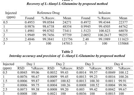
Table 1
Recovery of L-Alanyl L-Glutamine by proposed method
Injected (ppm)
Reference Drug Infusion
Found % Recov. Mean Found % Recov. Mean 0.5 0.4953 99.0584 24271 0.4972 99.4344 22377 1 0.9876 98.6738 48354 0.9945 99.4505 44762 1.5 1.4981 99.8702 73411 1.5123 100.823 68070 2 1.9949 99.7456 97759 2.0052 100.2617 90255 2.5 2.4846 99.3841 121756 2.48 99.1989 111623
3 3 100 147013 3 100 135068
Table 2
Interday accuracy and precision of L-Alanyl L-Glutamine by proposed method
Injected
(ppm) RSD Day 1 %Recov. RSD Day 2 %Recov. RSD Day 3 %Recov. RSD Day 4 %Recov. 0.5 0.0045 99.06 0.0032 99.43 0.0014 99.57 0.0049 100.12 1 0.0076 98.67 0.0009 99.45 0.0013 99.23 0.0016 100.26 1.5 0.0006 99.87 0.0007 100.82 0.0013 100.30 0.0011 99.91 2 0.0006 99.75 0.0004 100.26 0.0008 100.52 0.0004 99.96 2.5 0.0073 99.38 0.0008 99.20 0.003 99.42 0.0042 99.67 3 0.0008 100 0.0021 100 0.0036 100 0.0015 100
For intra-day precision, six concentration of each compound were analyzed on the same day. Each concentration of sample was injected 4 times a day. Table 3 summarizes the relative standard deviation (RSD). Generally acceptable repeatability of the results with in one day and day-to-day was
observed. Data of the relative retention times obtained in a series of four consecutive injections also showed acceptable repeatability when analyzed not only on the same day but also on four consecutive days.
Table 3
Recovery and regression characteristics of L-Alanyl L-Glutamine by proposed method
Conc. injected Recovered concentration
(ppm) Day 1 Day 2 Day 3 Day 4
0.5 0.4953 0.4972 0.4979 0.5006
1 0.9867 0.9945 0.9923 1.0026
1.5 1.4981 1.5123 1.5045 1.4986
2 1.9949 2.0052 2.0105 1.9993
2.5 2.4846 2.48 2.4856 2.4918
3 3 3 3 3
Correlation
3.2 System suitability and specificity
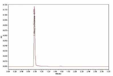
System suitability of the method was evaluated by analyzing the symmetry L-Alanyl L-Glutamine, peaks, theoretical plates of the column.
A typical chromatogram of L-Alanyl L-Glutamine is shown in figure 1.
Figure 1
3.3 Ruggedness.
Ruggedness of this method was evaluated on two different instruments of the same make in two different QC laboratories at Nirlife Healthcare Division, Nirma Ltd.
4. CONCLUSION
The proposed method was found to be simple, specific and highly accurate, required less
time consumption for analysis and this can be employed for the routine analysis.
5. ACKNOWLEDGEMENTS
The authors extend their sincere thanks to NIRLIFE HEALTHCARE, NIRMA LTD for providing the necessary chemicals, reagents and facilities.
6. REFERENCES
1. Young VR, Ajami AM. Glutamine: The emperor or his clothes? J Nutr 2001; 131(9 Suppl):2449S–2459S.
2. Souba WW: Nutritional support. N Engl J Med1997; 336:41–48.
4. Planas M, Schwartz S, Arbos MA, et al: Plasma glutamine levels in septic patients. JPEN J Parenter Enteral Nutr 1993; 17: 299–300.
5. Houdijk AP, Rijnsburger ER, Jansen J, et al: Randomised trial of glutamine- enriched enteral nutrition on infectious morbidity in patients with multiple trauma. Lancet 1998; 352:772–776.
6. Parry-Billings M, Evans J, Calder PC, et al: Does glutamine contribute to immunosuppression after burns? Lancet 1990; 336: S23–S25.
7. Gianotti L, Alexander JW, Gennari R, et al: Oral glutamine decreases bacterial translocation and improves survival in experimental gut-origin sepsis. JPEN J Parenter Enteral Nutr1995; 19:69–74. 8. Souba WW, Klimberg VS, Plumley DA, et al:
The role of glutamine in maintaining a healthy gut and supporting the metabolic response to injury and infection. J Surg Res 1990; 48:383–391.
9. O’Dwyer ST, Smith RJ, Hwang TL, et al: Maintenance of small bowel mucosa with glutamine-enriched parenteral nutrition. JPEN J Parenter Enteral Nutr1989; 13:579–585 10. Kudsk KA, Wu Y, Fukatsu K, et al:
Glutamine- enriched total parenteral nutrition maintains intestinal interleukin-4 and mucosal immunoglobulin A levels. JPEN J Parenter Enteral Nutr2000; 24:270–274. 11. Ameho CK, Adjei AA, Harrison EK, et al:
Prophylactic effect of dietary glutamine sup- plementation on interleukin 8 and tumour necrosis factor alpha production in trinitrobenzene sulphonic acid induced colitis. Gut1997; 41:487– 493.
12. Ziegler TR, Young LS, Benfell K, et al: Clinical and metabolic efficacy of glutaminesupplemented parenteral nutrition after bone marrow transplantation: A
randomized, double-blind, controlled study. Ann Intern Med1992; 116:821–828.
13. van der Hulst RR, van Kreel BK, von Meyenfeldt MF, et al: Glutamine and the preservation of gut integrity. Lancet 1993; 3 41: 1363–1365.
14. Jerkovich AD, Mellors JS, Jorgenson JW. LCGC 2003;21,7 : 660–611.
15. Wu N , Lippert JA, Lee ML, J. Chromotogr 2001; 1: 911.
16. Unger K K, Kumar D , Adam Th, Scumacher K, and Renker S, J. Chromatogr A 2000; 47: 892.
17. Swartz M E and Murphy B. Lab Plus Int 2004; 6:18.
18. Swartz M E, Murphy B. Pharm. Formulation Quality 2004; 6, 5: 40.
19. Van Deemter JJ, Zuiderweg EJ, Klinkenberg A.Longitudinal diffusion and resistance to mass transfer as causes of non ideality in chromatography. Chem. Eng. Sci. 1956; 5: 271–289
20. Zhang YH, Gong XY, Zhang HM, Larock RC, and Yeung ES. J. Comb. Chem. 2000; 2: 450–452.
21. Zhou C, Jin Y, Kenseth JR, Stella M, Wehmeyer KR and. Heineman WR. J. Pharmac. Sci 2005;94: 576-589.
22. Zhu J, Goodall DM, and Wren SAC, LCGC 2005; 23,1: 54–72.
23. Greibrokk T. and Andersen T. J. Chromatogr, A 2003; 1000: 743–755.
24. Gerber F , Krummen M, Potgeter H, Roth A , Siffrin C, and Spoendlin C. J. Chromatogr,A 2004; 1036: 127-133.
25. Tanaka N , Kobayashi H, Nakanishi K, Minakuchi H, and Ishizuka N. Anal. Chem.2001 ; 73: 420A–429A.
26. Wu N , Dempsey J , Yehl PM, Dovletoglu A, Ellison A, and Wyvratt J. Anal. Chim. Acta 2004 ; 523: 149–156.