T
T
h
h
e
e
r
r
a
a
n
n
o
o
s
s
t
t
i
i
c
c
s
s
2016; 6(1): 78-92. doi: 10.7150/thno.13242 Review
Clinical Application of Radiolabeled RGD Peptides for
PET Imaging of Integrin
α
v
β
3
Haojun Chen1,2, Gang Niu2, Hua Wu1, Xiaoyuan Chen2
1. Department of Nuclear Medicine, Xiamen Cancer Center, the First Affiliated Hospital of Xiamen University, Xiamen, China
2. Laboratory of Molecular Imaging and Nanomedicine, National Institute of Biomedical Imaging and Bioengineering, National Institutes of Health, Bethesda, Maryland
Corresponding authors: Gang Niu (niug@mail.nih.gov); Hua Wu (wuhua1025@163.com); Xiaoyuan Chen (shawn.chen@nih.gov)
© Ivyspring International Publisher. Reproduction is permitted for personal, noncommercial use, provided that the article is in whole, unmodified, and properly cited. See http://ivyspring.com/terms for terms and conditions.
Received: 2015.07.14; Accepted: 2015.10.06; Published: 2016.01.01
Abstract
Molecular imaging for non-invasive assessment of angiogenesisis is of great interest for clinicians because of the wide-spread application of anti-angiogenic cancer therapeutics. Besides, many other interventions that involve the change of blood vessel/tumor microenvironment would also benefit from such imaging strategies. Of the imaging techniques that target angiogenesis, radiolabeled Arg-Gly-Asp (RGD) peptides have been a major focus because of their high affinity and selectivity for integrin αvβ3--one of the most extensively examined target of angiogenesis. Since the level of
integrin αvβ3 expression has been established as a surrogate marker of angiogenic activity, imaging
αvβ3 expression can potentially be used as an early indicator of effectiveness of antiangiogenic
therapy at the molecular level. In this review, we summarize RGD-based PET tracers that have already been used in clinical trials and intercompared them in terms of radiosynthesis, dosimetry, pharmacokinetics and clinical applications. A perspective of their future use in the clinic is also provided.
Key words: Angiogenesis, RGD, PET, clinical translation
Introduction
Tumor angiogenesis is the process of forming new blood vessels from preexisting vasculatures, which has been well recognized as an essential hall-mark for tumor growth, invasion and metastasis [1]. Without the neovasculature to transport oxygen and nutrients, solid tumors cannot grow beyond 1–2 mm in size [2]. Once vascularized, the tumor begins to grow rapidly. The angiogenic process depends on migration and invasion of vascular endothelial cells and is mainly regulated by vascular endothelial growth factor receptors (VEGFRs) and cell adhesion receptor integrins [3].
Integrins, which contain two noncovalently bound transmembrane subunits (α and β subunits), are the bridges for cell-cell and cell-extracellular ma-trix (ECM) interactions [4]. Integrins play very im-portant roles in tumor angiogenesis and metastasis [5]. Several integrins, especially integrin αvβ3, are
sig-nificantly up-regulated on activated endothelial cells during angiogenesis but not on quiescent endothelial cells [6, 7]. Therefore, integrin αvβ3 represents a po-tential molecular marker for angiogenesis during imaging and therapy [4].
Majority of integrin-targeted imaging tracers are based on the tripeptide Arg-Gly-Asp (RGD) acid se-quence because of its high affinity and specificity for integrin αvβ3. These tracers were developed for vari-ous imaging strategies, including positron emission tomography (PET), single photon emission computed tomography (SPECT), molecular magnetic resonance imaging (mMRI), optical fluorescence, optical biolu-minescence, photoacoustic, and targeted con-trast-enhanced ultrasound [8-13]. Although the spa-tial and temporal resolutions of PET may not be as impressive as some other imaging modalities, its specificity and sensitivity are exquisite [14].
Moreo-Ivyspring
ver, quantitative analysis is relatively easy to accom-plish with PET data. Therefore, several RGD peptide based PET tracers have been investigated for clinical translation including [18F]Galacto-RGD, [18 F]Flucicla-tide, [18F]RGD-K5, [18F]FPPRGD2, [18F]Alfatide,
[68Ga]NOTA-RGD and [68Ga]NOTA-PRGD2. In this
review, we focus on these clinically available RGD-based PET tracers and intercompare them in terms of radiosynthesis, dosimetry, pharmacokinetics and clinical efficacy. We also noticed that several 99mTc- and 111In-labeled RGD peptide tracers have been used in clinical investigations. Due to the lack of direct comparison between SPECT and PET, these non-PET tracers are not included for discussion.
Radiosynthesis
For radionuclide imaging, strategies in radio-tracer design and synthesis are required to decrease the barriers involved in radiochemistry and allow more researchers and clinicians to pursue preclinical investigation or clinical practice [15]. The RGD peptides have been successfully radiolabeled with 18F
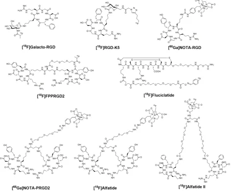
and 68Ga via different routes of chemistry and radiochemistry (Figure 1). The radiolabeling stategies of these compounds are summarized in Table 1.
[18F]Galacto-RGD was the first RGD PET tracer tested in human subjects [16]. This compound was designed by conjugating a sugar amino acid to the cyclic peptide c(RGDfK) [17]. Radiolabeling of the
glycopeptide was performed via acylation of the
amino methyl group at the C1-position of the sugar moiety using 4-nitrophenyl-2-[18F]fluoropropionate. The whole radiolabeling process requires 4 steps of radiosynthesis and 3 rounds of HPLC purification. Final HPLC with a semi-preparative column obtains [18F]Galacto-RGD with specific activities ranging be-tween 40 and 100 GBq/μmol and radiochemical p u-rity over 98%. The total synthesis time was about 200 ± 18 min (including final HPLC purification) with radiochemical yield of 29.5 ± 5.1% (decay corrected). According to Beer et al. [18], starting with 2,200 MBq of [18F]F-, 185 MBq of [18F]Galacto-RGD could be pre-pared.
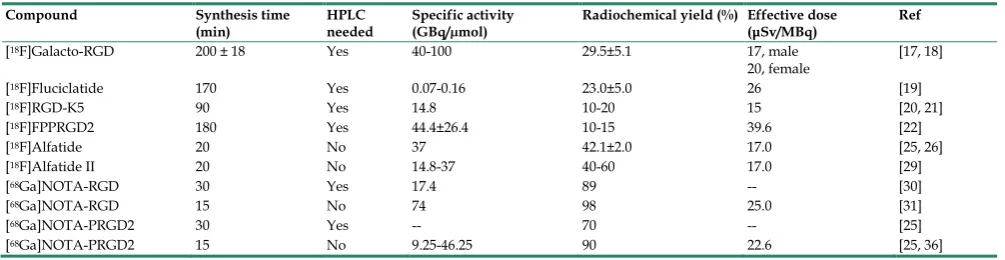
Table 1.Radiosynthesis and dosimetry of clinically available RGD-based PET Tracers
Compound Synthesis time
(min) HPLC needed Specific activity (GBq/µmol) Radiochemical yield (%) Effective dose (μSv/MBq) Ref
[18F]Galacto-RGD 200 ± 18 Yes 40-100 29.5±5.1 17, male
20, female [17, 18]
[18F]Fluciclatide 170 Yes 0.07-0.16 23.0±5.0 26 [19]
[18F]RGD-K5 90 Yes 14.8 10-20 15 [20, 21]
[18F]FPPRGD2 180 Yes 44.4±26.4 10-15 39.6 [22]
[18F]Alfatide 20 No 37 42.1±2.0 17.0 [25, 26]
[18F]Alfatide II 20 No 14.8-37 40-60 17.0 [29]
[68Ga]NOTA-RGD 30 Yes 17.4 89 -- [30]
[68Ga]NOTA-RGD 15 No 74 98 25.0 [31]
[68Ga]NOTA-PRGD2 30 Yes -- 70 -- [25]
[68Ga]NOTA-PRGD2 15 No 9.25-46.25 90 22.6 [25, 36]
*The radiolabeling procedures of several peptides were performed without HPLC purification (such as 18F-Alfatide, 68Ga-NOTA-RGD, 68Ga-NOTA-PRGD2), and the specific
activity is strongly dependent on the activity used for the synthesis.
[18F]Fluciclatide, also known as [18F]AH11185, was developed by GE Healthcare with a core peptide sequence of ACDRGDCFCG. The compound was optimized by the cyclization and introduction of two disulfide bonds to stabilize the molecule with mini-mal disruption of the RGD pharmacophore. In con-trast to Galacto-RGD, fluciclatide shows highest binding affinity for integrin αvβ5 (IC50 0.1 nM), fol-lowed by integrin αvβ3 (IC50 11.1 nM). Radiosynthesis of [18F]Fluciclatide was performed on an automated module (TRACERlab FX F-N; GE Healthcare) by coupling an aminooxy-functionalized precursor of Fluciclatide with 4-[18F]- fluorobenzaldehyde at pH 3.5 to form the oxime [18F]Fluciclatide. The whole procedure involves two reaction pots and two steps of HPLC purification, the reaction time was around 170 min. The specific activity of [18F]Fluciclatide, deter-mined by HPLC, ranging from 76-170 GBq/mmol, which is substantially less than that of [18F]Galacto-RGD [19].
[18F]RGD-K5 was developed by Siemens
Molec-ular Imaging Inc. Synthesis of [18F]RGD-K5 was ac-complished on an Explora RN automated synthesis module with average yields of 10-20% (decay cor-rected) and average synthesis time of 90 min [20]. Briefly, pentyne tosylate was reacted with K18F in the presence of Kryptofix 222, K2CO3 and acetonitrile (MeCN) at 110 ºC followed by distillation and clicking with RGD-K5-N3 in the presence of alcohol (EtOH)
and water. [18F]RGD-K5 was purified by RP-HPLC
(MeCN: water with 0.05% TFA) and reconstituted in 10% EtOH:Water via C18 trap and release. The specific activity of [18F]RGD-K5 was calculated to be at least 14.8 GBq/μmol [21]. Since RGD K5 is prepared by click chemistry, it may be suitable for (semi) auto-mated synthesis.
[18F]FPPRGD2, based on a PEGylated dimeric
RGD peptide (PEG3-Glu[c(RGDyK)]2), was the first dimeric RGD peptide approved by the Food and Drug Administration as an exploratory investigative new
drug (IND 104150) to test in human. Regarding its radiosynthesis, 4-nitrophenyl-2-[18F]fluoropropionate ([18F]NPE) was produced first by means of nucleo-philic 18F fluorination of methyl 2-bromo propionate, hydrolysis, and esterification at one-pot synthesis performed with a GE TRACERlab FXFN synthesizer. Subsequently, the conjugation between [18F]NPE and the RGD dimer was performed in a customized mod-ule to yield [18F]FPPRGD2 with a specific radioactivity level of 44.4 ± 26.4 GBq/μmol. Starting from [18F]F-, the total synthesis time of [18F]FPPRGD2 was about 180 min with the radiochemical yield of 10-15% (de-cay corrected) [22].
As shown, all these 18F-labeled RGD peptides suffered from multistep, time-consuming and low-yield synthetic procedures. Moreover, it is very challenging to make these radiosynthetic processes fully automatic, which in turn sets a high technical barrier for using these PET probes in the clinical set-ting. Application of chelation chemistry has led to the
discovery and development of [18F]fluoride-
aluminum complexes to radiolabel peptides [23, 24]. Chen’s group has successfully prepared [18F]AlF-NOTA-PRGD2 (donated as [18F]Alfatide). The whole radiosynthesis (including purification) could be accomplished within 40 min [25]. Under re-cently introduced kit formulation method, the total time can be reduced to 20 min, with a decay-corrected yield of 42.1 ± 2.0% and radiochemical purity of over 95% [26]. The specific activity of [18F]Alfatide was calculated to be at least 37 GBq/μmol.
However, the glutamic acid linked dimeric pep-tide with a free α-amine is not stable under acidic condition [27], presumably due to the neighboring amine participation in the hydrolysis. Consequently, a
much more stable new tracer [18F]NOTA-E[PEG4-
with radiochemical yield of 40-60% (corrected) and radiochemical purity > 95%. The specific activity was about 14.8-37 GBq/μmol at the end of synthesis based on the amount of peptide used and the amount of radioactivity trapped on the C18 column. Compare
with the radiosynthesis of [18F]Galacto-RGD,
[18F]Fluciclatide and [18F]FPPRGD2, this rapid radio-fluorination kit provides a strategy to simplify the radiolabeling procedure for RGD peptides.
Besides 18F, 68Ga is another popular posi-tron-emitting radioisotope which can be produced by use of a 68Ge/68Ga generator. To facilitate 68Ga label-ing, NOTA-RGD and NOTA-PRGD2 were produced by coupling SCN-Bz-NOTA to the RGD peptide c(RGDyK) or PEG3-E[c(RGDyK)]2 over the lysine or PEG3-NH2 side chain. Labeling with 68Ga was possible by incubation at room temperature for 10 min. This reaction condition is attractive, especially for the la-beling of heat-sensitive compounds. For [68Ga]NOTA-RGD, Kim et al. [30] reported labeling efficiency was around 90% (uncorrected) with the specific activity of 17.4 GBq/µmol (with HPLC puri-fication). In a later report from the same group,
[68Ga]NOTA-RGD was synthesized using a minor
modification for a clinical study (without HPLC puri-fication) [31]. In this study, the compound could be produced with a reaction time of 5 min followed by Sep-Pak purification, yielding in >98% of the product with a specific activity of 74 GBq/umol. Regarding the dimeric RGD compound, the radiolabeling of
[68Ga]NOTA-PRGD2 was reported by Chen group.
With HPLC purification, the radiochemical yield for
[68Ga]NOTA-PRGD2 was about 70% (uncorrected)
with the specific activity around 9.4 GBq/µmol [25]. To further simplify the labeling procedure, the sol-id-phase extraction method was used to prepare
[68Ga]NOTA-PRGD2. Through this method, the
radi-ochemical yield was 90% at 80 ºC with radiradi-ochemical purity over 95%. The specific activity ranges from 9.25-46.25 GBq/μmol at the end of synthesis based on the amount of peptide and radioactivity used. As the
labeling chemistry of [68Ga]NOTA-RGD/[68Ga]
NOTA-PRGD2 is performed by metal complexation, it is therefore more straightforward and less
time-consuming than [18F]Galacto-RGD,
[18F]Fluciclatide and [18F]FPPRGD2. Moreover, for the same peptide (such as NOTA-PRGD2), the labeling yield of forming [68Ga] complex is higher that of forming [18F]FAl complex, and the amount of peptide needed is also less [25]. Despite the short synthesis time for [68Ga]-labeled RGD, the short half-life (68 min) makes it difficult for commercial distribution.
Pharmacokinetics
Although being structurally different, all of clinically investigated RGD peptides, including
monomers and dimers, depict very similar in vivo
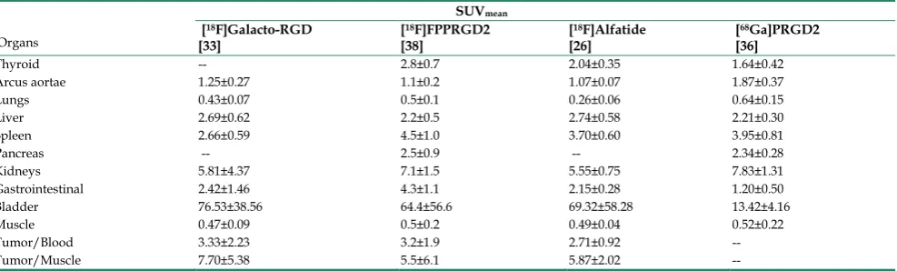
pharmacokinetic properties [21, 26, 32-36]. Tracer uptake of different RGD compounds ([18F]Galacto- RGD, [18F]FPPRGD2, [18F]Alfatide and [68Ga]PRGD2) in normal organs were summarized in Table 2 based on existing clinical data. The biodistribution data showed primary renal route of tracer clearance, with prominent tracer uptake in the kidneys and bladder (Figure 2). Concerning the intense tracer accumula-tion in the urine, image quality and analysis of lesions that adjacent to the urogenital tract and bladder are impaired. Therefore, before imaging, patients should be instructed to urinate to reduce the degree of tracer uptake in the bladder. Under certain circumstances, urinary catheter irrigation should be considered to lower the background signal.
Table 2. Tracer uptake of different RGD compounds in normal organs 60 min after injection (SUVmean, expressed as mean ± SD).
Organs
SUVmean
[18F]Galacto-RGD
[33] [
18F]FPPRGD2
[38] [
18F]Alfatide
[26] [
68Ga]PRGD2
[36]
Thyroid -- 2.8±0.7 2.04±0.35 1.64±0.42
Arcus aortae 1.25±0.27 1.1±0.2 1.07±0.07 1.87±0.37
Lungs 0.43±0.07 0.5±0.1 0.26±0.06 0.64±0.15
Liver 2.69±0.62 2.2±0.5 2.74±0.58 2.21±0.30
Spleen 2.66±0.59 4.5±1.0 3.70±0.60 3.95±0.81
Pancreas -- 2.5±0.9 -- 2.34±0.28
Kidneys 5.81±4.37 7.1±1.5 5.55±0.75 7.83±1.31
Gastrointestinal 2.42±1.46 4.3±1.1 2.15±0.28 1.20±0.50
Bladder 76.53±38.56 64.4±56.6 69.32±58.28 13.42±4.16
Muscle 0.47±0.09 0.5±0.2 0.49±0.04 0.52±0.22
Tumor/Blood 3.33±2.23 3.2±1.9 2.71±0.92 --
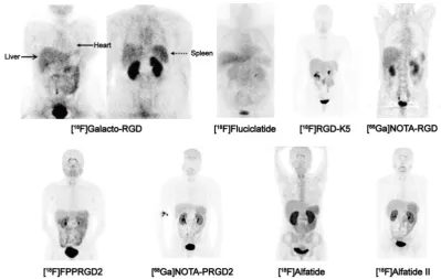
Figure 2. Biodistribution of clinically available RGD-based PET agents 1 h after intravenous administration in healthy volunteers, except for [18F]-Galacto-RGD PET,
which is from a patient with osteomyelitis. All images are coronal views. High tracer retention is notable in urogenital tract, due to predominant renal clearance. Intermediate uptake is found in the liver, spleen, and intestines. Reproduced with the permission from references [46][32][21][31] [34][36][26][35].
The RGD radioligand is retained in the tumor tissue for more than 60 min, whereas the background activity in the blood pool and muscle tissue is low and gradually decreased over time. For example, the blood and plasma clearance curves show that
ap-proximately 25% of [18F]FPPRGD2 remains in the
circulation at 30 min post-injection (p.i.), and less than 20% at 60 min [34]. Therefore, image acquisition at 40-60 min p.i. is recommended as earlier time point scans will have high nonspecific uptake which is likely to affect the image quality and ability to quan-tify integrin receptor level.
With such biodistribution patterns, RGD PET is well suited for detecting lesions in lungs, mediasti-num, head-and-neck area, thorax including the breast, skeletal system and the extremities. Respecting tu-mors in the liver, spleen, and intestines, the detection efficiency may be unsatisfactory due to the relatively high background activity in these organs. For exam-ple, at 1 h after injection, [18F]AH11185 shows normal
liver uptake with a SUVmean of 3.7-4.6 [37],
[18F]Galacto-RGD and [18F]RGD-K5 have a liver SU-Vmean of 4 and 2.7 at 1 h p.i., respectively [21, 33]. PEGylation of the RGD peptide, such as [18F]FPPRGD2, decreases lipophilicity and thereby decreases the hepatic uptake. However, the clinical study of [18F]FPPRGD2 still showed a SUVmean of 2.2 in the liver [38]. The relatively high physiological liver uptake usually makes it hard to identify liver metas-tases (SUVs from the liver lesions usually range from
1.4-3.9) [37]. On the other hand, RGD peptide is not able to cross the blood brain barrier (BBB), as tracer uptake in normal brain tissue is even lower than the tracer uptake in background tissue, like muscle [39]. In patients with high-grade glioma (grade III-IV), BBB is partially or completely disrupted, tu-mor/background ratios are normally remarkable within the area of brain [38, 40, 41].
Regarding the stability of these clinically inves-tigated RGD compounds, all of them were confirmed to be stable in vivo after intravenous injection. The radioligands were slowly metabolized to hydrophilic metabolites, with the mean percentage of intact tracer in blood at 120 min after injection ranging from 75-96% [18, 42, 43].
Radiation Dosimetry
As the patient is exposed to high-energy γ-rays during the PET scan, knowing the potential risk from radiation exposure is essential in assessing the clinical utility of PET, and this should be quantified and un-derstood so that risk-benefit ratios could be evaluated [44]. According to the literature, the effective dose from [18F]FDG PET/CT, with a diagnostic CT and 370 MBq administered FDG dose, is up to 32.18 mSv [45]. However, the effective dose from PET scan is only 6-7
mSv (16-18 μSv/MBq), which means the radiation
dose to patient from a PET scan is modest and most of the radiation comes from the CT scan.
[18F]Galacto-RGD PET scan was determined to be 17 μSv/MBq for male and 20 μSv/MBq for female [46], which is very similar to an [18F]FDG PET scan. The mean effective dose of a 370 MBq administered activ-ity of [18F]Fluciclatide was reported to be 9.6 mSv (26 μSv/MBq) [32], and was 8.3 mSv (15 μSv/MBq) for a 555 MBq injected dose of [18F]RGD-K5 [21], both of
which are comparable with [18F]FDG PET and
[18F]Galacto-RGD. For the dimeric RGD compound, the mean effective dose of [18F]FPPRGD2 was 15 mSv for a 370 MBq injected dose (39.6 μSv/MBq) [34]. The effecitive dose for [18F]Alfatide II was 17.0 μSv/MBq, determined from 5 healthy volunteers with a bladder void time of 60 min [35].
For [68Ga]NOTA-RGD and [68Ga]NOTA-PRGD2,
the mean effective doses were reported to be 25.0 μSv/MBq and 22.6 μSv/MBq [31], which are similar to [18F]Galacto-RGD and [18F]fluciclatide but less than [18F]FPPRGD2. Although 68Ga-labeled radiotracers have shorter physical half-life than 18F-labeled radio-tracers, this difference is balanced in terms of the ra-diation dose by the initial kinetic energy of positrons emitted (Emax=1899 keV for 68Ga vs. 633 keV for 18F).
The effective doses of all clinically tested RGD PET tracers range from 10-40 μSv/MBq (Table 1). This corresponds to the second risk category defined by 2007 International Commission on Radiological Protection (ICRP), which is greater than 1 mSv but not more than 20 mSv [47]. Overall, 18F-labeled RGD pep-tides are safe PET tracers with dosimetry profiles
comparable to [18F]FDG. Moreover, because
RGD-based compounds are mainly cleared out through the urinary tract, the effective dose could be further reduced by reducing the voiding interval. Under certain circumstances, diuretic agents may also be considered to lower the effective dose.
Clinical applications
Detection of malignant lesions and tumor staging
One of the applications of RGD PET in the clinic is for tumor detection and staging, as integrin αvβ3 is a well-established biomarker of neoangiogenesis. A wide variety of tumor types were evaluated in clinical studies, e.g. glioblastoma multiforme (GBM), squa-mous cell carcinoma of the head and neck (SCCHN), non-small cell lung cancer (NSCLC), breast cancer, melanoma, sarcoma, renal cancer, rectal cancer etc., with decent tumor/background contrast (Table 3). For [18F]Galacto-RGD, tracer accumulation in tumor lesions showed great heterogeneity, with mean SUVs
ranged between the background level to a maximum of 10 [16] (Figure 3A). Summarized from all the stud-ies reported, the sensitivity of [18F]Galacto-RGD PET for all lesions falls in the range of 59-92%. To be spe-cific, the sensitivity for primary lesions range between 83% and 100%, however, the sensitivity for metastatic lymph nodes (LNs) was only 33-54% and was 46-78% for distant metastases [16, 33, 39, 48-51]. Another
18F-labled RGD compound [18F]Fluciclatide, like
[18F]Galacto-RGD, showed heterogeneous tumor
up-take with SUVs from 1.4 to 40 (Figure 3B). The sensi-tivity for all lesions ranged from 88-94% and was 71-88% for distant metastases [37, 52]. These data demonstrated that the monomeric peptide tracer [18F]Galacto-RGD or [18F]Fluciclatide had only mod-erate sensitivity for metastatic lymph nodes and dis-tant metastases, insufficient for tumor staging.
Figure 3. (A) [18F]Galacto-RGD PET images in a patient with invasive ductal
breast cancer of left breast, axillary lymph-node metastases on the left side, an osseous metastasis to the sternum, and a pulmonary metastasis on the right side. Maximum-intensity projection of [18F]Galacto-RGD PET and planar images
show primary tumor, lymph-node (LN) metastases, and osseous metastasis with good tumor/background contrast. (B) [18F]Fluciclatide PET/CT images in a
Table 3.Tumor diagnostic value of clinically available RGD-based PET tracers
Tracer name Patient
num-ber Tumor type Detection rate (Sensitivity) SUVs Ref
[18F]Galacto-RGD 8 5 Melanomas 6/7 (86%), primary lesion 1.2-10.0 [16]
2 Sarcomas
1 RCC 1/1 (100%), distant metastases 7/8 (88%), all lesions
[18F]Galacto-RGD 19 10 Musculoskeletal 17/19 (89%), primary lesion 1.2-10.0 [48]
4 Melanoma 2 SCCHN 2 GBM 1 Breast cancer
[18F]Galacto-RGD 11 11 SCCHN 10/12 (83%), primary lesion 2.2-5.8 [49]
2/6 (33%), lymph nodes 12/18 (67%), all lesions
[18F]Galacto-RGD 16 10 NSCLC 13/14 (93%), primary lesion 0.3-6.8 [53]
2 RCC 7/13 (54%), lymph nodes 1 SCCHN 25/32 (78%), metastases 1 Rectal cancer
1 Breast cancer 45/59 (76%), all lesions
[18F]Galacto-RGD 12 12 GBM 11/12 (92%), primary lesion 0.8-2.8 [39]
[18F]Galacto-RGD 16 16 Breast cancer 12/12 (100%), primary lesion 1.4-8.7 [50]
3/8 (38%), lymph nodes 11/24 (46%), metastases 26/44 (59%), all lesions
[18F]Fluciclatide 7 7 Breast cancer 1/1 (100%), primary lesion 1.4-40.0 [37]
15/17 (88%), metastases 16/17 (94%), all lesions
[18F]Fluciclatide 17 11 RCC 10/10 (100%), primary lesion 1.8-10.0 [52]
6 Melanoma 5/7 (71%), metastases
[18F]RGD-K5 12 Breast cancer
15/17 (88%), all lesions
122/157 (77.7%), all lesion [51]
[18F]FPPRGD2 8 Breast cancer 8/8 (100%), primary lesion 2.4-9.4 [65]
4/5 (80%), lymph nodes 17/17 (100%), metastases 29/30 (97%), all lesions
[18F]FPPRGD2 15 Recurrent GBM 17/17 (100%), primary lesion 0.8-5.8 [41]
[18F]Alfatide 9 NSCLC 9/9 (100%), primary lesion 2.91±0.73 [26]
[18F]Alfatide II 30 Bone metastases 100%, osteolytic metastases
70%, osteoblastic metastases 100%, mixed bone metastases 98%, bone marrow metastases 92%, all metastases
0.95-13.87 [66]
[18F]Alfatide II 9 Brain metastases 20/20 (100%), brain metastases 1.8-10.0 [35]
[68Ga]NOTA-PRGD2 12 Glioma 10/12 (83.3%), primary lesion 0.5-2.31 [40]
As a matter of fact, the in vivo biodistribution pattern of RGD affects the detection efficiency of le-sions in different regions. The organs with moderate to intense RGD uptake are mostly located in the ab-domen and pelvis area, such as the liver, spleen, in-testines, kidneys and bladder, making the assessment of metastases rather difficult in these areas. As to the missed LNs or metastases located in the lung, medi-astinum, and thorax (area with low background ac-tivity), most of reported studies did not obtain tissue samples from these lesions. Therefore, it remains a question mark whether the failure to depict these le-sions is a result of technical problems (limited spatial resolution of a clinical PET scanner) or insufficient integrin expression in these lesions.
A direct comparison showed that both [18F]FDG PET and contrast enhanced CT detected more lesions than [18F]Galacto-RGD PET did [53]. Moreover, it is rather challenging for [18F]Galacto-RGD PET to
iden-tify lesions in the liver, due to the relatively intense background activity and therefore lack of contrast between tumor and adjacent normal liver tissue. These pilot data indicate that [18F]FDG PET may be superior to RGD PET for tumor staging. However, it is worth noting that the tumors with low or intermedi-ate [18F]FDG uptake, such as prostate cancer or car-cinoid tumors, RGD PET may demonstrate better re-sults for lesion detection and tumor staging than [18F]FDG PET. Moreover, since RGD peptide does not accumulate in normal brains, RGD PET may have higher sensitivity for detecting glioma and higher specificity for determining tumor demarcation, as compared to [18F]FDG PET.
integrin imaging [60-62]. However, a higher absolute tumor uptake of multimeric tracers does not neces-sarily result in improved imaging quality [59]. Indeed, the consequent higher background signals from te-trameric and octameric RGD peptides gave way to the more favorable dimeric scaffold for developing RGD peptide tracers [63].
[18F]FPPRGD2 was the first dimeric RGD peptide tracer applied in the clinic for PET imaging [64]. In a clinical study with 8 breast cancer participants [65], primary and metastatic lesions showed [18F]FPPRGD2 uptake with SUVs of 2.4-9.4 (mean, 5.6 ± 2.8) and 2.5-9.7 (mean, 5.0 ± 2.3) at 60 min p.i. (Figure 4A). No significant difference in SUVs was found between [18F]FPPRGD2 and [18F]FDG PET, for both primary lesions and metastases. Similar results were also re-ported in a recent study which compared [18F]FPPRGD2 with [18F]FDG PET in 35 patients [38].
The average tumor SUVs from [18F]FPPRGD2 PET is
higher than those from [18F]Galacto-RGD and
[18F]Fluciclatide PET, which is in accord with the pre-clinical animal studies [22]. Interestingly, in the case of lobular breast cancer, there was obvious tracer up-take in primary lesion and in sub-centimeter biop-sy-proven axillary lymph nodes and thoracic spine metastases, whereas these lesions were not detected by 18F-FDG PET. Furthermore, three biopsy-proven
inflammatory lymph nodes showed mild 18F-FDG
uptake but no [18F]FPPRGD2 accumulation.
There-fore, the overall sensitivity and specificity of
[18F]FPPRGD2 PET (95.7% and 100%, respectively)
were higher than those of [18F]FDG PET (87.0% and 57.1%, respectively). In a more recent study, [18F]FPPRGD2 PET was performed in 15 patients with
recurrent GBM, and compared with [18F]FDG and
brain MR. Surprisingly, [18F]FPPRGD2 PET showed a higher detection rate of recurrent GBM than brain MR (100.0% vs. 93.3%) and [18F]FDG (100.0% vs. 86.7%) [41]. With regard to another dimeric RGD peptide [18F]Alfatide, one clinical study investigated the fea-sibility of [18F]Alfatide PET for lung cancer detection (Figure 4B). Results showed that [18F]Alfatide PET was able to clearly identify all primary lesions (sensi-tivity is 100% for primary lesions) with desirable im-age contrast (mean tumor SUV, 2.9±1.0) [26]. In order
to evaluate the diagnostic value of dimeric RGD in metastatic lesions, [18F]Alfatide II PET was recently performed in patients with bone metastases [66] and brain metastases [35], respectively. For bone metasta-sis from 30 patients, [18F]Alfatide II PET/CT has sim-ilar detection efficiency as [18F]FDG PET/CT for de-tecting osteolytic and mixed bone metastases (100%
vs. 90%). However, [18F]Alfatide II is superior to [18F]FDG for osteoblastic lesions (70% vs. 53%) (Figure
5). For bone marrow metastatic lesions, the positive
rate of [18F]Alfatide II PET was also higher than that of [18F]FDG PET (98% vs. 77%). In all bone metastatic lesions, no significant difference of tracer uptake was observed between [18F]Alfatide II and [18F]FDG (4.27 ± 2.42 vs. 4.18 ± 2.58, P > 0.05). For brain metastases, all 20 brain lesions from 9 patients were visualized by [18F]Alfatide II PET, while only 10 by [18F]FDG PET, and 13 by CT. Despite the fact of overall higher tumor uptake of [18F]FDG over [18F]Alfatide II (10.0 ± 5.7 vs. 1.8 ± 1.1), [18F]Alfatide II demonstrated much higher tumor/background ratio (18.9 ± 14.1) than [18F]FDG (1.5 ± 0.5), which is consistent with the clinical data from [18F]FPPRGD2 study [41].
Overall, existing clinical studies of [18F]FPPRGD2 and [18F]Alfatide II showed that both tracers are able to detect integrin-positive tumors with good imaging contrast. These dimeric RGD peptides have comparable imaging properties and pharmaco-kinetics, and exhibit high sensitivity and specificity for tumor detection and staging. However, besides urogenital system, moderate to prominent tracer up-take in normal liver is also observed from both [18F]FPPRGD2 and [18F]Alfatide PET images. There-fore, it can be an issue when using RGD peptides for detecting tumors within the liver. Based on existing clinical data, the sensitivity of dimeric RGD PET for primary lesions ranges from 83.3-100% and 70-100% for metastatic lesions, the specificity of [18F]FPPRGD2 PET even reached 100% (only from 8 patients in one study). It is worth noting that [18F]Alfatide and [18F]Alfatide II own a much easier radiosynthetic
procedure than [18F]FPPRGD2, which would lower
the bar for large scale clinical trials. Up to now, only a small number of clinical investigations of dimeric RGD peptides were reported, and the sensitivi-ty/specificity between dimers and monomers was never compared in the same patients and will unlikely to be studied. Therefore, additional evaluation with large cohorts are needed to determine if multimeric strategy indeed provides a higher sensitivity and specificity for tumor detection and staging than the
monomeric RGD compounds and [18F]FDG.
Noninvasive quantification of tumor integrin αvβ3 expression
histological examination including intensity of im-munohistochemical staining against integrin αvβ3 and microvessel density (MVD) [39, 48-50, 52, 67]. More-over, immunohistochemistry has confirmed the lack of αvβ3 expression in RGD-negative tumors and
nor-mal tissue [48]. These findings highlight such nonin-vasive imaging techniques hold the potential for ap-propriate patient selection, especially for those who are going to receive anti-αvβ3 treatments.
Figure 4. (A) [18F]FPPRGD2 PET images in a patient with multicentric lobular carcinoma of the right breast. [18F]FPPRGD2 PET image shows the primary right breast
lesion, right axillary lymph node, and thoracic spine metastases (arrows; all proven by biopsy). (B) [18F]Alfatide PET images in a patient with non-small cell lung cancer.
Major organs and regions are labeled with arrows. Corresponding PET/CT fusion image at certain coronal slice further shows uptake of tumor and other organs. Reproduced with permission from references [65][26].
Figure 5. 2D projection images of [18F]Alfatide II (A) and [18F]FDG PET (B) of a patient with metastatic adenocarcinoma of unknown primary site. [18F]Alfatide II PET
demonstrated intense local accumulation of radioactivity in the bone metastatic lesions located in thoracic vertebras, sacrum and right scapula, and right clavicle with good background contrast, whereas[18F]FDG PET only showed moderate uptake in some thoracic vertebras and sacral lesions. The transaxial CT (C), [18F]Alfatide
II PET (D), and [18F]FDG PET(E) were presented to focus on the lesions at sacrum. There is also bone metastasis with abnormal [18F]Alfatide II uptake (G) but not
However, αvβ3 is not only expressed on neovas-culature, but also on the surface of some tumor cells as well, particularly in melanoma, glioma, and renal cell carcinoma [68]. Therefore, it is important to keep in mind that RGD PET does not necessarily reflect an-giogenesis in all tumor types because tumoral ex-pression of integrin αvβ3 was not excluded. For
exam-ple, tumor uptake of [18F]FPRGD2 were compared
with integrin αvβ3 expression and histological markers of angiogenesis in 27 renal cell carcinoma (RCC) pa-tients. In the group of clear cell RCC (ccRCC), the [18F]FPRGD2 PET signal correlated with integrin αvβ3 expression by tumor cells and IHC results confirmed high expression of αvβ3 on ccRCC cells, whereas in the papillary RCC (pRCC) group, the signal correlated with integrin αvβ3 level on tumor vessels [67]. These results demonstrated that RGD uptake reflects the expression of integrin αvβ3 in renal tumors but repre-sents angiogenesis only when tumor cells do not ex-press integrin αvβ3.
Up to now, several different tumor types have been systematically examined regarding their αvβ3 expression patterns as shown by RGD PET. In patients with SCCHN, immunohistochemistry showed pre-dominant αvβ3 expression on tumor vasculature but not on tumor cells, thus in SCCHN, RGD PET may be regarded as a surrogate marker of angiogenesis [49]. However, in malignant melanoma and ccRCC, it is not appropriate to use RGD PET to evaluate angio-genesis, because αvβ3 is also highly expressive on the surface of these tumor cells [52]. Consequently, the interpretation of RGD PET imaging strongly depends on the tumor type analyzed, which still needs further investigation.
Imaging αvβ3 expression with RGD PET has been advocated for lesion detection but is not consistent
with [18F]FDG PET. In one study which compared
[18F]Galacto-RGD with [18F]FDG PET, no correlation between FDG and RGD PET parameter in 18 patients with NSCLC and various other tumors was found (r = 0.157) [53]. Similar results were reported in a recent
study which compared 68Ga-RGD and [18F]FDG
PET/CT in different types of breast cancers [69]. This finding may be explained by the different accumula-tion mechanisms of the two compounds since FDG reflects the distribution of glucose uptake and phos-phorylation by tumor cells, but RGD binds to integrin αvβ3 expressed on the surface of tumor cells and acti-vated endothelial cells. It is also interesting to note that bone metastases showed a tendency of high RGD uptake [66], which corroborates with the observation that integrin αvβ3 is an indicator for bone metastases [70]. Therefore, [18F]FDG and [18F]RGD PET provide complementary molecular information about tumors.
Applications beyond oncology
Ischemic vascular diseases, including myocardi-al infarction (MI) and stroke, have been the leading causes of death and disability worldwide. Angiogen-esis is considered an integral part of the repair process after ischemic injury and has been a major focus of cardiovascular and neurovascular research [71]. In-tegrin αvβ3 may be an important theranostic target associated with post-MI and post-stroke repair pro-cesses after ischemic injury [72]. In one investigation, a patient with myocardial infarction underwent [18F]Galacto-RGD PET/CT scan two weeks after per-cutaneous coronary intervention [73]. Focal tracer retention was found in the infarcted area, which was defined by the extent of delayed enhancement from MRI and severely reduced myocardial blood flow from 13N-ammonia PET/CT. This signal may suggest that myocardial healing was taking place within the
infarcted area. More recently, [68Ga]NOTA-PRGD2
PET/CT was employed to evaluate integrin αvβ3-related repair in 23 post-MI and 16 post-stroke patients [74]. Patchy [68Ga]NOTA-PRGD2 uptake was found to be located at or immediately around the is-chemic regions in 20/23 MI patients and punctate multifocal uptake occurred in 8/16 stroke patients.
Moreover, [68Ga]NOTA-PRGD2 uptake related with
the disease phase and severity, suggesting that RGD PET can provide valuable semi-quantitative infor-mation about post-MI and post-stroke repair. Thus, noninvasive RGD PET appears to be promising for assessing therapies aiming to stimulate angiogenesis in ischemic vascular diseases.
angiogenic activity gradually decreased in a time-dependent manner and was estimated to nor-malize at approximately 6 months after surgery. Thus, the assessment of angiogenic activity using RGD PET is expected to be an effective tool for evaluating the efficacy of revascularization therapy in MMD pa-tients.
Atherosclerosis is the major reason for most clinical cardiovascular events, and inflammation and intraplaque angiogenesis are important features of atherosclerotic plaque progression and vulnerability [79-81]. In atherosclerotic lesions, both macrophages and activated endothelial cells highly express integrin αvβ3 [82]. Therefore, imaging integrin αvβ3 expression may be helpful to predict future risk of plaque rup-ture and allow evaluating anti-atherosclerotic therapy response. In a study involving patients with high grade carotid stenosis, Beer et al. [83] have demon-strated the feasibility of [18F]Galacto-RGD PET/CT to visualize atherosclerotic lesions and evaluate integrin αvβ3 expression in plaques. Focal [18F]Galacto-RGD uptake can be visualized in atherosclerotic lesions, and the uptake significantly correlated with intra-plaque αvβ3 expression, suggesting that visualization of integrin αvβ3 expression in human plaques is gen-erally possible with PET imaging by using appropri-ate RGD tracers.
Rheumatoid arthritis (RA) is one of the most common rheumatic disorders. RGD peptide tracers hold the potential for diagnosing and therapy re-sponse monitoring in RA because synovial angiogen-esis and pannus formation are major histopathologi-cal findings in patients with RA. One clinihistopathologi-cal study
used [68Ga]PRGD2 PET to evaluate synovial
angio-genesis and monitor response to treatment in patients with active RA [84]. High levels of [68Ga]PRGD2 ac-cumulation were found in the involved joints and tendon sheaths and diffuse distribution in the lining of the synovium. Additionally, in patients with in-tense [18F]FDG uptake in muscles caused by arthritic
pain, [68Ga]PRGD2 PET/CT was found to be better
able to evaluate disease severity than [18F]FDG
PET/CT. Moreover, the change of [68Ga]PRGD2
ac-cumulation was found to be in response to therapeu-tic intervention, and the changes in [68Ga]PRGD2 up-take significantly correlated with the changes in clin-ical disease activity index. From this study, RGD PET/CT appears to be an effective tool for identifying and assessing inflammatory synovial angiogenesis in RA patients.
Other perspective clinical applications
Patient risk stratification and patient selection for antiangiogenic therapySince integrin αvβ3 is an essential hallmark of tumor growth, invasion and metastasis, imaging in-tegrin αvβ3 expression with PET is potentially inter-esting for patient risk stratification and patient selec-tion for antiangiogenic therapy. In preclinical studies, expression of integrin αvβ3 has been reported to be associated with tumor aggressiveness and metastatic potential in malignant tumors [4]. For example, in-tegrin αvβ3 plays an essential role in the progression of malignant melanoma, during the transition of cells from radial growth phase to the vertical growth phase [85]. [18F]Galacto-RGD PET observed high variations of tumor uptake in melanoma patients [33]. For sar-coma and glioma, RGD uptake was positively corre-lated with the grade of tumor differentiation [40, 48]. Preclinical studies have also shown that expression level of integrin αvβ3 is associated with tumor aggres-siveness and metastatic potential of breast cancer [86], and integrin targeted therapy with Cilengitide showed promising results in breast cancer tu-mor-bearing mice [87]. Therefore, RGD PET may be effective for patient risk stratification. In addition, integrin αvβ3 imaging with RGD peptide tracers might also be useful in determining patient integrin status before starting therapies against the same target(such as Cilengitide). The current controversial findings and limited success especially in multicenter clinical trials with Cilengitide as monotherapy or as an adjuvant to the standard of care might suggest the importance of integrin imaging prior to patient recruitment. It is hopeful that companion integrin imaging and an-ti-integrin therapy, or integrin targeted theranostics, will likely have better outcomes. Patient stratification by integrin imaging will only enroll patients with medium to high integrin expression. Those patients with low levels of αvβ3 will be switched to alternative treatment regimens in the first place, avoiding inef-fective treatment.
Therapy response monitoring
early predicting antiangiogenic therapy response. This is particularly important as antiangiogenic ther-apy usually leads to a delay of tumor growth, rather than tumor shrinkage. As such, traditional imaging methods that only monitor morphologic changes will not be appropriate.
Currently, there are some preclinical studies on the use of RGD-based PET tracers for antiangiogenic therapy response monitoring. Unlike previous re-searches [88, 89], these studies mostly focus on the tumor models in which integrin αvβ3 is mainly ex-pressed on tumor vasculature, thus, the signal from RGD PET imaging only reflects αvβ3 expression on activated endothelial cells of the tumor vasculature. However, the results are still controversial. Some studies indicate that RGD PET is capable of measur-ing the changes of neovascular density and integrin expression during antiangiogenic therapy [90, 91]. However, other studies concluded that the tumor uptake of RGD peptide does not necessarily reflect changes of αvβ3 expression by intratumoral blood vessels early during the course of antiangiogenic therapy [92]. Thus, the efficacy of RGD PET imaging for tumor therapy monitoring relies heavily on the therapy protocol and tumor type analyzed.
Moreover, the findings regarding monitoring antiangiogenic therapies with RGD PET are confined to experimental animals. A well-defined clinical trial using αvβ3 imaging as a surrogate marker to assess response to antiangiogenic therapies is going to be the most important next step. This is difficult to carry out as many antiangiogenic drugs are now combined with chemotherapy, it is therefore not clear whether the change observed in the PET signals is due to the de-creased angiogenesis or other factors like apoptosis or necrosis induced by chemotherapy. Hopefully, through imaging αvβ3 expression, we’ll get a new tool at hand for guiding and tailoring antiangiogenic treatments to achieve individualized medicine.
Limitations of RGD compounds
As most RGD peptides have predominant renal clearance, this will impair detection of lesions in the urogenital tract. As such, tumors such as transitional cell carcinoma of bladder and urethra, localized prostate cancer and renal cancer are unlikely to be fully visualized by RGD PET. To a less extent this also applies to organs located in the abdominal area, such as spleen, liver and intestines. There is usually mod-erate to intense metabolic uptake in these organs (SUVs ranging from 2.5 to 4.0 at 1 h p.i.) [26, 33, 37, 65].
Furthermore, as we have mentioned before, RGD PET does not necessarily allow specific visualization of angiogenesis in all tumor types because tumoral
expression of integrin αvβ3 was not excluded. Thus, the interpretation of RGD PET is complex, and the potential of imaging integrin αvβ3 expression as a genuine biomarker of angiogenesis relies on the tu-mor type analyzed.
Another problem is the specificity of the signal obtained by RGD PET. This is because integrin αvβ3 can also be highly expressed on various of benign cells, such as smooth muscle cells, macrophages and osteoclasts. This has been confirmed by a number of clinical studies, e.g., myocardial infarction [93, 94], atherosclerosis [83], and cerebral infarct [95]. RGD-based compounds can also accumulate in some inflammatory lesions, such as cutaneous delayed-type hypersensitivity reaction [96], villonodular synovitis [16], hemangioma [53, 97] and oncocytoma [67]. This is because inflammatory lesions also involve neovas-culature with activated endothelial cells which ex-press integrin αvβ3. Therefore, like [18F]FDG PET, RGD PET may not be able to differentiate inflamma-tory disease from malignant tumors effectively.
As for image quantification of RGD PET, the parameters from static PET scans, like SUV, %ID/g, are being widely used. However, besides the tracer binding affinity, the ability to convert tissue uptake into target concentration is unavoidably affected by other factors such as heterogeneity of blood supply, interstitial fluid pressure and vascular permeability. According to preclinical researches from Chen’s group [98, 99], dynamic RGD PET followed by the three compartment kinetic modeling can be used to discern specific integrin binding in tissue as it deline-ates specific uptake from the non-specific accumula-tion of RGD tracers in the tumor region. Moreover, parametric imaging from dynamic PET with RGD tracer demonstrates advantages in distinguishing ef-fective from inefef-fective therapy early during vascu-lar-disrupting [100] or chemotherapeutic [101] treat-ment. Therefore, quantitative analysis according to dynamic PET provides more sensitive evaluation than static PET. In one pilot clinical study with 18 lesions from 7 breast cancer patients, the kinetics of [18F]fluciclatide has been established by using a two-tissue reversible binding model, the k3/k4 ratio was found to be a reasonable measure of specific binding, which suggests that this index could be used for estimating αvβ3 expression in cancer lesions [102]. More recently, multi-bed-position dynamic imaging
of [68Ga]NOTA-PRGD2 was successfully performed
Figure 6. Comparison of a patient with slight myocardial infarction (MI) and a patient with severe MI. Upper row: In a 58-year-old man at day 5 after the event, a small apical region with decreased [99mTc]MIBI perfusion (A, arrow) and [18F]FDG metabolism (B, arrow) showed mild [68Ga]NOTA-PRGD2 accumulation (C, arrow).
Lower row: In a 45-year-old woman on the 7th day after the event, an apical defect on [99mTc]MIBI perfusion images (D, arrow) and [18F]FDG metabolism images (E,
arrow) corresponded with moderate [68Ga]NOTA-PRGD2 uptake (F, arrows).Reproduced with permission from reference [74].
Conclusion
Great efforts have been made to develop radio-labeled RGD peptides for non-invasive determination of αvβ3 expression as well as monitoring tumor rep-sone to therapeutics involving angiogenesis. Some of them like [18F]Galacto-RGD, [18F]Fluciclatide,
[18F]RGD-K5, [68Ga]NOTA-RGD, [68Ga]NOTA-
PRGD2, [18F]FPPRGD2, and [18F]Alfatide II are cur-rently under clinical investigations for the identifica-tion of malignant lesions and quantitative assessment of integrin αvβ3 expression. Among them, [18F]Alfatide II and [68Ga]NOTA-PRGD2 substantiat-ed their advantages including easy preparation, fast labeling and superior in vivo pharmacokinetics than most of the existing monomeric RGD peptides. These tracers demonstrated the potential value of non-invasive techniques for patient risk stratification, patient selection for αvβ3-targeting therapies and monitoring of patients receiving such therapies. Therefore, to define the ultimate role of imaging αvβ3 expression in the clinic, large scale clinical trials using radiolabeled RGD tracers for therapy response as-sessment or patient prognosis evaluation are war-ranted.
Acknowledgment
The authors gratefully acknowledge the National Natural Science Foundation of China (81471684 and 81371596) and the Intramural Research Program, National Institute of Biomedical Imaging and Bioengineering, National Institutes of Health. Haojun Chen was partially funded by the China Scholarship Council (CSC).
Competing Interests
The authors have declared that no competing interest exists.
References
1. Carmeliet P. Angiogenesis in health and disease. Nat Med. 2003; 9: 653-60. 2. Folkman J. Angiogenesis in cancer, vascular, rheumatoid and other disease.
Nat Med. 1995; 1: 27-31.
3. Avraamides CJ, Garmy-Susini B, Varner JA. Integrins in angiogenesis and lymphangiogenesis. Nat Rev Cancer. 2008; 8: 604-17.
4. Niu G, Chen X. Why integrin as a primary target for imaging and therapy. Theranostics. 2011; 1: 30-47.
5. Chen X. Integrin targeted imaging and therapy. Theranostics. 2011; 1: 28. 6. Takada Y, Ye X, Simon S. The integrins. Genome Biol. 2007; 8: 215.
7. Zitzmann S, Ehemann V, Schwab M. Arginine-glycine-aspartic acid (RGD)-peptide binds to both tumor and tumor-endothelial cells in vivo. Cancer Res. 2002; 62: 5139-43.
8. Cai W, Niu G, Chen X. Imaging of integrins as biomarkers for tumor angiogenesis. Curr Pharm Des. 2008; 14: 2943-73.
9. Wu H, Chen H, Sun Y, Wan Y, Wang F, Jia B, et al. Imaging integrin αvβ3
positive glioma with a novel RGD dimer probe and the impact of antiangiogenic agent (Endostar) on its tumor uptake. Cancer Lett. 2013; 335: 75-80.
10. Wu H, Chen H, Pan D, Ma Y, Liang S, Wan Y, et al. Imaging integrin alphavbeta 3 and NRP-1 positive gliomas with a novel fluorine-18 labeled RGD-ATWLPPR heterodimeric peptide probe. Molecular Imaging Biol. 2014; 16: 781-92.
11. Iagaru A, Gambhir SS. Imaging tumor angiogenesis: the road to clinical utility. AJR Am J Roentgenol. 2013; 201: W183-91.
12. Haubner R, Beer AJ, Wang H, Chen X. Positron emission tomography tracers for imaging angiogenesis. Eur J Nucl Med Mol Imaging. 2010; 37 Suppl 1: S86-103.
13. Ye Y, Chen X. Integrin targeting for tumor optical imaging. Theranostics. 2011; 1: 102.
14. Massoud TF, Gambhir SS. Molecular imaging in living subjects: seeing fundamental biological processes in a new light. Genes Dev. 2003; 17: 545-80. 15. Hooker JM. Modular strategies for PET imaging agents. Curr Opin Chem Biol.
2010; 14: 105-11.
16. Haubner R, Weber WA, Beer AJ, Vabuliene E, Reim D, Sarbia M, et al.
Noninvasive visualization of the activated αvβ3 integrin in cancer patients by positron emission tomography and [18F]Galacto-RGD. PLoS Med. 2005; 2: e70.
17. Haubner R, Kuhnast B, Mang C, Weber WA, Kessler H, Wester HJ, et al. [18F]Galacto-RGD: synthesis, radiolabeling, metabolic stability, and radiation
dose estimates. Bioconjug Chem. 2004; 15: 61-9.
18. Beer AJ, Schwaiger M. PET imaging of αvβ3 expression in cancer patients.
Methods Mol Biol. 2011; 680: 183-200.
20. Kolb H, Chen K, Walsh J, Chen G, Gangadharmath U, Kasi D, et al. Synthesis and imaging of an 18F-labeled RGD peptide for detecting αvβ3 integrin
expression in vivo J Label Compd Radiopharm. 2009; 52: S67-S.
21. Doss M, Kolb HC, Zhang JJ, Belanger MJ, Stubbs JB, Stabin MG, et al. Biodistribution and radiation dosimetry of the integrin marker 18F-RGD-K5
determined from whole-body PET/CT in monkeys and humans. J Nucl Med. 2012; 53: 787-95.
22. Liu S, Liu Z, Chen K, Yan Y, Watzlowik P, Wester HJ, et al. 18F-labeled galacto
and PEGylated RGD dimers for PET imaging of αvβ3 integrin expression. Mol
Imaging Biol. 2010; 12: 530-8.
23. McBride WJ, Sharkey RM, Karacay H, D'Souza CA, Rossi EA, Laverman P, et al. A novel method of 18F radiolabeling for PET. J Nucl Med. 2009; 50: 991-8. 24. McBride WJ, D'Souza CA, Sharkey RM, Karacay H, Rossi EA, Chang CH, et al.
Improved 18F labeling of peptides with a fluoride-aluminum-chelate complex.
Bioconjug Chem. 2010; 21: 1331-40.
25. Lang L, Li W, Guo N, Ma Y, Zhu L, Kiesewetter DO, et al. Comparison study of [18F]FAl-NOTA-PRGD2, [18F]FPPRGD2, and [68Ga]Ga-NOTA-PRGD2 for
PET imaging of U87MG tumors in mice. Bioconjug Chem. 2011; 22: 2415-22. 26. Wan W, Guo N, Pan D, Yu C, Weng Y, Luo S, et al. First experience of
18F-Alfatide in lung cancer patients using a new lyophilized kit for rapid
radiofluorination. J Nucl Med. 2013; 54: 691-8.
27. Lang L, Ma Y, Kiesewetter DO, Chen X. Stability analysis of glutamic acid linked peptides coupled to NOTA through different chemical linkages. Mol Pharm. 2014; 11: 3867-74.
28. Guo J, Lang L, Hu S, Guo N, Zhu L, Sun Z, et al. Comparison of three dimeric
18F-AlF-NOTA-RGD tracers. Mol Imaging Biol. 2014; 16: 274-83.
29. Wu C, Yue X, Lang L, Kiesewetter DO, Li F, Zhu Z, et al. Longitudinal PET imaging of muscular inflammation using 18F-DPA-714 and 18F-Alfatide II and
differentiation with tumors. Theranostics. 2014; 4: 546-55.
30. Jeong JM, Hong MK, Chang YS, Lee YS, Kim YJ, Cheon GJ, et al. Preparation of a promising angiogenesis PET imaging agent: 68Ga-labeled
c(RGDyK)-isothiocyanatobenzyl-1,4,7-triazacyclononane-1,4,7-triacetic acid and feasibility studies in mice. J Nucl Med. 2008; 49: 830-6.
31. Kim JH, Lee JS, Kang KW, Lee HY, Han SW, Kim TY, et al. Whole-body distribution and radiation dosimetry of 68Ga-NOTA-RGD, a positron emission
tomography agent for angiogenesis imaging. Cancer Biother Radiopharm. 2012; 27: 65-71.
32. McParland BJ, Miller MP, Spinks TJ, Kenny LM, Osman S, Khela MK, et al. The biodistribution and radiation dosimetry of the Arg-Gly-Asp peptide
18F-AH111585 in healthy volunteers. J Nucl Med. 2008; 49: 1664-7.
33. Beer AJ, Haubner R, Goebel M, Luderschmidt S, Spilker ME, Wester HJ, et al.
Biodistribution and pharmacokinetics of the αvβ3-selective tracer
18F-galacto-RGD in cancer patients. J Nucl Med. 2005; 46: 1333-41.
34. Mittra ES, Goris ML, Iagaru AH, Kardan A, Burton L, Berganos R, et al. Pilot pharmacokinetic and dosimetric studies of 18F-FPPRGD2: a PET
radiopharmaceutical agent for imaging αvβ3 integrin levels. Radiology. 2011;
260: 182-91.
35. Yu C, Pan D, Mi B, Xu Y, Lang L, Niu G, et al. 18F-Alfatide II PET/CT in
healthy human volunteers and patients with brain metastases. Eur J Nucl med Mol Imaging. 2015 in press. DOI 10.1007/s00259-015-3118-2
36. Zheng K, Liang N, Zhang J, Lang L, Zhang W, Li S, et al. 68Ga-NOTA-PRGD2
PET/CT for integrin imaging in patients with lung cancer. J Nucl Med, 2015 Oct 1. pii: jnumed.115.160648. [Epub ahead of print]
37. Kenny LM, Coombes RC, Oulie I, Contractor KB, Miller M, Spinks TJ, et al. Phase I trial of the positron-emitting Arg-Gly-Asp (RGD) peptide radioligand
18F-AH111585 in breast cancer patients. J Nucl Med. 2008; 49: 879-86.
38. Minamimoto R, Jamali M, Barkhodari A, Mosci C, Mittra E, Shen B, et al. Biodistribution of the 18F-FPPRGD2 PET radiopharmaceutical in cancer
patients: an atlas of SUV measurements. Eur J Nucl Med Mol Imaging. 2015, in press. DOI 10.1007/s00259-015-3096-4.
39. Schnell O, Krebs B, Carlsen J, Miederer I, Goetz C, Goldbrunner RH, et al.
Imaging of integrin αvβ3 expression in patients with malignant glioma by [18F]
Galacto-RGD positron emission tomography. Neuro Oncol. 2009; 11: 861-70. 40. Li D, Zhao X, Zhang L, Li F, Ji N, Gao Z, et al. 68Ga-PRGD2 PET/CT in the
evaluation of Glioma: a prospective study. Mol Pharm. 2014; 11: 3923-9. 41. Iagaru A, Mosci C, Mittra E, Zaharchuk G, Fischbein N, Harsh G, et al.
Glioblastoma multiforme recurrence: An exploratory study of 18F-FPPRGD2
PET/CT. Radiology. 2015, in press. DOI: http://dx.doi.org/10.1148/radiol.2015141550.
42. Gaertner FC, Kessler H, Wester HJ, Schwaiger M, Beer AJ. Radiolabelled RGD peptides for imaging and therapy. Eur J Nucl Med Mol Imaging. 2012; 39 Suppl 1: S126-38.
43. Zhou Y, Chakraborty S, Liu S. Radiolabeled Cyclic RGD peptides as radiotracers for imaging tumors and thrombosis by SPECT. Theranostics. 2011; 1: 58-82.
44. Brix G, Lechel U, Glatting G, Ziegler SI, Munzing W, Muller SP, et al. Radiation exposure of patients undergoing whole-body dual-modality
18F-FDG PET/CT examinations. J Nucl Med. 2005; 46: 608-13.
45. Huang B, Law MW, Khong PL. Whole-body PET/CT scanning: estimation of radiation dose and cancer risk. Radiology. 2009; 251: 166-74.
46. Beer AJ, Haubner R, Wolf I, Goebel M, Luderschmidt S, Niemeyer M, et al. PET-based human dosimetry of 18F-galacto-RGD, a new radiotracer for
imaging αvβ3 expression. J Nucl Med. 2006; 47: 763-9.
47. The 2007 Recommendations of the International Commission on Radiological Protection. ICRP publication 103. Annals of the ICRP. 2007; 37: 1-332.
48. Beer AJ, Haubner R, Sarbia M, Goebel M, Luderschmidt S, Grosu AL, et al. Positron emission tomography using [18F]Galacto-RGD identifies the level of
integrin αvβ3 expression in man. Clin Cancer Res. 2006; 12: 3942-9.
49. Beer AJ, Grosu AL, Carlsen J, Kolk A, Sarbia M, Stangier I, et al. [18F]galacto-RGD positron emission tomography for imaging of αvβ3
expression on the neovasculature in patients with squamous cell carcinoma of the head and neck. Clin Cancer Res. 2007; 13: 6610-6.
50. Beer AJ, Niemeyer M, Carlsen J, Sarbia M, Nahrig J, Watzlowik P, et al.
Patterns of αvβ3 expression in primary and metastatic human breast cancer as
shown by 18F-Galacto-RGD PET. J Nucl Med. 2008; 49: 255-9.
51. Doss M, Kolb HC, Zhang JJ, Bélanger MJ, Stubbs JB, et al. Biodistribution and radiation dosimetry of the integrin marker 18F-RGD-K5 determined from
whole-body PET/CT in monkeys and humans. J Nucl Med. 2012 May;53(5):787-95..
52. Mena E, Owenius R, Turkbey B, Sherry R, Bratslavsky G, Macholl S, et al. [18F]fluciclatide in the in vivo evaluation of human melanoma and renal
tumors expressing αvβ3 and αvβ5 integrins. Eur J Nucl Med Mol Imaging.
2014; 41: 1879-88.
53. Beer AJ, Lorenzen S, Metz S, Herrmann K, Watzlowik P, Wester HJ, et al.
Comparison of integrin αvβ3 expression and glucose metabolism in primary
and metastatic lesions in cancer patients: a PET study using 18F-galacto-RGD
and 18F-FDG. J Nucl Med. 2008; 49: 22-9.
54. Chen X, Tohme M, Park R, Hou Y, Bading JR, Conti PS. Micro-PET imaging of
αvβ3-integrin expression with 18F-labeled dimeric RGD peptide. Mol Imaging.
2004; 3: 96-104.
55. Wu Y, Zhang X, Xiong Z, Cheng Z, Fisher DR, Liu S, et al. microPET imaging
of glioma integrin αvβ3 expression using 64Cu-labeled tetrameric RGD
peptide. J Nucl Med. 2005; 46: 1707-18.
56. Zhang X, Xiong Z, Wu Y, Cai W, Tseng JR, Gambhir SS, et al. Quantitative PET
imaging of tumor integrin αvβ3 expression with 18F-FRGD2. J Nucl Med. 2006;
47: 113-21.
57. Wu Z, Li ZB, Chen K, Cai W, He L, Chin FT, et al. microPET of tumor integrin
αvβ3 expression using 18F-labeled PEGylated tetrameric RGD peptide
(18F-FPRGD4). J Nucl Med. 2007; 48: 1536-44.
58. Li ZB, Cai W, Cao Q, Chen K, Wu Z, He L, et al. 64Cu-labeled tetrameric and
octameric RGD peptides for small-animal PET of tumor αvβ3 integrin
expression. J Nucl Med. 2007; 48: 1162-71.
59. Li ZB, Chen K, Chen X. 68Ga-labeled multimeric RGD peptides for microPET
imaging of integrin αvβ3 expression. Eur J Nucl Med Mol Imaging. 2008; 35: 1100-8.
60. Thumshirn G, Hersel U, Goodman SL, Kessler H. Multimeric cyclic RGD peptides as potential tools for tumor targeting: solid-phase peptide synthesis and chemoselective oxime ligation. Chemistry. 2003; 9: 2717-25.
61. Poethko T, Schottelius M, Thumshirn G, Hersel U, Herz M, Henriksen G, et al. Two-step methodology for high-yield routine radiohalogenation of peptides:
18F-labeled RGD and octreotide analogs. J Nucl Med. 2004; 45: 892-902.
62. Boturyn D, Coll JL, Garanger E, Favrot MC, Dumy P. Template assembled cyclopeptides as multimeric system for integrin targeting and endocytosis. J Am Chem Soc. 2004; 126: 5730-9.
63. Liu Z, Liu S, Wang F, Liu S, Chen X. Noninvasive imaging of tumor integrin expression using 18F-labeled RGD dimer peptide with PEG4 linkers. Eur J Nucl
Med Mol Imaging. 2009; 36: 1296-307.
64. Chin FT, Shen B, Liu S, Berganos RA, Chang E, Mittra E, et al. First experience with clinical-grade ([18F]FPPRGD2): an automated multi-step radiosynthesis
for clinical PET studies. Mol Imaging Biol. 2012; 14: 88-95.
65. Iagaru A, Mosci C, Shen B, Chin FT, Mittra E, Telli ML, et al. 18F-FPPRGD2
PET/CT: pilot phase evaluation of breast cancer patients. Radiology. 2014; 273: 549-59.
66. Mi B, Yu C, Pan D, Yang M, Wan W, Niu G, et al. Pilot Prospective Evaluation of 18F-Alfatide II for detection of skeletal metastases, in comparison with 18F-FDG PET/CT. Theranostics. 2015; 5: 1115-1121..
67. Withofs N, Signolle N, Somja J, Lovinfosse P, Nzaramba EM, Mievis F, et al.
18F-FPRGD2 PET/CT imaging of integrin αvβ3 in renal carcinomas:
correlation with histopathology. J Nucl Med. 2015; 56: 361-4.
68. Montet X, Montet-Abou K, Reynolds F, Weissleder R, Josephson L. Nanoparticle imaging of integrins on tumor cells. Neoplasia. 2006; 8: 214-22. 69. Yoon HJ, Kang KW, Chun IK, Cho N, Im SA, Jeong S, et al. Correlation of
breast cancer subtypes, based on estrogen receptor, progesterone receptor, and HER2, with functional imaging parameters from 68Ga-RGD PET/CT and 18F-FDG PET/CT. Eur J Nucl Med Mol Imaging. 2014; 41: 1534-43.
70. Zhao Y, Bachelier R, Treilleux I, Pujuguet P, Peyruchaud O, Baron R, et al.
Tumor αvβ3 integrin is a therapeutic target for breast cancer bone metastases.
Cancer Res. 2007; 67: 5821-30.
71. Khurana R, Simons M, Martin JF, Zachary IC. Role of angiogenesis in cardiovascular disease: a critical appraisal. Circulation. 2005; 112: 1813-24. 72. Giancotti FG, Ruoslahti E. Integrin signaling. Science. 1999; 285: 1028-32. 73. Makowski MR, Ebersberger U, Nekolla S, Schwaiger M. In vivo molecular
imaging of angiogenesis, targeting αvβ3 integrin expression, in a patient after
acute myocardial infarction. Eur Heart J. 2008; 29: 2201.
74. Sun Y, Zeng Y, Zhu Y, Feng F, Xu W, Wu C, et al. Application of 68Ga-PRGD2
PET/CT for αvβ3-integrin imaging of myocardial infarction and stroke. Theranostics. 2014; 4: 778-86.
![Figure 3. (A) [good tumor/background contrast. (B) [18F]Galacto-RGD PET images in a patient with invasive ductal breast cancer of left breast, axillary lymph-node metastases on the left side, an osseous metastasis to the sternum, and a pulmonary metastasis](https://thumb-us.123doks.com/thumbv2/123dok_us/8720865.1743794/6.612.317.556.310.609/background-contrast-galacto-invasive-metastases-metastasis-pulmonary-metastasis.webp)


