ABSTRACT
Background and Objective: Normal hemoglobin (Hb) is formed of a heme group and a protein group known as globin. Globin is made of four polypeptide chains and in hemoglobinopathies, the structure of one of these four polypeptide chain becomes abnormal. Cellulose acetate method is a common way to differentiate haemoglobinopathies. Inability to identify the components of Hb low concentrations and incapability to isolate all Hb types are among the disadvantages of this method. The aim of this study was to report the prevalence of hemoglobinopathies in the North of Iran by capillary electrophoresis method.
Methods: All patients with suspected hemoglobinopathies, referred by physicians for electrophoresis, have been studied in a private center in the city of Gorgan, Iran. The level of HbA2, HbA, HbF and other Hb was recorded.
Results: Overall, 725 blood samples were analyzed using the capillary method. HbE was reported in 2 patients, HbH was observed in 2 patients and Hb Barts was reported in 3 patients. Using the capillary method, among patients with the SDG area, only 4 of 38 (10.52%) had HbS and the majority of them (89.48%) had HbD.
Conclusion: HbD is the most common hemoglobinopathy in the North of Iran.
Keywords: Hemoglobinopathy; Hemoglobin D; Capillary Electrophoresis; Iran.
Hamid Reza Joshaghani(PhD)
Laboratory Science Research Center, Golestan University of Medical Sciences, Gorgan, Iran
Saeid Parvizi (MD)
Department of Gastroenterology and Hepatology, Golestan University of Medical Sciences, Gorgan, Iran
Khodaberdi Kalavi(PhD)
Laboratory Science Research Center, Golestan University of Medical Sciences, Gorgan, Iran.
Naser Behnampour(PhD)
Department of Biostatistics, Health faculty, Golestan University of Medical Sciences, Gorgan, Iran
Hadi Joshaghani
Medical Student, Medical School, Shahid Beheshti University of Medical Sciences, Tehran, Iran
Aliasghar Ayatollahi(MLD)
Department of Medical Laboratory, Golestan University of Medical Sciences, Gorgan, Iran
Nader Hashemi(Msc)
Department of Medical Biotechnology Laboratory Science Research Center, Golestan University of Medical Science, Gorgan, Iran
Sahar Alijanpour
department of medical genetics, Faculty of advanced medical technologies, Golestan university of medical sciences, Gorgan, Iran
Corresponding author: Hamid Reza Joshaghani
Tel: +981732423093
Email: [email protected]
Address: HamidrezaJoshaghani, School of Para-Medicine, Falsafi Building, Hirkan Boulevard, Gorgan, Iran
Received : 13 Oct 2015
Revised: 10 Nov 2015
Accepted: 24 Nov 2015
Hemoglobin D is the most Common Hemoglobinopathy in the North of Iran
This paper should be cited as: Joshaghani HR, Parvizi S, Kalavi Kh, Behnampour N, Joshaghani H, Ayatollahi AA, Hashemi N, Alijanpour S.[Hemoglobin D is the most Common Hemoglobinopathy in the North of Iran]. mljgoums. 2015; 9(5):28-32
Medical Laboratory Journal, Nov, Dec 2015; Vol 9: No 5 Original Article
electrophoresis method, high performance liquid chromatography (HPLC) could also be applied for the separation of HbA2 from HbE.(6) the disadvantages of using HPLC methods include false reduction of HbA2 in HbD-Punjab, false increase in HbS of patients (heterozygous and homozygous) and interaction of HbA2 with other Hb-like HbE.(7) The accurate diagnosis and quantitative determination of normal and abnormal Hb forms are of great clinical importance. Since normal HbF and A are changed frequently in patients with
thalassemia and hemoglobinopathies,
determination of Hb levels can be useful for the diagnosis of these patients. On the other
hand, the high prevalence of
hemoglobinopathies in Golestan province (according to the official statistics of 1995) highlights the need of accurate and safe diagnostic methods. Therefore, this study aimed to report the prevalence of hemoglobinopathies in the North of Iran using the capillary electrophoresis method. MATERIAL AND METHODS
All patients with suspected
hemoglobinopathies, referred by physicians for electrophoresis, have been studied in a private center in the city of Gorgan, Iran, in 2013. Hb electrophoresis was performed by Sebia minicap (France). The level of HbA2 and HbA, HbF and other Hb were recorded for each individual. The distorted and suspected results, including the results that the electrophoresis system could not determine the exact type of Hb, were excluded from the study. The blood samples (n=725) were obtained from 378 females and 347 males. The blood samples were then analyzed using the capillary method to detect hemoglobinopathies.
RESULTS
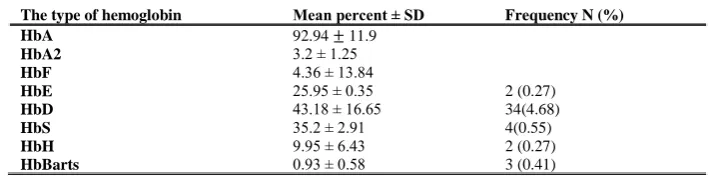
The participants were 378 females and 347 males. The prevalence of HbD, HbS and HbE were 34(4.68), 4(0.55), 4(0.55), 2 (0.27) %, respectively (Table 1).
INTRODUCTION
Normal hemoglobin (Hb) is composed of a protein group known as globin and a heme group that is identical in all types of human Hb. In hemoglobinopathies, the structure of one of the four polypeptide
chains that compose globin become
abnormal. These abnormalities are often caused by the replacement of a single amino acid. Hemoglobinopathies can be divided into two subcategories: Thalassemia and structural disease. (1) The structure of Hb is different in the blood of infants and adults. In Infant blood, HbF level is about 70 to 80 % and HbA 20 to 30%. However, in adults, HbA, HBA2 and HbF are 97, 2.5 and 0.5 %, respectively. (2) According to the World Health Organization (WHO), at least 5.2 % of the world’s population is the carrier of a defective form of Hb, while the HbS form makes about 40% of all cases. In addition, about 20% of the world's population has alphathalassemia. It is estimated that about 332,000 infants per year are born worldwide with various type of hemoglobinopathies, while sickle cell anemia and major thalassemia are responsible for about 275,000 (83%) and 56,000 (17%) of these cases, respectively. Almost 3.4% of annual mortalities among under-5 children is caused by Hb defects.(3) Due to prenatal diagnosis or abortion, the prevalence of birth with major beta-thalassemia (β-TM) in Iran has declined from 39.38 to 2.68 cases per 100,000 live births from 2005 to2010.(4) Cellulose acetate method is a
common way to differentiate
haemoglobinopathies due to its many advantages including, ease of use, rapid separation of HbA, F, S and C portability and long-term maintenance. However, lack of Hb components identification with low concentrations and inability to isolate all Hb types, especially unstable Hb, are among its disadvantages. Also HbF and A observed together when not be separated due to low levels of each against the high level of another (5). In addition to using capillary
The type of hemoglobin Mean percent ± SD Frequency N (%)
HbA 92.94 ± 11.9
HbA2 3.2 ± 1.25
HbF 4.36 ± 13.84
HbE 25.95 ± 0.35 2 (0.27)
HbD 43.18 ± 16.65 34(4.68)
HbS 35.2 ± 2.91 4(0.55)
HbH 9.95 ± 6.43 2 (0.27)
HbBarts 0.93 ± 0.58 3 (0.41)
Table 1- The mean and standard deviation of types of hemoglobin and Frequency distributions in the capillary electrophoresis
Medical Laboratory Journal, Nov, Dec 2015; Vol 9: No 5
1.1% of neonates. In England, one in every 2,000 newborns has sickle cell anemia (10). In the Middle East, the highest incidence is seen in Saudi Arabia where the frequency of carriers is 4.2% and 0.26% are patients. Eastern provinces have the highest prevalence where 17% are carriers and 1.2% are patients. The prevalence of this disease in India has increased from 9.4% to 22.2%. (11) In the present study HbE was reported in 2 cases (25%) by the capillary method. In contrast, HbE was not reported in the acetate method. Meanwhile, HbA2 was reported very high in two patients in the cellulose acetate method, which was probably due to the inability of this method to distinguish HbE from HbA2. HbE is composed of two alpha chains and two beta chains. A mutation in the beta chain of the Hb can cause mild chronic hemolytic anemia, which is often seen in Southeast Asia (Thailand, Myanmar, Cambodia, Laos and Vietnam) with an average prevalence of 30 to40%. Also, the prevalence of carriers in Northeastern India and Northeastern Thailand is about 60% and 50 to70%, respectively. The HbE mutation found in the European patients and East Asian patients are divergent, which indicates that the origin of their genetic mutation is different. The prevalence of HbE is very low among white and black people (12). Alpha–thalassemia is one form of thalassemia that involves HbA1and HbA2 genes. Alpha– thalassemia is the result of the impaired production of one, two, three, or four alpha-globin chains. Thus, the alpha-alpha-globin chain decreases and leads to formation of four beta chains (HbH) and four gamma chains (HbBarts) in adults and infants, respectively (13). Since Hb Barts has four gamma-chains with low solving capability, it accumulates in red blood cells. In addition, its intense desire for oxygen prevents oxygen delivery to tissues (14). As indicated by our results, HbH was reported in 2 patients via the capillary method with average Hb of 9.95%. Also, Hb Barts was reported in 3 patients with a mean of 0.93%. Kim and coworkers in 2011 compared the capillary electrophoresis method with cellulose acetate
method regarding the detection of
hemoglobinopathies. The study was performed in two groups where one group had normal CBC and another group had hypo-chromic microcytic anemia (103 patients). n the group with hypo-chromic microcytic s anemia, 29 patients showed reduced HbA2, 2 cases had increased HbA2, 3 cases had increased HbF DISCUSSION
Generally, HBS, G and D migrate closely together in the cellulose acetate method, making them difficult to differentiate. According to the worldwide prevalence of this condition, it is mainly considered as HbS. In the capillary method, HbD was recorded in 34 patients, HbS in 4 patients and HbG was not found. Among all the evaluated patients with SDG status, only 4 of 38 (10.52%) were found with HbS, while the majority of the cases (89.48%) had HbD. HbD was the fourth most common variant of Hb among its seven types. The most common and popular is the HbD Punjab, named for its high prevalence in the Punjab region of India and Pakistan. Hb is also highly prevalent in the Xinjiang region of China. Studies have shown that HbD is responsible for more than 55% of all Hb variants in these regions. A person with both HbD and HbA is a carrier of HbD that increases the risk of HbD disease and beta-thalassemia disease. HbD disease can cause mild hemolytic anemia with mild to moderate splenomegaly. (8) HbS, which is an Hb variant, often is seen in patients with sickle cell anemia and is the result of a point mutation in the beta-chain. Sickle cell anemia is most prevalent in tropical regions, particularly desert of South Africa, India and the Middle East. It is also seen in Europe due to high rate of immigration from these areas to the European countries. Nevertheless, 75% of all sickle cell anemia cases are found in Africa. In a recent report by the WHO, approximately 2% (150,000 children) of all neonates in Nigeria are affected by sickle cell anemia. Sickle cell trait prevalence is 10 to 40% in Nigeria and tropical Africa, one to two percent in North Africa and less than one percent in South Africa. Almost 90,000 Americans are affected by sickle cell anemia while the prevalence of this disease in America is estimated as 1in 5,000 often observed in migrants from the desert of South Africa. According to the report of the National Institutes of Health in America, one in every 500 African-American children and one in every 36,000 Spanish –American children are born with sickle cell anemia. Most new born with sickle cell anemia are detected in America by routine screening tests (9). In Europe, the problem in France. In 2010, 341 newborns were born with Sickle cell anemia and 8744 heterozygote carrier that it was equivalent of Winichagoon et al. investigated the common Hb variants in Thailand using the capillary
30/ Hemoglobin D is the most. . .
Medical Laboratory Journal, Nov, Dec 2015; Vol 9: No 5
immigration from North Africa and South African desert. This has caused Sickle cell anemia to be considered as a serious health electrophoresis method. Overall, 459 adults consisted of healthy subjects, carriers of thalassemia and different genotypes of thalassemia were entered in this study. In HbE carriers, HbA 2 wasincreased by 3.5 ± 0.4% confirming that HbE is the silent phenotype of beta-thalassemia (18). Keren and coworkers in 2008 compared Sebia capillary electrophoresis
with HPLC methods to study
hemoglobinopathies among 297 individuals. HbA level was similar in both methods, but the HbA2 level was higher in the capillary electrophoresis method. Also HbS was reported higher in this method compared with the HPLC method. In the capillary electrophoresis method, HbA2 was not from HbC, but it was from HbE. The results of the study showed the benefits of both methods in hemoglobinopathies (19). Evaluation of patients with SDG by the capillary method showed that only 4 of 38 patients (10.52%) had HbS and the majority of the patients (89.48%) had HbD. Because of the inability of the cellulose acetate method to differentiate HbS, G and D, was unclear which of Hb was considered, but due to the worldwide prevalence of this status, it was mainly considered as HbS.
CONCLUSION
The cellulose acetate method is not suggested as an appropriate method for accurate screening of hemoglobinopathies. Due to the close binding of HbS, D and G fractions and also E, A2 and O-Arab, the use of capillary method is recommended.
ACKNOWLEDGMENT
We would like to thank Kavosh medical
laboratory in Gorgan and Mr. Seyed
Mohammad HedayatMofidi. CONFLICT OF INTEREST
The authors declare that they have no conflict of interest.
and 2 cases showed an increase in both Hb. (15)Higgins et al. in 2009 studied the capillary electrophoresis method for quantitative evaluation of HbA2 in patients with and without beta-thalassemia, and assessed heterozygous patients for HbE, HbS, HbC and HbD Punjab. With capillary system, HbA2 was lower in patients with beta-thalassemia and without thalassemia. The results of this study demonstrate that the capillary 2 method is superior to the Variant II method for HbA2
quantified measurement (16). Mais and
coworkers in 2009 studied the rate of HbA2 association with HbE in the capillary electrophoresis method. The available samples with HbE were studied for evaluation of
hemoglobinopathies using the capillary
electrophoresis and HPLC methods. Fifty-two adult heterozygote patients were compared with 209control subjects in terms of HbE. The mean of HbA2 in patients with HbE was 4.3% that was significantly higher than the control group (2.6%). Seven heterozygous samples were also analyzed or HbE. The mean of HbA2 was 4.4% in this group, which was significantly higher than the heterozygous group. (6) In Thailand, Srivorakun and colleagues in 2009 examined fetal blood using the capillary electrophoresis method for hemoglobinopathies. In this study, 47 blood samples were studied by cordocentesis
in18-28 weeks using the capillary
electrophoresis system. Of these, 47 samples, 20 cases were at risk of Hb Bart's hydrops fetalis.
DNA analysis, detected four cases of
homozygous alpha-thalassemia. Hb analysis with capillary electrophoresis indicated a high level of Hb Barts (3.81-4.78%) and HbH (0.8-4.1%) and lower level of embryonic HbS. There was no case of HbF and HbA found in this study. The results of this study indicated capillary electrophoresis efficient for the assessment of prenatals evere thalassemia (17). In 2008, highest incidence is seen in France due to its African Caribbean population growth and
Medical Laboratory Journal, Nov, Dec 2015; Vol 9: No 5
11. Powars DR, Elliott-Mills DD, Chan L, Niland J, Hiti AL, Opas LM, et al. Chronic renal failure in sickle cell disease: risk factors, clinical course, and mortality". Ann Intern Med. 1991; 115(8): 614-20.
12. Green NS, Fabry ME, Kaptue-Noche L, Nagel RL. "Senegal haplotype is associated with higher HbF than Benin and Cameroon haplotypes in African children with sickle cell anemia". Am. J. Hematol 1993; 44(2): 145–6.
13. Ass KA, Lane PA, Fernhoff PM . "US newborn screening system guidelines II: follow-up of children, diagnosis, management, and evaluation". J Pediatr 2000; 137 (37): S1– S46.
14. Chernoff AI, Minnich V, Nanakorn S and et al. Studies on hemoglobin E. I. The clinical, hematologic, and genetic characteristics of the hemoglobin E syndromes. J Lab Clin Med. 1956; 47: 455–489.
15. Kim JE, Kim BR, Woo KS, Kim JM, Park JI, Han JY. Comparison of Capillary Electrophoresis with Cellulose
Acetate Electrophoresis for the Screening of
Hemoglobinopathies. The Korean journal of laboratory medicine 2011; 31(4): 238.
16. Higgins TN, Khajuria A, Mack M. Quantification of HbA2 in Patients With and Without β-Thalassemia and in the Presence of HbS, HbC, HbE, and HbD Punjab Hemoglobin Variants Comparison of Two Systems. American journal of clinical pathology. 2009; 131(3): 357-62.
17. Srivorakun H, Fucharoen G, Sae‐Ung N, Sanchaisuriya K, Ratanasiri T, Fucharoen S. Analysis of fetal blood using capillary electrophoresis system: a simple method for prenatal diagnosis of severe thalassemia diseases. European journal of haematology 2009; 83(1): 57-65.
18. Winichagoon P, Svasti S, Munkongdee T, Chaiya W, Boonmongkol P, Chantrakul N and et al. Rapid diagnosis of thalassemias and other hemoglobinopathies by capillary electrophoresis system. Translational research 2008; 152(4): 178-84.
19. Keren DF, Hedstrom D, Gulbranson R, OuCN, Bak R. Comparison of SebiaCapillarys capillary electrophoresis with the Primus high-pressure liquid chromatography in the evaluation of hemoglobinopathies. American journal of clinical pathology 2008; 130(5): 824-31.
REFERENCES
1. Rogers BB, Wessels RA, OuCN, BuffoneGJ. High-performance liquid chromatography in the diagnosis of hemoglobinopathies and thalassemias. Report of three cases.
Am J ClinPathol. 1985; 84(5): 671-4.
2. Roa D, Turner EA, AguinagaMDP. Reference ranges for hemoglobin variants by HPLC in African Americans. Ann Clin Lab Sci 1995; 25(3): 228-235.
3. Wang J, Zhou S, Huang W, Liu Y, Cheng Ch, Lu X, Cheng J. CE-based analysis of hemoglobin and its applications in clinical analysis. Electrophoresis 2006; 27(15): 3108-3124.
4. Haghpanah S, Nasirabadi S, Rahimi N, Faramarzi H, Karimi M. Sociocultural challenges of beta-thalassaemia major birth in carriers of beta-thalassaemia in Iran. J Med Screen. 2012; 19(3): 109-11. doi: 10.1258/jms.2012.012038.
5. Wajcman H, Moradkhani K. Abnormal haemoglobins: detection & characterization. The Indian Journal of Medical Research 2011; 134(4): 538.
6. Mais DD, Gulbranson RD, Keren DF. The range of hemoglobin A2 in hemoglobin E heterozygotes as determined by capillary electrophoresis. American journal of clinical
pathology. 2009; 132(1): 34-8. doi:
10.1309/AJCPP50JIXXZVLSS.
7. Joutovsky A, Hadzi-Nesic J, Nardi MA. HPLC retention time as a diagnostic tool for hemoglobin variants and hemoglobinopathies: a study of 60000 samples in a clinical diagnostic laboratory. ClinChem. 2004; 50(10): 1736-1747. 8. Tyagi S, Marwaha N, Parmar V, Basu S. Sickle cell hemoglobin-D Punjab disease (Compound Heterozygous state). Ind J Hematol Blood Transf 2000; 18: 31-2.
9. Balgir RS. Community expansion and gene geography of sickle cell trait and G6PD deficiency, and natural selection against malaria: experience from tribal land of India.
Cardiovascular & Hematological Agents in Medicinal Chemistry. 2012; 10(1): 3-13.
10. Platt OS, Brambilla DJ, RosseWF, Milner PF, Castro O, Steinberg MH, et al. Mortality in sickle cell disease. Life expectancy and risk factors for early death. N Engl J Med. 1994; 330(23): 1639-44.
32/ Hemoglobin D is the most. . .
Medical Laboratory Journal, Nov, Dec 2015; Vol 9: No 5