Research Report
The Comparison of Clinical and Surgical Staging of Cervical Cancer:
A Retrospective Study on Patients at Dr. Cipto Mangunkusumo General Hospital,
Jakarta, Indonesia
Perbandingan Hasil Pemeriksaan Klinis Pre dan Post Operatif Staging Kanker Serviks: Sebuah Studi Retrospektif terhadap Pasien Rumah Sakit Umum Pusat Nasional
Dr. Cipto Mangunkusumo, Jakarta, Indonesia
Bram Pradipta, Catherine, Tricia D. Anggraeni, Kartiwa H. Nuryanto
Oncology Gynecology Division Department of Obstetrics and Gynecology
Medical Faculty of Indonesia University/ Dr. Cipto Mangunkusumo Hospital
Jakarta
Abstract
Objective: To evaluate the accuracy of clinical examination in determining the stage of operable cervical cancer and the extent of the disease.
Method: The study involved 58 subjects from outpatient, emer-gency unit, and ward of Department of Obstetrics and Gynecology Dr. Cipto Mangunkusumo Hospital, from January 2008 to Decem-ber 2010 with a diagnosis of cervical cancer. Patients who were di-agnosed with cervical cancer up to stage IIA were included and pa-tients lost to follow-up, receiving preoperative neo-adjuvant che-motherapy, and died before getting treatment were excluded. The outcomes evaluation were postoperative clinical staging, including the presence of enlarged lymph nodes, parametrial involvement, and tumor size. Lymph nodes, parametrial, and the tumor size were as-sessed from the surgery and pathological anatomy results.
Result: The age distribution of 58 subjects ranged from 25 to 70 years (mean 48.39 years, SD 8.82). Squamous cell carcinoma was the most frequent type (44.9%), followed by adenocarcinoma (24.1%). Errors in preoperative clinical staging compared with post-operative was 40% in stage IA1, 9.52% in stage IB1, 17.65% in stage IB2, and 7.14% in stage IIA. Sensitivity, specificity, positive predictive value, and negative predictive value for preoperative clinical examination of lymph nodes were 11.1%, 100%, 100%, and 85.96%. Sensitivity, specificity, positive predictive value, and ne-gative predictive value for preoperative clinical examination of pa-rametrial involvement were 37.5%, 100%, 100%, and 90.90%. Sen-sitivity, specificity, positive predictive value, and negative predic-tive value for preoperapredic-tive clinical examination of the tumor size were 91.84%, 88.89%, 97.83% and 66.67%.
Conclusion: Clinical examination has limitation, especially in determining lymph nodes and parametrial involvement. Other diag-nostic modalities in determining the extent of the disease is ne-cessary. Enforcement of the right diagnosis in patients with cervi-cal cancer is needed to determine the appropriate treatment.
[Indones J Obstet Gynecol 2011; 35-1: 25-9]
Keywords: staging, cervical cancer, preoperative, postoperative
Abstrak
Tujuan: Untuk mengevaluasi ketepatan pemeriksaan klinis da-lam menentukan staging kanker serviks yang operable dan penye-barannya.
Metode: Penelitian melibatkan 58 subjek pasien poliklinik, Ins-talasi Gawat Darurat dan Public Wing Obstetri dan Ginekologi RSUPN Dr. Cipto Mangunkusumo, Jakarta, sejak Januari 2008 sampai Desember 2010 dengan diagnosis kanker serviks. Kriteria inklusi adalah pasien poliklinik, Instalasi Gawat Darurat dan Pu-blic Wing Obstetri dan Ginekologi RSUPNCM yang didiagnosis kanker serviks secara klinis mencapai staging IIA. Kriteria eksklusi adalah pasien kanker serviks yang lost to follow up, mendapatkan neo adjuvant chemotherapy preoperatif, dan meninggal sebelum mendapatkan terapi. Luaran yang diteliti adalah staging klinis pascaoperatif termasuk pembesaran kelenjar getah bening, keter-libatan parametrium dan ukuran tumor. Kelenjar getah bening, parametrium dan ukuran serviks dinilai dari hasil operasi dan patologi anatomi.
Hasil: Dari 58 subjek penelitian ini, didapatkan rentang usia dari 25 sampai dengan 70 tahun dengan rerata usia 48,39 thn (SD 8,82). Karsinoma sel skuamosa merupakan tipe histopatologi ter-banyak (44,9%) dan diikuti oleh adenokarsinoma dengan 24,1%. Kesalahan staging secara klinis preoperatif dibandingkan dengan pascaoperatif didapatkan pada stadium IA1 sebesar 40%, IB1 sebe-sar 9,52%, IB2 sebesebe-sar 17,65% dan IIA sebesebe-sar 7,14%. Sensitivitas, spesifisitas, nilai duga positif, dan nilai duga negatif dari pemerik-saan klinis kelenjar getah bening preoperatif adalah 11,1%, 100%, 100%, dan 85,96%. Sensitivitas, spesifisitas, nilai duga positif, dan nilai duga negatif dari pemeriksaan klinis parametrium preoperatif adalah 37,5%, 100%, 100%, dan 90,90%. Sensitivitas, spesifisitas, nilai duga positif, dan nilai duga negatif untuk pemeriksaan klinis ukuran serviks preoperatif adalah 91,84%, 88,89%, 97,83% dan 66,67%.
Kesimpulan: Pemeriksaan secara klinis memiliki keterbatasan terutama dalam menentukan keterlibatan KGB dan parametrium. Diperlukan modalitas diagnosik tambahan dalam menentukan ke-terlibatannya. Penegakan diagnosis yang tepat pada pasien dengan kanker serviks diperlukan untuk menentukan perencanaan terapi yang tepat.
[Maj Obstet Ginekol Indones 2011; 35-1: 25-9]
Kata kunci: staging, kanker serviks, preoperatif, pascaoperatif
INTRODUCTION
Cervical cancer is a malignant disease with mortality and morbidity rate higher than 280,000 and 500,000 women respectively each year worldwide. This fact puts cervical cancer as the second largest cancer in the world and ranked first in developing countries in-cluding Indonesia.1-4 80% of these cases occur in
de-veloping countries where more than two-thirds of the cases are found in an advanced stage that cause low survival rates. Asia has nearly 1390,4 million women aged 15 years or more, at risk to develop cervical cancer each year. It is estimated there are 265,884 women who are diagnosed with cervical cancer and 142,735 died because of it each year.5 In Indonesia,
it is estimated there are 15,050 women who are diag-nosed with cervical cancer and 7,566 die of it annu-ally. According to WHO data in 2004, 40% cases of cervical cancer in the world found in Southeast Asia.6
Cervical cancer is atypical in nature and does not have certain symptoms and signs in early develop-ment, thus requiring every woman to continue to make early diagnosis by cytological examination of Papanicolaou tests (Pap). Pap tests have been done routinely in developed countries and give good results in decreasing the incidence of cervical cancer by 50 - 60%.7-9 Routine examination are difficult in
devel-oping countries like Indonesia because of difficulties in access to the service center that has a laboratory and professional health personnel, the price of Pap tests that are relatively expensive and the need for repeated visits to the health center.9-11 These
difficul-ties make lots of women in Indonesia reluctant to do the screening. Whereas routine screening of early sta-ge cervical cancer are more easily diagnosed and with proper management will reduce the incidence of cer-vical cancer.10,11
The high incidence of cervical cancer in develop-ing countries is due to a lack of effective programs that can do the screening and immediate treatment of pre-cancerous lesions adequately before the lesions develop into invasive cancer. Like in Indonesia, the existing screening programs using Pap tests are often ineffective, it is because Indonesia’s population are more than 237.56 million people, with geographical factors that consists of 17,000 islands is not balanced when compared to the availability of facilities and human resources necessary to perform screening.12
Based on data obtained from the Indonesia Patholo-gist Society13, the number of anatomical pathologist
until the year 2009 throughout Indonesia is 297 peo-ple and the number of its cytotechnician is only amounted to less than 100 people. While according to the Indonesia Obstetrician and Gynecologist Asso-ciation (POGI) the number of obstetrics and gyneco-logy specialists is currently about 2350 people.14 This
imbalance is causing the low screening coverage in Indonesia.
To diagnose advanced stage of cervical cancer is not difficult. The problem is how to diagnose cervical cancer in early stage with highest accuracy as possi-ble.8,15-17 A multi-institutional study by the
Gyneco-logic Oncology Group (GOG) has confirmed that cli-nical staging is often inaccurate in determining the extent of disease in patients with cervical cancer.18
Clinical staging is inaccurate in 50% of the patients. Most of patients will experience "upstaging" at the time of surgical exploration, usually due to a hidden node metastasis.19 The greatest difficulties in the
cli-nical evaluation of patients with cervical cancer are the assessment of parametrial and pelvic sidewall in-vasion, beside the evaluation of lymph node and dis-tant metastasis.20
Condition of the lymph node is one of the key to determine the prognosis and treatment of cervical can-cer. However, because it is not possible to detect clinically pelvic and para-aortic lymph node metasta-sis, surgery such as lymphadenectomy may reveal me-tastatic spread. Lately, the use of magnetic resonance imaging (MRI) and computed tomography (CT) to de-termine the status of the lymph nodes also have been increased, but not one of these methods incorporated into the FIGO staging of cervical cancer. Sentinel node biopsy and positron emission tomography (PET) also has started being used.21
Invasion of the parametrial is also an important factor in the pre-operative evaluation of cervical can-cer that significantly influences staging and treat-ment.22 It is important to determine whether the tumor
extends into the parametrial. The degree of parame-trial tumor extension has been assessed mainly by clinical examination. Clinical staging, however, is of-ten inaccurate. A discrepancy of 34 - 39% has been reported between clinical and surgical staging.23
Clinical staging is limited, surgical staging, even though invasive, has significant benefit in identify pa-tients with metastasis. Identification in these papa-tients will determine the most accurate therapy.24
METHODS
This retrospective study was conducted on 58 cervical cancer patients in Dr. Cipto Mangunkusumo General Hospital, Jakarta, Indonesia from January 2008 until December 2010. The study involved 58 subjects from outpatient, emergency unit, and ward of Obstetrics and Gynecology Dr. Cipto Mangunkusumo Hospital, from January 2008 to December 2010 with a diagno-sis of cervical cancer. Patients who were diagnosed with cervical cancer up to stage IIA were included and patients lost to follow-up, receiving preoperative neo-adjuvant chemotherapy, and died before getting treatment were excluded.
Cervical cancer staging is determined by clinical examination and grouped based on the International Federation of Gynecologic and Obstetrics (FIGO) cer-vical cancer staging. Parametrial and lymph nodes status are also examined in clinical examination sta-ging.
The outcomes evaluated were postoperative clini-cal staging, including the presence of enlarged lymph nodes, parametrial involvement, and tumor size. Lym-ph nodes, parametrial, and the tumor size were as-sessed from the surgery and pathological anatomy re-sults. The research data is processed using SPSS soft-ware 16.00. The statistical analysis used in this study is descriptive analysis and diagnostic comparative between clinical examination staging, lymph nodes and parametrial condition pre and postoperatively.
RESULT
In this study, there were 58 subjects with age varied 25 - 70 years with a mean age of 48.39 years (SD 8.82) who were diagnosed with cervical cancer and fulfilled inclusion criteria. Of the 58 study subjects, majority was diagnosed with stage IB1 (36.2%) both by clinical staging and postoperatively (36.2%). Whereas in clinical staging stage IB2 and IIA had 29.3% each and both was a second rank; in surgical staging, stage IB2 and IIA were a second and third rank with 29.3% and 24.1%, respectively.
Errors on cervical cancer staging were seen in all stages. Error rate are 40%, 9.52%, 17.65% and 7.14% consecutively to diagnose stage IA1, IB1, IB2 and IIA. (Table 1)
Based on the histopathology results, the majority of cervical cancer are keratinized squamous cell car-cinoma and adenocarcar-cinoma 25.9 and 24.1%, respec-tively. Unkeratinized squamous cell carcinoma (19%) and Adenosquamosa carcinoma (13.8%) also held a big number. Other types like endometrioid adenocar-cinoma, clear cell carcinoma and small cell carcinoma are also found in small numbers.
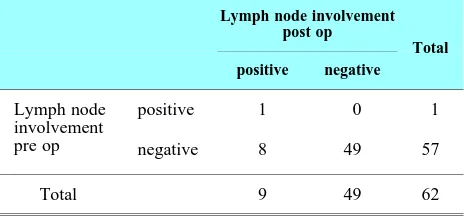
Lymph nodes that being assessed are pelvic lymph nodes based on clinical examination. Based on exami-nation, there is only 1 patient that has lymph nodes involvement. Sensitivity, specificity, positive predic-tive value and negapredic-tive predicpredic-tive value for lymph nodes examination are 11.11%, 100%, 100% and 85.96%. (Table 2)
Table 2. Lymph nodes comparison pre and post operatively.
Lymph node involvement post op
Total positive negative
Lymph node involvement pre op
positive 1 0 1
negative 8 49 57
Total 9 49 62
Parametrial are the connective tissue of the pelvic wall that extend laterally from the anterior fornix and between the broad ligament layers sensitivity, speci-ficity, positive predictive value and negative predic-tive value for parametrial involvement are 37.5%, 100%, 100% and 90.90%. (Table 3)
Table 3. Parametrial involvement pre and post operatively.
Parametrial involvement post op Total
positive negative
Parametrial involvement
pre op
positive 3 0 3
negative 5 50 55
Total 8 50 58
Cervical length were also compared and grouped into 2 categories which are less and more than 4 cm. Sensitivity, specificity, positive predictive value and negative predictive value for cervical length exami-nation are 91.84%, 88.89%, 97.83% and 66.67%. (Table 4)
Table 4. Cervical length comparison pre and post operatively.
Post op result
Total Less than
4 cm
More than 4 cm (bulky)
Cervical length
Less than 4 cm
45 1 46
More than 4 cm (bulky)
4 8 12
Total 49 9 58
DISCUSSION
Staging of cervical cancer is a clinical staging, using staging system from FIGO, that is also approved by American Joint Committee on Cancer (AJCC). The use of MRI, CT-scan, and PET-CT can help to deter-mine the most appropriate therapy, but are not used to determine the staging of cervical cancer.25
Table 1. The preoperative and post operative clinical staging.
Post-operative diagnosis
Total Stad IA1 Stad IB1 Stad IB2 Stad IIA Stad IIB
Pre-operative Diagnosis
Stad IA1 3 0 0 0 0 3
Stad IB1 2 19 0 0 0 21
Stad IB2 0 2 14 1 0 17
Stad IIA 0 0 3 13 1 17
From 58 patients in our study, we found a normal distribution of age with mean 48.39 years old. This is consistent with the literature that mention the in-creased incidence of cervical cancer in the age range from 25 - 34 years old and showed a peak at age 35 - 44 years old in Dr. Cipto Mangunkusumo General Hospital and 45 - 54 years old in Indonesia.26,27
From histopatology results, it is showed that the majority type are squamous cell carcinoma with kera-tin and adenocarcinoma, respectively 25.9 and 24.1%. Based on the literature, 70 - 80% of cervical cancer are squamous cell carcinomas type with 90% of early lesions originated from the transformation zone squamo-columnar junction of cervical epithelial cells. Changes from normal cells to dysplastic cells can last for 7 - 10 years.28 Squamous cell carcinoma without
keratin accounted for 19%, so that makes the percent-age of all squamous cell carcinoma was 44.9%.
Cervical cancer staging is still performed clini-cally; therefore, there should not be any mistakes in determining the stage of cervical cancer. In our study, each stage has a different percentage of error. The error obtained in making the diagnosis in stage IA1 was 40%, IB1 was 9.52%, IB2 was 17.65%, and IIA was 7.14%. In stage IB1, errors of diagnosis were found with 2 of 21 patients diagnosed with IB1, turned out to be at stage IA1 in surgical staging. In stage IB2, errors of diagnosis were found with down-staging of 2 patients who are supposedly in stage IB1 and upstaging with 1 patient who are supposedly in stage IIA in surgical staging out of 17 patients who are diagnosed as satge IB2. In stage IIA, errors of diagnosis were found with downstaging of 3 patients who are supposedly in stage IB2 and upstaging with 1 patient who are supposedly in stage IIB in surgical staging out of 17 patients who are diagnosed as stage IIA. Of 17 patients with clinical diagnose of stage IB2, there were 2 cases that undergo downstaging to IB1 and 1 case went upstage to IIA. Of 17 patients with clinical diagnose of stage IIA, 3 cases were found to be at stage IB2 in surgical staging and 1 case were found to be at stage IIB in surgical staging. The biggest error in making the clinical staging in our study was found at stage IA1 with 2 of 5 cases that are supposedly IA1 are diagnosed as a stage IA2. This unseemingly big error may be caused by some reasons. A few numbers of cases in our study, diffe-rent skills of the clinician who staged clinically and operatively could be the causes of the big errors.
The therapy for cervical cancer in stage IB and IIA is hysterectomy radical with pelvic lymphadenecto-my, therefore, the mistake in staging between them can still be "accepted". But, there were differences in error in diagnosing patients between staging IIA and IIB. At stage IIB, the therapy is radiotherapy {exter-nal radiation, continued by intracavitary radiotherapy (high dose rate or low dose rate)} and chemoradia-tion. Radiotherapy can be given to primary treatment, preoperative adjuvant, and palliative.
Lymph node metastasis are a risk factor that influ-ences the prognosis. Sentinel lymph node can be used to detect the lymph nodes with sensitivity 83.3%, positive predictive value 97.1%, and negative predic-tive value 16.6%.17,29-31 Our study showed that
clini-cal examination of lymph nodes has sensitivity
11.11%, specificity 100%, positive predictive value 100%, and negative predictive value 85.96%.
Parametrial involvement was examined clinically and confirmed by pathology anatomy examination. Clinical examination of parametrial involvement has sensitivity of 37.5%, specificity of 100%, positive predictive value of 100%, and negative predictive value of 90.90%.
The tumor size can be classified into bulky (> 4cm) and non-bulky (≤ 4cm) tumor. The tumor size was examined clinically and confirmed by surgical stag-ing. The sensitivity, specificity, positive predictive va-lue, and negative predictive value for clinical exami-nation of tumor size were 91.84%, 88.89%, 97.83% and 66.67%.
Further analysis will be done toward this research to take into account the time between diagnosing and treating the cervical cancer, as it has not been ac-counted before.
CONCLUSION
The error rate are 40%, 9.52%, 17.65% and 7.14% consecutively to diagnose stage IA1, IB1, IB2 and IIA. Sensitivity, specificity, positive predictive value and negative predictive value for lymph nodes exami-nation are 11.11%, 100%, 100% and 85.96%. Sensi-tivity, specificity, positive predictive value and nega-tive predicnega-tive value for parametrial involvement are 37.5%, 100%, 100% and 90.90%. Sensitivity, speci-ficity, positive predictive value and negative predic-tive value for cervical length examination are 91.84%, 88.89%, 97.83% and 66.67%. Supportive examina-tions would help clinician to obtain an optimal result of staging.
REFERENCES
1. Ferlay J, Shin H, Bray F, Forman D, Mathers C, Parkin D. Estimates of worldwide burden of cancer in 2008: GLO-BOCAN 2008. Int J Cancer. 2010
2. Bosch F, Qiao Y, Castellsagué X. The epidemiology of human papillomavirus infection and its association with cervical cancer. Int J Gynecol Obstet. 2006; 94(Supplement 1): 8-21
3. Domingo E, Noviani R, Noor M, Corazon A, Ngelangel D, Limpaphayom K. Epidemiology and Prevention of Cer-vical Cancer in Indonesia, Malaysia, the Philippines, Thai-land and Vietnam. Vaccine 2008; 26: 71-9
4. Indonesia DKR. Profil Kesehatan Indonesia 2008. 2009 5. WHO. Human Papillomavirus Infection and Cervical
Can-cer WHO, 2005
6. WHO. Global Burden of Disease 2004 Part 3: Disease In-cidence, Prevalence, and Disability, 2004
7. Sjamsuddin S. Pencegahan dan deteksi dini kanker serviks. Cermin Dunia Kedokteran 2001; 133: 8-13
8. Young R. Gynecologic Malignancy. In: Braunwald E, Fauci A, Hauser S, Jameson J, Kasper D, Longo D, editors. Harrison’s Principles of Internal Medicine. New York: McGraw-Hill; 2005: 556-8
9. Schiffman M, Castle P. The promise of global cervical-can-cer prevention. New Eng J Med. 2005; 353(20): 2101-4 10. Improving screening coverage rates of cervical cancer
pre-vention programs: A Focus on Communities, 2004 11. Crum C. The Beginning of the End for Cervical Cancer?
Eng J Med 2002; 347(21): 1703-5
12. BPS. Hasil Sensus Penduduk Indonesia 2010 Data Agregat per Propinsi. Jakarta: BPS. 2010
13. Data Anggota. Ikatan Anggota Patologi Indonesia Jakarta: Sekretariat IAPI; 2010
14. POGI. Daftar POGI Cabang Jakarta: POGI; 2010 15. Andrijono. Kanker Serviks. Divisi Onkologi Departemen
Obstetri Ginekologi RSCM; 2004
16. Moore D. Cervical cancer. Obstet Gynecol. 2006; 107(5): 1152-61
17. Levenback C. Cervical cancer. In: Barakat R, Bevers M, Gershenson D, Hoskins W, editors. Handbook of Gyne-cologic Oncology. London: Martin Dunitz Publishers Ltd; 2002: 231-47
18. Lagasse LD, Creasman WT, Shingleton HM, Ford JH, Blessing JA. Results and complications of operative staging in cervical cancer: Experience of the Gynecologic Onco-logy Group 1980; 9(1): 90-8
19. Hacker NF. Operative Treatment of Cervical Cancer. Clini-cal and operative staging of cerviClini-cal cancer. Baillière’s Clinical Obstetrics and Gynaecology, 1988; 2(4): 747-59 20. Chung HH, Kang S-B, Cho JY, Kim JW, Park N-H, Song
Y-S. Can preoperative MRI accurately evaluate nodal and parametrial invasion in early stage cervical cancer? Japan J Clin Oncol 2007
21. Selman TJ, Mann C, Zamora J, Appleyard T-L, Khan K. Diagnostic accuracy of tests for lymph node status in pri-mary cervical cancer: a systematic review and metaanalysis. CMAJ. 2008: 855-62
22. Jena A, Oberoi R, Rawal S, Das SK, Pandey) KK. Para-metrial invasion in carcinoma of cervix: role of MRI meas-ured tumor volume. Bri J Radiol. 2005; 78: 1075-7 23. Sironi S, Belloni C, Taccagni GL, DelMaschio A.
Carci-noma of the cervix: Value of MR Imaging in Detecting Parametrial Involvement. AJR. 1991: 156
24. Monaghan JM. Operative Treatment of Cervical Cancer. Management decision making using clinical and operative staging in cervical cancer. Baillière’s Clinical Obstetrics and Gynaecology. 1988; 2(4): 737-46
25. National Comprehensive Cancer Network (NCCN). NCCN Guidelines on Cervical Cancer, 2011
26. Aziz MF. Gynecological cancer in Indonesia. J Gynecol Oncol. 2009; 20(1): 8-10
27. Aziz MF, Mangunkusumo R. Epidemiology Cancer of the Cervix. CME on Gynaecological Oncology, 2000 28. Shin B, Dubeau L. Cell cycle abnormalities in squamous
cell carcinoma of the cervix. CME J Gynecol Oncol. 2001; 6: 167-72
29. Krivak T. Cervical and Vaginal Cancer. Novak’s Gynecology. Philadelphia: Lippincott Williams and Wilkins; 2002: 1223-5 30. Hacker N. Cervical Cancer. In: Berek J, Hacker N, editors. Practical Gynecologic Oncology Philadelphia: Lippincott Williams and Wilkins; 2005: 337-95