Abstracts of the Fourth European Congress of Andrology and
23rd Congress of the French Speaking Society of Andrology
Andrologie 2006, 16, N~ 319-393
Posters
(PO 001 to PO 116)
PO 001
Obesity and male reproduction function
S. PFLIEGER-BRUSS 1, F. WEMBER1, R.H.
BODEKER2, W.B. SCHILL1, H.C. SCHUPpE1
1 Centre of Dermatology and Andrology, and 2 Institute forMedical Informatics, Justus Liebig University, Giessen, Germany (Hans-Christian. Schuppe@derma. med. uni-
giessen.de)
Objective
: Reproductive function may be affected by environmental and occupational exposures as well as changing lifestyle. However, whether or not these factors contribute to an increasing risk of male reproductive health problems including poor semen quality is stirring an ongoing debate. On the other hand, obesity is becoming increasingly prevalent worldwide and is now considered to be one of the most important health concerns in many countries. Notably, the impact of the woman's body mass index (BMI) on fecundity is well established, whereas only few reports addressed the potential reproductive hazards of obesity in males. Therefore, this study aimed at investigating the relationship between BMI and parameters of reproductive function in men attending an infertility clinic.Patients and Methods :
The retrospective study included a total of 496 men undergoing andrological examination for infertility work-up. Apart from infertility, inclusion criteria comprised age (18-50 years), BMI >18.5 kg/m2, uneventful andrological history, and normal genital examination. Patients with genetic or congenital abnormalities, genital disorders, systemic disease, therapeutic use or abuse of drugs, as well as seminal signs of inflammation or obstruction were excluded. Semen analysis had been performed according to WHO guidelines extended by biochemical markers and microbiology. For statistical analysis, patients were categorized according to BMI considering cigarette smoking and positive seminal bacteriology as confounders.Results
: A normal BMI (>18.5 to <25 kg/m2) was found in 209 men (42.1%), 287 patients (57.9%) showed a BMI >25 kg/m 2. Compared to the general population in Germany, the prevalence of obesity was markedly increased, with the highest difference among the subgroups of men aged 25-29 years and 30-34 years (39.7 vs 61.2% and 48.3 vs 62.3%, respectively). Semen analysis revealed normozoospermia in 35.4% of men with normal weight, but only in 25.4% of those with a BMI >25 kg/m2. However, this trend was not significant and also the confounding factors showed no marked effect on semen quality. A significant negative correlation could be observed between BMI and serum testosterone, whereas gonadotrophin levels remained unaffected.Conclusions
: The results confirm previous reports describing a decrease of serum testosterone with increasing BMI. The high prevalence of obesity among men attending an infertility clinic suggests that this factor may contribute to male reproductive health problems, although the trend towards deterioration of semen quality with increasing BMI is not significant. With regard to the alarming prevalence of obesity among adolescents, this issue might become increasingly important in clinical andrology in order to prevent at least some cases of male subfertility.PO 002
Incidence of dysspermia categories in a
contemporary diagnostic setting
D.A. ADAMOPOULOS, S. NICOPOULOU,
C. MICHALAKIS, A. PAPPA, E. KOUKKOU, E. VENAKI
Andro/ogy Clinic, Endocrine Dept., E/ena Venize/ou Hosp., 11521Athens, Greece
Objective :
To classify sperm disturbances in relation to possible aetiological factors using the standard clinical approach, employing all available diagnostic tools andfollowing WHO's guidelines for categorization (W.H.O., 2000).
Design :Analysis of the clinical material of an Endocrinology Department embedded Andrology Clinic of a busy inner-city hospital in Athens for classification of sperm disturbances was made using the diagnostic categories proposed by W.H.O. but expanded to include new classes not previously reported (e.g. epididymopathies, multifactorial causes, etc.).
Materials-Methods : A total of 774 cases investigated for couple subfertility and found to be dysspermic have been selected from a large database from which normozoospermic men have been excluded. The diagnostic classification was based on meticulous history taking, physical examination, semen analysis (W.H.O., 1999 criteria,) imaging techniques, endocrine tests (including inhibin-B), and when needed, testicular biopsy and chromosome and molecular analysis.
Results : On the basis of the factors identified these cases were groupped into 3 major categories: (a) single-factor group (37.3%), (b) two-factor group (34.0%) and (c) three or more factors group (28.27%).
In the single-factor group, the incidence recorded was in the following order: 1. Idiopathic OTA (40.6%), 2. varicocele (18.7%), 3. epididymopathy (12.8%), 4. environmental (8.0%), 5. infections (5.3%), 6. acquired testicular damage (4.8%), 7. congenital anomalies (3.2%), 8. systemic causes (2.1%), 9. endocrine causes (1.6%), 10. sexual-ejaculatory dysfunction (1.3%) and four other diagnostic groups with less than 1% or no representation. In the two-factors group, epididymopathy (31.3%), varicocele (26.5%) and environmental (20.6%) factors were the most frequently encountered components of the various combinations. Finally, in the three or more factors group, the main components were environmental (24.8%), varicocele (19.2%) and epididymopathy (19.0%).
As it is obvious, in single factor aetiology idiopathic OTA was the principal category whereas in all multifactorial combinations varicocele, environmental factors and epididymopathy were dominating the field.
Conclusions : The diagnostic categories formulated in this analysis differ significantly from the relevant data presented from the much larger series from other centers (Nieschlag, 2001). In our material, multiple factors have been recognized in the great majority of the cases and the single-factor category was clearly in a minority. It appears that the detailed diagnostic work-up with the introduction of new investigational parameters was instrumental in explaining, at least in part, the marked differences between the two series. On the other hand, one should also count the differences between the relevant populations in terms of environment, prevailing health conditions, socioeconomic status, etc. As it becomes obvious, studies charting each population's patterns of dysspermia should be available in each different population as a prerequisite for a sound reproductive policy for the male.
References :
1. World Health Organization WHO : Laboratory Manual for the Examination of Human Semen and Sperm-Cervical Mucus Interaction. 4th ed., Cambridge, CUP, 1999.
2. World Health Organization WHO : Manual for the standardized investigation, diagnosis and management of the infertile male. Rowe P.J. et al. eds. Cambridge, CUP, 2000.
3. Nieschlag, E. : Classification of andrological disorders. In: Nieschlag E., Behre H.M. eds. Andrology, Male Reproductive Health and Dysfunction. Berlin, Springer, 2001.
PO 003
Interests of the post coital test in the investigation of the infertile couples
N. ABID, N. BEN JAMAA, M. AJINA, A. SAAD Service of Cytogenetic and Biology of Reproduction, university Hospital F Hached Sousse, Tunisia. Email :
mounir.ajina @rns.tn
Objective : to establish the profile of the cervical mucus and the sperm to infertile couples of the Tunisian centre.
Design and place : Service of Cytogenetic and Biology of Reproduction, University Hospital F Hached Sousse, Tunisia.
Methods : Our study concerns 49 infertile couples which benefited of at least: a postcoital test (PCT) and a spermogram. Test was realized in meadow ovulatory period among the 11- th and the 12-th day of the cycle after an average sexual intercourse of 8 hours and a 3 days average sexual abstinence.
The exo cervical mucus is taken with an a bit long crowbar, we measures the filance between the two branches of the crowbar; on the other hand the endocervical mucus is taken by means of an aspiglaire. The endo and exo cervical mucus are examined under microscope between blade and small strip.
Results : The majority of the couples (63%) benefited from a hormonal treatment before the taking of the cervical mucus. Only 18% of the patients benefited during the PCT of a double taking endo and exocervical. Post coital test was positive in 89% of cases and negative in 11% of cases. Only 12% of the patients have cervical infertility and a single patient has an azoospermia.
Conclusion : post-coftal test is a test of reliable diagnosis to infertile couples because according to our results the majority of the patients who benefited from PCT are porters of cervical infertility.
References :
2. S. Hammamah et al. ; Medecine et biologie de la reproduction, 2eme edition Masson, 2004, p 152.
3. Clinical Guideline 11 : Fertility: assessment ant treatment for people with fertility problems. 1.4.6 Post-coital testing of cervical mucus.
correlated with the grade of the venous reflux. Doppler ultrasonography is an essential diagnostic tool before surgery and a valuable measure of postoperative results.
PO 005
PO 004
The place of Doppler ultrasonography in evaluating male infertility
V. CAUNI1, D. DINU 2, C. PERSU 1, P. GEAVLETE1
1 Department of Urology, Saint John Emergency C/in/ca/ Hospital, Bucharest, Romania 2 "C.I. Parhon" Institute of
Endocrinology, Bucharest, Romania ([email protected])
Introduction and Objectives : Male infertility may be the cause of several disorders, so that identifying a correct diagnosis and an appropriate treatment may represent a challenge for the clinician. Our goal was to evaluate in a retrospective study the correlations between spermatic parameters, scrotal abnormalities and the resistive index in infertile males, as determined by Doppler ultrasonography.
Patients and methods : Our study group consists of 56 infertile males, aged between 21 and 46 years old, and 40 fertile males, as a control group. The evaluation protocol included andrological examination, hormonal profile, and determination of antispermatic antibodies, prostatic and testicular Doppler ultrasonography.
Results : Testicle hypotrophy was diagnosed in 40.7% cases, 26% patients presented cysts or calcifications of the epydidimus, 32% had chronic prostatitis, 12% had spermatocystitis, 10% had testicular tumors. 29.64% of the infertile patients had olygospermia. Varicocele was present in 71.42% of cases, and in 31.57% cases the varicocele was bilateral- stage I varicocele in 26.6%, stage II in 40% and stage III in 33.4%. Doppler ultrasound combined with the Valsalva maneuver identified three grades of reflux : grade I in 22.2% cases, grade II in 35.5% cases and grade III in 42.3% cases. The mean value of the resistive index in the control group was 0.56, in patients with varicocele the mean value was 0.46, and in patients with azoospermia the mean value was 0.90. The severity of the spermatogenetic dysfunction was better correlated with the grade of reflux than with the clinical stage of varicocele.
Conclusions : Varicocele is the cause of infertility in about 50% of the patients studied. The severity of olygospermia is
Epidemiologic features of varicocele in a large cohort of Greek men with infertility
P.D. KANTARTZI, D.C. GOULIS, P.K. ILIADOU, C. TSAMETIS, D.G. GOULIS, J. BONTIS, J. PAPADIMAS
Unit of Reproductive Endocrinology, 1st Department of Obstetrics & Gynaecology, Aristotle University of
Thessaloniki, Greece
Objective : Varicocele is a common cause of male infertility and one of the most controversial issues in the field of Andrology. The main aim of this study was to analyze the epidemiologic, clinical, hormonal and sperm parameters in a large cohort of infertile men with varicocele in a northern Greek population. A secondary aim was to detect changes of these parameters in men who underwent surgical repair of varicocele.
Design : Retrospective, epidemiologic, descriptive, clinical study.
Materials and Methods : We accessed medical records of 925 infertile men that were examined in our outpatient clinics between 1991 and 2005 ; 429 (46%) of them having either a clinical varicocele or a surgically repaired varicocele were included in the study. Studied parameters included age of male and female partners, type and duration of infertility, testicular volume, side and grade of varicocele, FSH, LH, prolactin, testosterone and sperm parameters before and after the surgical repair where available. Of the 429 men, in 272 (64%) varicocele was the only cause of infertility, whereas other additional causes included infection (n=77 ; 18%), idiopathic non-obstructive azoospermia (INOA) (n=40 ; 9%), cryptorchidism (n=16 ; 4%), obstruction (n=7 ;2%) and other causes (n=17 ; 3%).
Results : Studied parameters are illustrated in the table. Results are given as mean + standard deviation or percentage.
In the subgroup of men (n=87) who underwent surgical repair of varicocele, no statistically significant changes were found in sperm and hormonal parameters after the operation. The lack of statistical significance remained when we analyzed separately men with varicocele only and men with varicocele plus INOA.
All men Varicocele only Varicocele and INOA Age of men (years) 33.5 • 6.3 33.5 • 6.2 32.7 • 6.5 Age of women (years) 33.3 + 5.6 33.1 • 6.1 32.2 • 4,3 Type of infertility
pdmary 75% 75% 84%
secondary 25% 25% 16%
Duration of infertility (years) 3.8 _+ 3.6 3.5 • 3.0 4.2 • 4.1 Right testis volume (mL) 21 _+ 5 22 • 3 15 • 7 Left testis volume (mL) 20 • 6 21 • 4 12 + 7 Varicocele side
left 61% 63% 54%
d@ht 4% 2% 22%
bilateral 35% 35% 24%
Varicocele grade
grade I 11% 10% 9%
grade II 85% 87% 82%
~lrade III 4% 3% 9%
FSH (mlU/mL) 9.3 • 9.5 6.9 _+ 4.0 16,4 • 14,7 LH (mlU/mL) 6.8 + 4.8 6.0 • 4.0 9.3 • 5.7 Prolactin (nglmL) 8.4 _+ 6.3 7.3 + 5.2 10.7 • 6.6 Testosterone (ng/dL) 505 • 221 533 • 228 427 + 241 Sperm volume (mL) 3.9 + 1.8 3.9 _+ 1.7 3.5 + 1.5 Sperm number (106/mL) 32.3 + 42 36.5 • 46,2 7.5 • 17.0 Sperm motility (%) 30.5 + 22 33.3 • 22.4 15.3 + 19,5 Sperm morphology (%) 28.8 • 21 30.2 • 21.2 17.8 • 17.9
Conclusions : Varicocele is a very common finding in infertile men, although an etiologic relationship between varicocele and male infertility is difficult to be established. According to our findings, surgical repair does not seem to be generally effective, thus it should be applied only in a meticulously selected group of men.
Presenting author: Prof. J. Papadimas, 1st Department of Obstetrics & Gynaecology, "Papageorgiou" General Hospital, Periferiaki Odos, Nea Efkarpia, 56403, Thessaloniki, Greece. e-mail: [email protected]
Material and Methods : 22 infertile men, aged 20 - 45 years old, with varicocele and normal hormone levels, were included. The evaluation protocol included an andrological examination, the evaluation of hormonal status, two spermograms in a three months interval, antispermatic antibody measurement, sperm cultures, cytogenetic exam and testicular pulse Doppler ultrasound. Venous reflux associated with the Valsalva maneuver was also determined. Patients were reassessed 2, 4 and 6 months after surgery for varicocele, by seminal liquid analysis, antispermatic antibodies measurement. Also, ultrasonography was routinely performed.
Results : Antispermatic antibodies were present in 77.8% of the infertile patients, significantly correlated with the grade of the venous reflux and less dependant on the clinical stage of the varicocele. The spermograms showed a wide range of abnormalities, from astenospermia to severe oligo- asthenoteratozoospermia. These abnormalities were more severe in patients with intratesticular varicocele and high grade venous reflux. After surgery, the testicle hypotrophy stopped its progression in all patients. Ultrasound evaluation 2 months after surgery showed the disappearance of the venous reflux at the Valsalva maneuver in 90% of cases. All patients who induced spontaneous pregnancies were under 35 years old.
Conclusions : Varicocele induces autoimmune processes, with an increase in spermatic antibodies. Surgery for varicocele significantly increased the fertility potential in men under 35 years old. Future research is needed in order to reveal the interdependent mechanisms involved in spermatogenesis abnormalities and decreased spermatic motility noted in males with varicocele.
PO 006 PO 007
The varicocele - a frequent cause of male infertility
Effect of varicocele on the formation of ant• antibodies " A S A "
V. CAUNI1, D. DINU 2, C. PERSU1, C. DUMITRACHE 2, P. GEAVLETE 1
1 Department of Urology, Saint John Emergency Clinical Hospital, Bucharest, Romania 2 "C.I. Parhon" Institute of
Endocrinology, Bucharest, Romania (caunivictor@yahoo. com)
ABD ALLAH M. ATTIA, ALAA H. MARAEE, KHALED A. ALl, HAYAM M. HANOUT, AZZA G.A. FARAG* AND
EMAN N. EL SHAFEY
Dermatology, Andrology & S. T.Ds and Clinical Pathology Depts*, Minoufiya University Corresponding author :
Hyperlink "mailto:yasienhossam@yahoomail. com" yasienhossam@yahoomail, com.
Introduction : Although the pathophisiology of the varicocele is a well known matter, its correlation with male infertility and with the optimal timing of surgery in men with associated spermatogenesis abnormalities and decreased spermatic motility is still controversial. Our objective is to asses the consequences of the varicocele on the reproductive function.
The exact etiopathology through which varicocele can affect fertility potential is still unknown.
The study included two groups: thirty evident varicocele (grades II and III) infertile patient (group I) and fifteen varicocele-free fertile, age matched volunteers as a control group (group II).
Both patients and controls were subjected to; standard semen analysis, expressed prostatic secretion examination; excluding those having chronic prostatitis and detection of both serum and seminal plasma ASA by ELISA.
The results showed that; the varicocele patients have highly significant low sperm density and percent of actively motile sperm (p<0.001) and significantly higher level of abnormal forms (p < 0.05) compared to controls.
The results also showed that; in varicocele group; both serum and seminal plasma ASA are negatively correlated with sperm density (r=-0.732 and -0.66 - p < 0.001) respectively, and percent of actively motile sperms (r=-0.739 and -0.771 - p < 0.001) respectively, whereas serum and seminal plasma ASA are positively correlated (r=0.685, p<0.001), denoting that seminal plasma ASA are derived from the systemic circulation.
In conclusion; varicocele can precipitate to the formation of ASA. The latter can be considered as one of the etiopathogenic mechanisms through which varicocele can affect the fertility potential.
Results : The semen analysis found an improvement of all the parameters for all the patients without normalization except for the morphology. The post operative outcome was statistically significant for the density. The majority of the patients (76% to 92%) had abnormal preoperative values of semen except for the morphology (28%). Semen analysis parameters were improved for 48% to 64% of patients depending on the parameter. For patients with abnormal semen, the lower the mean preoperative value of a parameter, the higher it becomes postoperatively. The post operative pregnancy rate was 31,42%. The fertile patients were those with a younger age, a shorter duration of infertility, constantly improved semen analysis parameters compared to infertile patients.
Conclusion : Values and improvement of semen analysis parameters are more favourable in patients with young age and those with bilateral varicocele and a secondary infertility.
Keywords: Fertility, Semen analysis, Varicocele repair.
PO 009
PO 008
Varicocelectomy and scrotal temperatures in infertile men with varicocele
Primary varicocele and fecundity : Post surgical assessment of sperm and fecundity parameters
L. NIANG, I. LABOU, O. ALl, M. JALLOH, R.KANE, M. NDOYE, S.M.GUEYE
Service d'Urologie et d'Andrologie H6pital Gen6ral Grand Yoff, Dakar- Etoile [email protected]
T. ALMONT1, E. HUYGHE 1,2, p. PLANTE 1,2, P. THONNEAU 1, L. BUJAN1, R. MIEUSSET 1
1 EA 3694 "Recherche en fertilit6 humaine - Sante de la reproduction dans les PVD ", HSpital Paule de Viguier, 330
av de Grande-Bretagne, TSA 70034, 31059 Toulouse Cedex, France 2 Service d'Urologie -Andrologie, HSpital
Rangueil, Toulouse, France
Objectives : To assess the post operative evolution of male infertility (semen analysis parameters) and the fertility outcomes.
Materials and Methods : We underwent a retrospective study including 50 patients with a varicocele operated according to Palomo procedure in the department of Urology of H6pital General de Grand Yoff. The parameters studied were related to semen analysis (density, mobility in the first hour and the vitality) and the spermocytogramme (count of normal spermatozoa). This analysis was done once before the operation and twice after the operation (between the 3rd and 8th month and from the 9th month).
Objectives : Increased scrotal temperature is one of the various factors either associated with or considered as a cause of impaired spermatogenesis in infertile men with varicocele. Some studies have reported a reduced scrotal temperature after varicocelectomy. We investigated whether this finding was true, and if such a reduction in temperature persisted over time after surgery.
Materials and Methods : This retrospective study included 2 groups of infertile patients with a left varicocele: an operated group of 116 patients, and a non-operated group of 40 patients included in an intrauterine insemination programme (IUI). Varicocele was diagnosed by clinical examination (grade I: Valsalva positive ; grade Ih palpable; grade IIh visible). In both groups clinical examination was performed before surgery
or before inclusion in the IUl programme (TO) and again at 3 months (T3) in both groups, and at 6 months (T6) after surgery in the operated group. Scrotal temperatures were measured on each side with a special thermometer with the patient naked and in a supine position for at least 10 minutes.
Results : Comparison of scrotal temperature values at TO and T3 revealed no significant change in left and right mean temperatures in the non-operated group, while both left and right mean temperatures decreased significantly in the operated group.
In the operated group, comparison of scrotal temperature values at TO, T3 and T6 indicated that left and right mean temperatures were significantly lower at T3 and T6, with no difference between T3 and T6 values.
Considering a value <35.1~ as the upper limit of normal scrotal temperature, an abnormal temperature (>35.1 ~ was observed in 51% (59/116) of the operated group before surgery. In this subgroup of patients with abnormal temperature before surgery, temperature was normal in 86% (51/59) 3 months after surgery. In the subgroup of patients with normal temperature before surgery, scrotal temperature was still normal in 91% (52/57) but abnormal in 9% (5/57).
Conclusions : Surgery induced a reduction in mean scrotal temperature in a group of infertile patients with a left varicocele. This reduction was indeed the result of surgery, as mean scrotal temperature was unchanged in the non-operated group of infertile patients with a left varicocele. Moreover, this reduction seems to be durable as temperature values were still decreased at 3 and 6 months after surgery. However, considering individual scrotal temperatures, only 51% of the patients in the operated group had an abnormal value before surgery.
Support : None.
PO 010
Prevalence of hypospermia and hyperspermia and their relationship with genital tract infection
in tunisian infertile men
N. ABID1, N. CHAKROUN 1, A. SELLAMI1, A. BAHLOUL2, T. REBAI1, L. AMMAR-KESKES 1
1 Laboratory of Histology-Embryology, Faculty of M6decine, Sfax-Tunisia. 2 Research Unit "male infertility",
Habib Bourguiba hospital, Sfax-Tunisia. Correspondance to Pr. Ammar-Keskes Leila, e-mail: Ikeskes@ yahoo.fr
their possible associations to other spermatic abnormalities and their possible relationship with the genital tract infection.
Design and Setting : Our retrospective study concerned 2332 spermiograms performed between 1995 and 2005 into the laboratory of Histology of Medicine University of Sfax, among patients consulting for couple infertility.
Materials and Methods : The spermiograms were carried out according to the standardized method of WHO. We distinguished three great groups according to the semen volume : Gn (normospermia), GH (hyperspermia) and Gh (hypospermia) ; from Gh we individualized one subgroup called Ghl (severe hypospermie : volume<lml). For all these groups, we determined the average values of semen parameters and compared them using the Student test; the frequencies of semen abnormalities (azoospermia, asthenospermia, necrospermia, oligospermia, leucocytospermia and teratospermia) were also determined and compared, using the Chi-2 test. The threshold of significance of p value was<0.05.
Results : The total prevalence of the hypospermia was 18.3%; severe hypospermia was found in 3.7% of cases. The frequency of the hyperspermia was only 6.8%. Many significant differences were found between Ghl, Gn and GH, mainly concerning motility, total spermatozoa count and vitality, which were lower in Gh and Ghl groups, comparatively with Gn and GH groups, as well as the average rate of coiled tails and the frequencies of semen abnormalities; in fact the frequencies of asthenospermia, necrospermia, leucocytospermia, oligospermia and azoospermia were higher in Gh and Ghl, comparatively with Gn and GH. In addition, in Gh and Ghl, azoospermia was more frequently associated to abnormal pH than in Gn and GH ; thus basic pH (>8.5) was associated to azoospermia, respectively in 32.5% and 46.15% of Gh and Ghl patients, versus 19.44% and 16.6% of Gn and GH patients; in the same way, azoospermia was associated to an acid pH in 4.6% and 15.38% of Gh and Ghl patients, versus 0% of Gn and GH patients.
Conclusion : It seems that hypospermia is associated to an active genital infection responsible for many spermatic disturbances and for the appearance of biological inflammation signs (leucocytospermia). But, our results led to suggest that hyperspermia is not related to an evolutive genital tract infection, since the average values of the principal semen parameters, as well as the frequencies of semen abnormalities were comparable with those found in the normospermic group. We could also suggest in the light of these results that hyperspermia would be associated to a recent infection that did not yet induced semen quality disruption. In addition, we suggest that among azoospermic patients, hypospermia is related to a prostatic pathlogy (prostatitis with high pH in seminal plasma) than to a seminal vesicule deficiency (low pH in seminal plasma).
PO 010 bis PO 011
Prevalence of asymptomatic inflammatory
prostatitis in young healthy men in Estonia
Genital tract infectious and inflammatory
pathology and male infertility
P. KORROVITS1,2, K. AUSMEES 2, R. MANDAR 1
M. PUNAB2
1 Department of Microbiology, University of Tartu, Estonia 2 Andrology Centre, Tartu University Hospital, Estonia
Hyperlink "mailto:[email protected] "
A. SELLAMI-BEN HAMIDA1,2, L. AMMAR-KESKES1,2,
N. ABID2, T. REBAI2, N.M. MHIRI1, A. BAHLOUL1
1 Research Unit "male infertility", Habib Bourguiba Hospital, Sfax Tunisia 2 Laboratory of Histology and
Embryology; Faculty of Medicine, Sfax Tunisia Correspondance to Pr. Ammar-Keskes Leila, e-mail :
Ikeskes@ yahoo.fr
Objective
: The aim of our study was to determine the prevalence of asymptomatic inflammatory (NIH category IV) prostatitis in young healthy men in Estonia.Materials
and Methods
: The study group consisted of 562 men (291 Estonians, 271 Russians) aged 16-25 years (mean age 18.8 years). Cytologic examination of their ejaculate (using Bryan-Leishman stained slides) was performed. In addition, all subjects were clinically examined for possible pathologies in genital region and basic semen parameters (volume, concentration and motility). Subjects with any clinical symptoms of inflammation were excluded.Results
: The prevalence of asymptomatic inflammatory prostatitis (>1 million WBC (white blood cells) per ml in sperm, according to WHO guidelines) was 6.0%, but when we used lower threshold suggested by our previous studies (>0.2 million WBC/ml), the prevalence was 19.0%. No difference between the two ethnic groups were found when seminal parameters and inflammatory markers were compared. In this study the preliminary analysis did not show any significant effect of leukocytospermia on sperm quality. We did not detect any seasonal differences in the prevalence of asymptomatic inflammatory prostatitis.Conclusions
: Asymptomatic inflammatory prostatitis is common among healthy young males, suggesting the need for further studies in order to investigate pathogenetic mechanisms of the disease. Our study also suggests that more attention should be paid to evaluation and treatment of asymptomatic inflammatory prostatitis in young men. Prevalence data we found should be taken into account when estimating the total prevalence of all forms of chronic prostatitis, both symptomatic and asymptomatic.Support
: The study was supported by Estonian Science Foundation (grant no 5701) and EU 6th FP project QLRT-2001- 0291.Objective
: To assess the prevalence of genital tract infection and inflammation on male infertility and to elucidate the importance of both clinical, biological and ultrasonographic investigations in the diagnosis of the male chronic genital tract infection.Design :
Retrospective study.Setting :
Research Unit "male infertility", Habib Bourguiba Hospital, Sfax Tunisia and Laboratory of Histology and Embryology; Faculty of Medicine, Sfax Tunisia.Materials and Methods
: A total of 220 male partners of infertile couples were evaluated by the study of their medical file, biological investigations (semen analysis and culture) and ultrasonographic examination. Our cohort was subdivided into two groups : (G1, n= 49) included patients with genitourinary tract infection/inflammation; and (G2: n=171) included patients without genitourinary tract infection/inflammation. The presence of genital tract infection/inflammation was attested by the presence of bacteriospermia or leukocytospermia or by the existence of several clinical and/or ultrasonographic and/or other biological abnormal parameters. Statistical analysis was performed using Statview software. Frequencies were compared by the chi-square test and means values were compared by Student's unpaired t test. Level of significance was fixed at p<0.05.Results
: Genital infection and/or inflammation was detected in 22.3% in our patients (49/220). In history taking, epididymo- orchitis and urethral discharge were found with significantly higher prevalence in G1 (24.4% and 18.3%, respectively) than in G2 (5.8% and 5.8%, respectively) ; p values were 0.004 and 0.01, respectively. Urinary tract infection was detected in 16.3% of patients in G1 and 1.1 % in G2 (p<0.001). Semen culture was negative in 38.8% of patients in GI. Semen parameters were altered in G1 with significant lower mean values of motility (total and rapid progressive spermatozoa), of vitality and of morphology (essentially tail defects) in G1 than in G2. Hypospermia was significantly more frequent in G1 than in G2 (32.5% vs 15.4%, p=0.02). The prevalence of leukocyospermia (>0,5millions/ml) wasalso significantly higher in G1 compared with G2 (24.1% vs 2.6%, p =0.01). By ultrasonography examination, genital male tract abnormalitites (cysts, nodules) and accessory gland calcifications were founded more frequently in G1 (21.8% and 11.9%, respectively) than in G2 (2.8% and 0.7%, respectively), with respective p values : <0,001 and 0.003.
Conclusions : Our results show that genital male tract infection and/or inflammation occurs frequently in infertile patients. Negative semen culture has low sensitivity for discrimination between patients with and without infection. To establish the diagnosis of infection or silent genital tract inflammation, it would be necessary to confront the clinical, biological and ultrasonographic investigation results.
PO 012
Bacteria trigger production of reactive oxygen intermediates and lipid sperm membrane
peroxidation in in vitro model
M. FRACZEK 1, A. SZUMALA-KAKOL2,
P. JEDRZEJCZAK3, M. KAMIENICZNA 1, M. KURPISZ1 1 Institute of Human Genetics, Polish Academy of Sciences, Poznan, Poland ; 2 Unit of Microbiology, Hospital
Medical College, Poznan, Poland; 3 Clinic of Infertility and Reproductive Endocrinology, University of Medical Sciences, Poznan, Poland ([email protected])
Objective : The relationship between the presence of infectious factor in semen and sperm fertilizing potential remains to be actively studied. Microorganisms invasion results in the development of inflammatory process and is usually accompanied by the oxidative stress. As the role of particular groups and species of microbes invading, colonizing or infecting the male reproductive tract is still poody understood, we aimed to assess an in vitro effect of the five most often isolated aerobic, anaerobic and atypic bacterial strains from semen of our infertile patients. These are : Escherichia coil, Staphylococcus haemolyticus, Streptococcus oralis, Bacteroides ureolyticus and Ureaplasma urealyticum. We have studied their influence on the level of reactive oxygen intermediates (ROI) generated in co-incubated suspensions of white blood cells (WBCs) and spermatozoa as well as quantitating sperm membrane malondialdehyde (MDA).
Design : An in vitro model of semen infection.
Materials and Methods : The venous blood samples were obtained from 10 healthy adults and WBCs were isolated using a density gradient centrifugation. Spermatozoa were isolated by swim-up technique ('swim-up' fraction) and
discontinuous Percoll gradient (90% and 47% Percoll fractions) from 10 healthy, normozoospermic volunteers. The measurement of ROI secretion by WBCs previously incubated with bacterial strains before and after addition of spermatozoal fractions was determined by chemiluminescence in the presence of luminol. MDA concentrations were studied by using a high-performance liquid chromatography (HPLC) in sperm lysates after incubation of sperm with bacteria and/or WBCs.
Results : The presence of bacteria in co-incubated suspensions of sperm and WBCs was connected with reduced ROI scavenging potential of sperm, especially of spermatozoa with the best seminological parameters selected by swim-up procedure. An increase of detected MDA after incubation of sperm cells with bacterial strains was observed (which was a natural consequence of non neutralized ROI in co-incubated mixture of WBCs and spermatozoa) although no statistic difference was found. The presence of leukocytes generally was associated with elevated levels of MDA levels, in 'swim- up' fraction in particular, and the greatest effect was exerted by B. ureolyticus and S. haemolyticus (p<0.01 and p<0.05, respectively, in comparison to spermatozoa incubated with leukocytes only). Comparisons between MDA content in spermatozoal fractions with no leukocytes and after leukocytes incubation with selected bacterial strains exhibited significant differences when Str. oralis, S. haemolyticus and U. urealyticum were applied (p<0.05 and p<0.01).
Conclusions : The results obtained in this study constitute another evidence indicating that bacteria which invade, colonize or infect the male reproductive tract are important inducers of the oxidative stress in semen which may play a crucial role in male gamete dysfunction through the peroxidative damage of sperm membrane lipids. Our data support the hypothesis about cooperation between bacteria and WBCs in triggering both structural and functional defects in human germ cells. Possibly extended imbalance between pro-and antioxidative activities in semen, primarly caused by an infectious factor may lead to limited fertilizing ability of spermatozoa as a consequence of enhanced ROI reactivity with cell components.
PO 013
Costimulatory molecules, chemokine receptors
and proinflammatory cytokines in dendritic cell
population in normal and chronically inflamed rat
testis
(auto)antigens and are in a status to migrate to the local nymph nodes for T cell activation. They are in a ready migratory state, but functionally immature.
Our data provide the first firm evidence for the existence of DC in the testis and in conjunction with the previous characterization of autoantigens a new tool for the investigation of the underlying causes of male factor immunological infertility.
M. FIJAK1, L. LUSTIG 2, W. VON WULFFEN3,
R. IOSUB1, V.A. GUAZZONE2, E. SCHNEIDER1,
A. MEINHARDT1, C. RIVAL2
1 Department of Anatomy and Cell Biology, Justus-Liebig- University of Giessen, Aulweg 123, 35392 Giessen,
Germany 2 Center for Research in Reproduction, University of Buenos Aires, Argentina 3 Department of
Pulmonary and Critical Care Medicine, University of Giessen Lung Center (UGLC), Germany Hyperling "mailto:Monika. Fijak@anatomie. med. uni-giessen, de"
Monika. Fijak@anatomie. med. uni-giessen, de
Dendritic cells (DC) are potent antigen presenting cells and presentation of self antigens by DC is likely to play an important role in the initiation of autoimmunity and its progression. Our recent characterization of testicular autoantigens in a model of chronic testicular inflammation, i.e. experimental autoimmune orchitis (EAO) prompted us to investigate the presence the DC in normal and EAO rat testis.
DC in the testes were quantified by immunohistochemistry using the DC specific antibodies Ox-62 and CD11c followed by stereological analysis. The number of DC in EAO testes (ca. 7xl05/testis) increased significantly compared to adjuvant and untreated control rats (ca. lxl05/testis).
The activation state of the DC is crucial in determining the outcome of antigenic challenge viz the development of either T cell immunity or tolerance. To better understand the role of DC in testicular inflammation, we performed a detailed analysis of different maturation markers such as costimulatory molecules, chemokine receptors and cytokines.
We analyzed the expression of CD80, CD86 and MHC class II molecules on DC by flow cytometry in testicular single cell suspensions. Moreover, we have isolated testicular DC from adjuvant control and EAO adult rats by magnetic beads separation followed by FACS sorting and determined the expression of mRNAs for chemokine receptors (CCR2 and CCR7), ILl0 and IL12.
Our preliminary results showed no significant differences in the expression of CD80, CD86 and MHC II between the investigated groups, but a significantly upregulated expression of CCR7 and a strong decrease of IL12 mRNA in the EAO group. The CCR2 mRNA level in EAO animals was not significantly changed comparing to adjuvant controls. These data suggest that the DC in EAO testis have already processed
PO 014
Uropathogenic E.coli but not commensale E.coli
infection activates Toll-like receptor-4 dependent
signaling pathways in rat testicular cells
S. BHUSHAN 1, S. TCHATALBACHEV2, J. KLUG 1,
T. CHAKRABORTy1, C. PINEAU3, A. MEINHARDTI#
Dept. of Anatomy and Cell Biology1, Dept of Microbiology2,Justus-Liebig-University of Giessen, Germany. INSERM U.625, Rennes, France3. #Email :
Andreas. Meinhardt@anatomie. med. uni-giessen, de
Immunological infertility such as infection, inflammation and autoimmunity account for at least 12-13% of all cases of male infertility. Uropathogenic Escherichia coil (UPEC) are the most common cause of urogenital tract infection, ultimately leading to infertility by germ cell loss. Toll-like receptor (TLR1- 13) family recognize conserved microbial structures such as bacterial lipopolysaccharide, peptidoglycan and viral double- stranded RNA, and activate MyD dependent/independent signaling pathways that result in innate immune responses against microbial infections. All TLRs activate a common signaling pathway that culminates in the activation of NF- kappa B transcription factors as well as the mitogen-activated protein kinases (MAPKs) ERK, p38 and JNK.
The so-called "immunological privilege" of the testis is believed to arise from the need to prevent immune responses against the autoantigens of the meiotic and haploid germ cells, which first appear at the time of puberty, long after the establishment of self-tolerance. Paradoxically, the testis has an active defense mechanism which is illustrated by the obvious capacity for inflammatory responses to local and systemic infection. However, the testicular defence to infection, particularly to bacteria, are poorly defined on the molecular level.
In a first step, we investigated the mRNA expression pattern of TLRs in isolated testicular cells by RT-PCR. Most somatic and germ cell types expressed at least one TLR, with Sertoli
cells (SC) synthesizing some, peritubular cells (PTC) most, and testicular macrophages (TM) all TLRs. Isolated rat TM, SC, and PTC were infected with human uropathogenic E.coli (UPEC) and non-pathogenic commensale E.coli (NPEC). Following Westem blot analysis TLR-4 protein levels increased 2h after UPEC infection in TM and SC. In contrast, TLR-4 levels in PTC increased only after 6h. Furthermore, we have also investigated the activation of the MAP Kinases (p38, JNK and ERK1/2) following UPEC infection in cultured testicular cells. Phospho-p38 levels were strongly enhanced after 30 min in TM, after 60 min in SC and after 120 min in PTC. ERK1/2 was phosphorylated transiently after 30 min in TM and at 60 min in SC, whereas PTC showed no activation. Only in SC JNK was activated following UPEC infection.
The transcription factor NF-kappa B plays a central role in immunological processes and several diseases. A primary level of control for NF-kappa B is through interactions with an inhibitor protein called I kappa B. Activation of NF-kappa B to move into the nucleus is controlled by the subsequent degradation of I kappa B, a process known to prevent apoptosis and induce proinflammatory cytokine production. TM and SC displayed no degradation of I kappa B alpha after UPEC infection, whereas in PTC I kappa B alpha degradation starts after lh. This indicates that TM and SC are driven to apoptosis after UPEC infection, which is supported by preliminary results from apoptosis assays. Addition of NPEC to testicular cells showed no effect in any of the above mentioned experiments, but peritoneal macrophages as control were sensitive to both UPEC and NPEC. These results suggest that testicular cells are much more sensitive to UPEC infection than to other E. coli strains. Moreover, SC and TM are significantly more sensitive to UPEC infection than PTC and that apoptosis of SC can be an underlying cause for the observed loss of spermatogenesis during bacterial infection of the testis and excurrent ducts.
PO 015
Increasement of apoptotic spermatozoa in infertile men with inflammatory changes in
seminal plasma
comparison to infertile men with no signs of inflammatory changes by flowcytometric staining of Annexin-V.
Thereby we could demonstrate that spermatozooas from infertile men with inflammatory changes showed increased Annexin-V binding. In the subsequent ejaculate (1 hour after first ejaculation) we could demonstrate a further significant increase of apoptotic spermatozooas. Interestingly, we could detect a high number of apoptotic spermatozooas after swim- up. The rate of apoptotic spermatozooas was again higher within the inflammatory group of infertile men. Furthermore, we could demonstrate that apoptosis of spermatozooas was stable over period of up to three hour and did not change significantly in both group. This suggests that apoptotic spermatozooas in the swim-up fraction were motile and initialized the apoptotic process before the procedere.
Taken together our data demonstrate that infertile men with inflammatory changes in the seminal plasma show an increase rate of apoptotic spermatozooas which might hamper fertilisation. Furthermore our data might open a rational basis for antiinflammatory therapy to reduce apoptosis.
PO 016
Bacteriospermia and in vitro fertilization
B. GODOUET-GETTI 1, Y. JASATIS 1, N. MOUSSET-SlMEON1, B. CLAVIER2,
B. MACE1, N. RIVES3
1 Reproductive Biology Laboratory - CECOS, Rouen University Hospital, Rouen, France 2 Department of Obstetrics and Gynecology, Rouen University Hospital,
Rouen, France. 3 Reproductive Biology Laboratory - CECOS, Centre d'lnvestigation Clinique Inserm 0204, Rouen University Hospital, Rouen, France Tel. : 02 32 88
82 25 Fax : 02 35 98 20 07 e-mail : HYPERLINK "mailto:nathalie. rives@chu-rouen, fr" nathalie, fives@chu-
rouen.fr
J.P. ALLAM, F. FROHNHOFFS, I. OLTERMANN, G.HAIDL
University Hospital Bonn, Department of Dermatology
Objective : It is more than likely that apoptotic sspermatozooas may hamper successful fertilisation. It is well known that inflammatory cytokines such as TNF-alpha may induce apoptosis.
So we investigated apoptotic spermatozooas in infertile men with inflammatory changes in the seminal plasma in
fertilization rate, (ii) number and quality of the embryos obtained as well as (iv) pregnancy rate and evolution.
Materials and Methods : Our study was performed on 514 consecutive cycles of IVF or ICSI. A spermoculture was carried out on each semen sample collected the day of oocyte puncture. Volume, pH, sperm count as well as motility (a+b) constituted sperm parameters studied and were evaluated after liquefaction according to the World Health Organization. The number of oocytes collected, inseminated and/or injected and fertilized as well as the number and the quality of the embryos represented oocyte and embryonic parameters. The follow-up of the pregnancies thus obtained was also evaluated. Sperm preparation and the techniques of IVF and ICSI were standardized. Statistical analysis was carried out using software STAVIEW for WINDOWS (Abaccus Concept).
Results : Data collection relates to 514 cycles including 262 IVF (50.97%) and 252 ICSI (49.03%). Bacteriospermia was detected in 21.21% semen samples. Bacteria most frequently found were Ureaplasma urealyticum (33.9% of the positive cultures), followed by Chlamydia trachomatis (11%), Streptococci other than B (10.1%) and yeasts (1.8%). Among the various studied sperm parameters, only motility (a+b) appeared significantly decreased (p<0,0125)in men presenting a positive culture. No visible sign of infection was observed in IVF or ICSI culture dishes during this period. No significant difference was observed by comparing (i) the rates of fertilization in the infected group (49.68%) versus not infected (50.41%) as well as (ii) the number and the quality of the embryos obtained. On 125 pregnancies obtained, 25 (20%) were in the infected group but without finding any statistically significant difference in the rate of pregnancy between the two groups (infected 22.9% versus not infected 24.94%). The same applies to the frequency of pregnancy complications as well as fetal and neonatal pathologies.
Conclusion : Bacteriospermia does not constitute a priori a major factor susceptible to disturb the principal parameters of IVF or ICSI. The systematic practice of a spermoculture before Assisted Reproductive Technique (ART) procedure can be called in question. On the other hand, taking into account the probable role of certain bacteria in sperm motility alteration, it appears essential to carry out a spermoculture in any assessment of mate infertility and this before proposing ART.
PO 017
Simian Immunodeficiency Virus in the male macaque genital tract: target organs and cells during primary and chronic infection and impact
of early-initiated antiretroviral treatment
A. LE TORTOREC 1, A.P. SATIE1, H. DENIS 1, B. JEGOU1, R. LE GRAND2, N. DEJUCQ-RAINSFORD 1
1 Inserm, U625, GERHM, univ. Rennes I, Rennes, F- 35042 France ; 2 CEA, Service de Neurovirologie,
Fontenay-aux-roses, F-92265 France
Objectives : Despite semen being the main vector of HIV dissemination, the sources of production of the viral particles contaminating this fluid remain unclear. Numerous studies have demonstrated compartmentalization of HIV strains between blood and semen, strongly suggesting viral production within the male genital tract (MGT). Moreover, men under antiretroviral treatment may still shed HIV in semen despite undetectable blood viremia (reviewed in (1,2)). In this context, determining the susceptibility of the MGT to HIV infection and the impact of antiretroviral treatments within this body site is of prime importance. Our study used SIV-infected macaques as an animal model to research this issue.
Methods : Macaques inoculated intravenously with SIVmac251 were sacrificed either during primo-infection at 14 days post-inoculation (n=4) or during the chronic phase (n=7). In addition, some animals were submitted to antiretroviral treatment initiated either 4h (n=4) or 7 days post-infection (n=3) for a 2 week period before sacrifice. Testes, prostates, epididymis and seminal vesicles were recovered and the presence of SIV RNA and DNA determined in nested RT- PCR and PCR, respectively. Immunohistochemistry and in situ hybridization were performed to appreciate the distribution of immune and SIV infected cells.
Results : In chronic and primary-infected untreated monkeys, viral RNA and DNA were detected in all the genital organs, with a frequency positively correlated with plasma viral load. Notably, elevated levels of detection of SIV nucleic acids were observed during primo-infection. Infection was consistently more frequent in the accessory glands than in the testes. The accessory glands often presented varying degrees of HLA-DR+ inflammatory infiltrates, observed as early as 14 days post-infection, and mainly composed of cytotoxic T lymphocytes (CD3+TIA-I+). Using combined in situ
hybridization for SIV gag RNA and immunohistochemistry for specific cell markers, we demonstrated the presence of SIV RNA co-localizing with CD68+ myeloid cell in the prostate of chronic and primary infected macaques. Two out of four animals treated as early as 4h post-infection and four out of four animals treated 7 days post-infection still displayed SIV
DNA in the accessory glands, in spite of a drastic decrease of the plasma viral load.
Conclusions : SlV infection of the male genital tract occurs early, the virus being detected as soon as 14 days post- infection. Infection of the MGT is associated with high blood viremia and triggers an inflammatory response. Early initiated treatment applied for a short duration does not prevent the viral spread to the male genital organs.
References :
1. Dejucq-Rainsford N., Jegou B. : Viruses in semen and male genital tissues : consequences for the reproductive system and therapeutic perspectives. Current Pharm. Design, 2004, 10 : 557-575.
2. Dejucq N., Jegou B. : Viruses in the mammalian male genital tract and their effects on the reproductive system. Microbiol. Mol. Biol. Rev., 2001, 65 : 208-231.
Fundings : Inserm, ANRS, Region Bretagne, Sidaction, Organon.
PO 018
Azoospermia and normal follicle stimulating hormone (FSH) levels - role of 0~-glucosidase as
discriminator between patients with or without obstruction
F. TUTTELMANN, F.M. WERNY, T.G. COOPER, M. SIMONI, E. NIESCHLAG
/nstitute of Reproductive Medicine of the University of Mdnster, Germany (frank.tuette/[email protected])
Objective : Although c~-glucosidase determination is included in the WHO Handbook for Semen Analysis (1), its value in the infertility workup is still under debate (2). We reinvestigated the diagnostic power of (z-glucosidase to distinguish between obstructive (OA) and non-obstructive azoospermia (NOA).
Design : Retrospective analysis of patients' data retrieved from the institute's database Androbase9 (3).
Materials and Methods : 164 healthy volunteers with normal sperm concentration (> 20 x 106/ml) formed the control group. The group with proven OA consisted of 86 patients with either diagnosis of vasectomy (n = 55) or CBAVD (n = 31). 79 patients with azoospermia and normal FSH levels (< 7 U/I) and no apparent clinical reason for their azoospermia (such as Klinefelter syndrome, post-chemotherapy, tumours, etc.) were subdivided into two study groups according to an .c~-
glucosidase threshold level derived from its frequency distribution in the control group. All men underwent routine andrological workup. Testicular biopsies were available from 32 patients.
Results : The 2.5-percentile (16 mU/ejaculate) of (z- glucosidase levels in the control group was chosen as the cut- off value to categorise the study group. 33 patients had o~- glucosidase levels < 16 mU/ejac. (suspected OA) and 46 were above this threshold (suspected NOA). The suspected OA group was indistinguishable from the group with proven OA in all relevant parameters evaluated. In contrast, the group with suspected NOA differed significantly from the proven OA group in regard to semen volume, fructose and c~- giucosidase, which were higher, while testicular volume and age were lower. No differences in hormone levels were found except that the control group had higher LH and lower FSH levels. All patients of the suspected OA group with available histology had elongated spermatids in their biopsy (16/16) while in the suspected NOA group several patients with spermatogenic arrest were identified (6/16 ; 10 had elongated spermatids) giving a specificity of 100% (pos. predictive value : 100%) and sensitivity of 62% (neg. predictive value : 38%) for an c~-glucosidase level of 16 mU/ejac.
Conclusions : The c~-glucosidase level of 16 mU/ejac., which is slightly lower than the WHO cut-off level of 20 mU/ejac. derived from proven fathers, provides a discriminator to help counselling patients with azoospermia and normal FSH levels with regard to the chance of finding sperm in a biopsy (specificity : 100%). However, the sensitivity of 62% is rather poor. Fructose and zinc do not improve the diagnosis.
Support : No external support. References :
1. WHO Laboratory Manual for the Examination of Human Semen and Semen-Cervical Mucus Interaction. 4th edition, Cambridge, Cambridge University Press, 1999.
2. Krause W., Bohring C. : Why do we determine alpha- glucosidase activity in human semen during infertility work- up ? Andrologia, 1999, 31 : 289-294.
PO 019 PO 020
Measurement of steroid hormones concentration in peripheral and spermatic blood in infertile patients with non-obstructive azoospermia : A
prospective comparative study
G. PASQUIER 1 , L. SIBERT 1 , Y. JASATIS 2, N. MOUSSET-SIMEON2, B. MACE 2, N. RIVES3 1 Urology, Rouen University Hospital, Rouen, France 2 Reproductive Biology Laboratory - CECOS, Rouen
University Hospital, Rouen, France
3 Reproductive Biology Laboratory - CECOS - Centre d'lnvestigation Clinique Inserm 0204, Rouen University
Hospital, Rouen, France
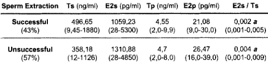
Objective and design : The serum levels of steroid hormones (testosterone, estradiol) were compared between spermatic vein blood and peripheral blood, in patients with non obstructive azoospermia (NOA), according to the results of surgical sperm retrieval. The aim of our study was to identify predictive factors of successful testicular sperm recovery in patients with non obstructive azoospermia.
Materials and Methods : A Prospective and comparative study (supported by a Rouen Hospital Grant and approved by the local ethical committee) was performed in a population of of 30 patients with NOA. For each subject who underwent testicular sperm extraction, serum levels of testosterone (T) and estradiol (E2) were determined in spermatic vein blood (Ts and E2s) and in peripheral blood (Tp and E2p). Differences in hormone levels between the two groups of patients with either successful or unsuccessful sperm recovery were analysed using non parametric variance analysis and Mann- Whitney U-test.
Results 9 Data are presented as the mean values with ranges.
Sperm Extraction I s (nglml) E2s (pg/ml) Tp (ng/ml) E2p (pg/mt) E2s ITs Successful 496,65 1059,23 4,55 21,08 0,002 a
( 4 3 % ) (9,45-1880) (28-5300) (2,0-9,9) (9,0-30,0) (0,001-0,005)
Unsuccessful 3 5 8 , 1 8 1310,88 4,7 26,47 0,004 a (57%) (12-1126) (28-4850) (2,0-8,0) (16,0-39,0) (0,001-0,009)
Conclusion : The ratio E2s/Ts appears significantly increased in the group with failure of testicular sperm retrieval (p=0,018). These data suggest that aromatase activity is higher when haploid germ cells are absent of the testis. The mechanisms of action of estrogens in the spermatogenesis remain to be clarified, especially with studies using another marker like tissue marker (cytochrome P450 aromatase activity, aromatase gene (CYP 19) expression).
A long term quality control for a serum inhibin B assay
A. MAHMOUD, F. COMHAIRE, KAUFMAN J.M. University Hospital Ghent - Andro/ogy Laboratory
Ghent, Belgium
Objective : Inhibin B in serum is a well-established marker for Sertoli cell function and spermatogenesis. The present study evaluates the reproducibility of an enzyme linked immunosorbant assay (ELISA) for inhibin B.
Materials and Methods : Two pooled blood sera with low (PL) and high activities of inhibin B, were included in each of 57 assays for inhibin B (Serotec, Oxford, UK) and the assay was performed at our laboratory according to manufacturer's instructions.
Pooled serum samples were processed in duplo allowing for the calculation of inter assay co-efficient of variation (CV) and the averages of the two readings in the different assays was used to calculate the inter assay CV. In total data on 3 different pairs of pools used in 57 runs for the period 1998- 2002 was available for analysis.
Results : The average intra-assay coefficient of variation (CV) for all the inhibin B runs performed at our laboratory was 6.9 % for the serum pools with low inhibin B levels (PL) and 4.4 % for those pools with high inhibin B activities (PH). The inter-assay CV ranged from 19 to 34 % (see table 1).
Table 1 : Intra- and inter-assay coefficient of variation (CV) for inhibin B measurement.
PL1 P H 1 P L 2 P H 2 P L 3 P H 3
Number of 24 24 22 22 11 11 runs
Average intra-assay
cv (%)
6.8 4.0 8.0 5.6 5.2 2.7
Inter-assay 24.3 1 9 . 2 20.0 34.0 28.0 30.2 CV (%)
Average inhibin B (n~/dl)
84.5 193.7 8 2 . 4 269.1 63.8 222.2
Conclusions : The high interassay variability might be explained by the use of different batches of inhibin B kits over the period of 5 years. The within batch interassay CVs is much lower (data not shown). The high inter-assay CVs over a long period of time indicates, however, that comparing results from different studies where different batches of inhibin B kits were used is not recommended. Support : none
PO 021
Inhibin B as a predictor before testicular sperm extraction - valuable completion or cost factor ?
F. REIHER1, O. RAg 1, T. LINDENMEIR1, I. NICKEL 2, J. KLEINSTEIN2, E. ALLHOFF1
1 Klinik und Poliklinik f(Jr Urologie, Otto-von-Guericke- Universit&t Magdeburg ASR, Deutschland, 2 Klinik f(~r Reproduktionsmedizin und gyn&kologische Endokrinologie, Otto-von-Guericke-Universit~t Magdeburg
ASR, Deutschland
Email : FRANK.REIHER@MEDIZIN. UNI- MAGDEBURG. DE
Introduction : Inhibin B as a predictive marker for a successful testicular sperm extraction (TESE) is discussed controversely in the literature. The aim of this study was to evaluate the predictive value of Inhibin B as a reliable marker for spermatogenesis in patients (pats.) with nonobstructive azoospermia (NOA).
Materials and Methods : In 78 pats. (33+4.8 years) undergoing TESE the following parameters were evaluated: (a) testicular volume; (b) serum follicle hormone (FSH) and (c) serum Inhibin B level. These parameters were correlated to the TESE-results (recovery of spermatozoa).
Results : In 49 of the 78 pats. (62%) spermatozoa were successfully retrieved (TESE-positive). The Serum inhibin B level was 103,5+12,5 pg/ml in the TESE-positive group. Compared to a control group (healthy probands) there was a significant difference regarding serum Inhibin-B levels between the TESE positive group and control group (103.5 vs. 183.6 pg/ml; p<0.05). We observed also a statistically significant difference of the serum Inhibin-B levels between controls and pats. with no evidence of spermatozoa in TESE (92.8 vs. 183.6 pg/ml; p<0.05). No statistically significant difference exists for the Serum Inhibin B levels in TESE- positive and TESE-negative pats. (p>0.05).
In contrast, a significant difference in serum FSH was evident. In TESE-positive pats. the serum-FSH level was 14.6+1.9 IE/I, in TESE-negative pats. 20.3+1.9 IE/I (p<0.05).
In pats. with normal serum FSH 82% of sperm retrieval attempts were successful, whereas in the group with pathologic serum FSH only in 42% of attempts sperms were retrieved (p<0.05). Additionally, spermatozoa recovery correlated also with testicular volume (p<0.05).
Conclusions : Serum FSH and testicular volume are valuable marker to estimate the expecting results of a testicular sperm extraction. In our hands Serum inhibin-B wasn't able to give further information predicting the possible evidence of spermatozoa.
No differences in the serum Inhibin B levels were observed in TESE-positive and TESE-negative pats.. But TESE-positive and TESE-negative pats. had significant lower serum Inhibin B levels compared to the control group, p<0.05.
In pats. with normal serum FSH 82% of sperm retrieval attempts were successful, 27/6 vs 22/23, p<0.05.
In TESE-positive pats. the serum-FSH level was with 14.6+1.9 IE/I significantly lower compared toTESE-negative pats. 20.3+1.9 IE/I (p<0.05).
PO 022
Testicular Fine Needle Aspiration Biopsy in the investigation of the subfertile male with
azoospermia and severe oligo-terato-asthenospermia
TH. MIKOS, P. POLICHRONOU, G. GRIMBIZIS, A. PAPANICOLAOU, E. ATHANASIOU, P. SEVASTIADOU,
D.G. GOULIS, B.C. TARLATZIS, J. BONTIS, J. PAPADIMAS
Unit of Reproductive Endocrinology, 1st Department of Obstetrics & Gynecology, Aristotle University of
Thessaloniki, Greece
Objective : To describe our experience with testicular Fine Needle Aspiration Biopsy (FNA) in the investigation of subfertile men with azoospermia and severe oligo-astheno-teratospermia (OAT).
Patients and Methods : From 1999 to 2005, 1087 subfertile men were studied at the outpatient clinics of a referral Andrology center in Northern Greece. All men underwent clinical, sperm, basal and dynamic hormonal evaluation. Genetic studies were applied as appropriate. Azoospermia and severe OAT were found in 78 (7.9%) and 27 (2.7%) men, respectively. Testicular FNA underwent 99 (94.2%) of these men. In addition, 15 men underwent Testicular Sperm Extraction (TESE) at a time later than FNA.
complete Sertoli-Cell only Syndrome (SCOS) (n = 22, 22%), and insufficient sample (n = 3, 2%). The cytological diagnoses in the subgroup of 51 men with INOA were: normal spermatogenesis (n = 1,2%), moderate hypospermatogenesis (n = 7, 14%), severe hypospermatogenesis (n = 18, 35%), incomplete maturation arrest (n = 2, 4%), complete maturation arrest (n = 9, 18%), complete Sertoli-Cell only Syndrome (SCOS) (n = 13, 25%), and insufficient sample (n = 1, 2%). Finally, during the TESE procedure, sperm was found in 7 men whereas not found in the remaining 8 men ; of them, the positive result was predicted during the FNA procedure in 6 men (Positive Predictive Value - PPV: 86%) and the negative result was predicted in 6 men (Negative Predictive Value - NPV: 75%.).
Conclusion : FNA plays an important role in both the diagnostic and therapeutic approach of subfertile man. In the diagnostic approach, a single testicular FNA according to our results has 51% possibility of detecting sperm in subfertile men with INOA. Aspirated spermatozoa can then be used for assisted reproduction techniques, such as Intra-Cytoplasmic Sperm Injection (ICSI). In the therapeutic approach, FNA has 86% PPV and 75% NPV of predicting TESE results, further facilitating the ICSI procedure.
Presenting author : Prof. J. Papadimas, 1st Department of Obstetrics & Gynaecology, "Papageorgiou" General Hospital, Periferiaki Odos, Nea Efkarpia, 56403, Thessaloniki, Greece. e-mail : [email protected]
PO 023
Presence and quality of testicular spermatozoa in azoospermic men according to region of their
residence in Slovenia
I. VIRANT-KLUN, S. DROBNIC, L. BACER- KERMAVNER, J. MIVSEK, T. TOMAZEVIC Department of Obstetrics and Gynaecology, University
Medical Centre Ljubljana, Slovenia E-mail : Hyperlink "mailto:irma. [email protected]"
irma. virant@ k clj. si
Objective : Little is known about the effect of geographical factors on the appearance and type of azoospermia, and manifestation of testicular cancer.
Design : The aim of this preliminary study was to compare the presence and quality (motility) of testicular spermatozoa, and outcome of ICSI in patients with azoospermia according
to 8 Slovenian geographical regions of their residence. Additionally, the incidence of testicular cancer in their medical history was compared among the regions.
Material and Methods : In this study 192 diagnostic testicular biopsies from 192 infertile men with azoospermia attending our Department were included. Each biopsy specimen was checked for the presence of spermatozoa. In patients with spermatozoa sperm motilility was evaluated (motile and non- motile spermatozoa). The testicular tissue was cryopreserved for later in vitro fertilization. The men were divided in 8 groups by the geographical region of their residence: I. Capital Ljubljana (n =71), I1. Region of_tajerska (n = 30), Ill. Region of Gorenjska (n = 25), IV. Region of Primorska (n = 20), V. Region of Dolenjska (n = 13), VI. Region of Koro_ka (n = 12), VII. Region of Severna Primorska (n = 9), VIII. others - Idrijsko-Cerkljansko, Posavje, Zasavje (n = 12). All groups of men were statistically compared according to the presence and motility of testicular spermatozoa, ICSI outcome, and the incidence of testicular cancer in their medical history.
Results : The mean male age was 36 years (min. 25 - max. 56 years) with no statistically significant difference among regions. There was no statistically significant difference among regions in the proportion of men with testicular sperms (1. 65%, II. 57%, II1.72%, V. 77%, VI. 67%, VIII. 70%), except for the Primorska region ( I V ) a significantly lower proportion of men (35% ; P < 0.05) due to a higher incidence of Sertoli Cell Only Syndrome and maturation arrest. Also a higher incidence of testicular cancer in the medical history was registered in the men from the region of Primorska than from other regions (15% vs 4%, P < 0.05). We found no significant differences in the proportion of men with motile sperms. In researched group of men 68 ICSI cycles were performed and 16 pregnancies (33.5% pregnancy rate per cycle) resulted (1. 23%, 11.27%, Ill. 37.5%, V. 25%, Vl. 20%, VIII. 25%), none to a couple from the Primorska region (0%). Ten children were born (7 girls and 3 boys), 2 pregnancies are still on-going, and 4 pregnancies ended in a spontaneous abortion.
Conclusions : In this preliminary study, performed in a small group of azoospermic men, we found that the Primorska region (towns Koper, Piran, Portoro_, Izola, Ankaran) situated at the coast of the Northern Adriatic Sea (Gulf of Trieste, Gulf of Koper, Gulf of Piran) shows negative effects on spermatogenesis and on incidence of testicular cancer. This study will be extended to a bigger group of azoospermic men to exclude possible bias (like genetic abnormalities and