Multilineage Differentiation Activity by the Human Umbilical
Vein-Derived Mesenchymal Stem Cells
Mehdi Kadivar
1, Shohreh Khatami
*1, Yousef Mortazavi
2, Mohammad Taghikhani
3and
Mohammad Ali Shokrgozar
41
Dept. of Biochemistry, the Pasteur Institute of Iran, Tehran;2Dept. of Pathology, Zanjan Medical School, Zanjan;
3
Dept. of Clinical Biochemistry, School of Medical Sciences, Tarbiat Modarres University, Tehran; 4National Cell Bank of Iran (NCBI), the Pasteur Institute of Iran, Tehran, Iran
Received 26 October 2005; revised 12 April 2006; accepted 22 May 2006
ABSTRACT
Background: Mesenchymal stem cells (MSC) are a very promising transplantable stem cell source for a variety of cell replacement therapies. As the main source of MSC is bone marrow (BM), most of studies have been done on BM-derived MSC (BM-MSC). Umbilical cord (UC)-derived MSC (UC-MSC) which are recently introduced, is one of the good alternative source for these cells. The objective of this study was to isolate and characterize UC-MSC from human UC veins and studying of their potential to differentiate into various cell types such as fat, bone, cartilage and neuronal cells. Methods:In this way, a cell population was isolated from 20 UC veins using a solution of 0.1% collagenase type IV. After identification of isolated cells by immunocytochemical and flow cytometry methods, these cells were exposured with adipogenic, osteogenic, chondrogenic and neurogenic agents. Resulted differentiated cells were detected using specific staining for each lineage and room temperature (RT)-PCR. Results: Immunophenotypically, this cell population was positive for CD73, CD166, CD105 markers and -smooth muscle actin and negative for CD31, CD34, CD49d markers, von Willebrand factor and smooth muscle myosin. Exposure of these cells to adipogenic, osteogenic, chondrogenic and neurogenic agents resulted in morphological changes followed by lineage-specific staining for each differentiated cell type. RT-PCR analysis showed that these differentiated cells express fat, bone, cartilage and neuronal markers, respectively. Conclusion: Altogether, these findings indicate that UC-MSC possess morphological, immunophenotypical and cell differentiation capacities similar to BM-MSC and so they can be used more extensively in cell based therapy protocols and in vitro differentiation study models. Iran. Biomed. J. 10 (4): 175-184, 2006
Keywords: Mesenchymal stem cells (MSC), Cell differentiation, Umbilical cord (UC)
INTRODUCTION
esenchymal stem cells (MSC) are multipotent precursors that are capable of differentiating into various cell types of mesodermal origin, including chondrocytes, osteocytes, adipocytes and stromal cells [1]. Although the differentiation potential of adult stem cells was initially believed to be restricted to its tissue of origin, a great deal of work accumulated recently on the issue of stem cell plasticity. This means that these precursor cells are able to originate differentiated cells of other organs and tissues, such as hepatic, renal, neural and cardiac cells [2].
On the basis of these characteristics, many investigators have focused on the application of MSC in cell based therapy protocols. Although bone marrow (BM) is the main source of MSC (BM-MSC), it posses a high risk for viral infection and a significant drop in proliferative/differentiation capacity and in cell number with age [3]. Both of these can limit the use of BM-MSC as an ideal source of MSC. In this regard, most attention should be focused on tissues containing cells with higher proliferative potency and differentiation capacity and lower risk for viral contamination. In addition to BM, MSC can be obtained from other sites in the adult, fetus [4], amniotic fluid [5], or cord blood
M
cells [6]. MSC are also enriched in preterm cord blood, decreasing in number with gestational age [7]. Recently, many groups succeeded in isolating MSC from umbilical cord blood (UCB) [8-10], whereas controversial results were obtained by others who suggested that cord blood is not a source for MSC [11, 12 ]. What seems to be validated is that, UCB is enriched in pluripotent MSC in the middle of gestation and these cells home after they leave circulation and excess of them possibly deposit in placenta/umbilical cord (UC) stroma, including blood vessels [3].
Recently, instead of using the cord blood, Romanov et al. [3] and Covas et al. [13] obtained MSC starting from cells detached from the UC vein, in a manner similar to that initiating human umbilical vein endothelial cell (EC) cultures. These UC vein-MSC are easier to get in comparison with BM-MSC [14] and without noted problems which are present in BM-MSC. Although it is appear that BM-MSC and UC-MSC are functionally similar to each other, a few studies have been done on UC-MSC. To extend the characterization of the MSC derived from umbilical cord veins, here we report the isolation and characterization of MSC from the endothelium layer of the human UC veins and after that we use these UC-MSC cells as an in vitromodel system for studying of their differentiation and plasticity potential. Using lineage-specific staining and room temperature (RT)-PCR analysis, we showed that under proper stimulation, UC-MSC can express fat, bone, cartilage and neuronal markers.
MATERIALS AND METHODS
Isolation and culture of UC-MSC. Umbilical
cords (n = 20) were obtained after normal or caesarian term deliveries from healthy infants under aseptic conditions and were carried to the lab in filtered PBS (pH 7.4) containing penicillin (300 U/ml), streptomycin (300 µg/ml), gentamycin (150 µg/ml) and fungizone (1 µg/ml) and were processed within 6-12 h. The umbilical veins were canulated and washed twice with PBS containing 100 U/ml heparin. The distal ends were clamped and veins were filled with 0.1% collagenase type IV in M199 medium supplemented with antibiotics. After clamping the proximal ends, UC were incubated at 37ºC for 20 min. The veins were washed with PBS, followed by gentle massaging of the cords. The suspension of EC/subendothelial cells were collected and centrifuged at 600g for 10 min. The pellet was
resuspended in DMEM supplemented with 100 U/ml penicillin, 100 µg/ml streptomycin and 15% FBS. Then, the cells were plated onto 25 cm² tissue culture flasks at a concentration of 10³ cells/cm² were incubated at 37ºC, 5% CO2. Non-adherent cells
were removed after 3 days by changing the medium. Adherent cells were kept in culture and were fed with fresh medium every 3 days. These cells were kept until the outgrowth of fibroblastoid cells about 2 weeks later [3, 13]. At that time, cultures were harvested with PBS (pH 7.4) containing trypsin (0.05%) and EDTA (0.02%) and passaged (without dilution) into a new flask for further expansion.
Immunocytochemical characterization of MSC.
Cells in primary culture were fixed for 15 minutes at RT with 1% paraformaldehyde in PBS. After several washes with PBS and PBS-1% BSA, cells were incubated for 1 h with the following primary antibody: (von Willebrand factor (vWF; 1:100, Sigma, USA), platelet/EC adhesion molecule-1 (PECAM-1; 1:50, Sigma, USA). As well as above, some of the expanded UC-MSC in second or third passages, were fixed with 4% paraformaldehyde in PBS containing 0.1% Triton X-100, washed with PBS and PBS-1% BSA, and then incubated for 1 h with anti-α smooth muscle actin (ASMA; 1:400, Sigma, USA) and anti-myosin smooth muscle (MySM; 1:500, Sigma, USA). After rinsing with PBS, cells were incubated with anti mouse IgG secondary antibody conjugated with horse raddish proxidase (1:300, Amersham, USA). Diamino benzidine (DAB) substrates for peroxidase were used to visualized the antibody binding.
Flow cytometry. Expanded UC-MSC were
detached from the culture flask by the use of PBS (pH 7.4) containing trypsin (0.05%) and EDTA (0.02%), washed once with DMEM and once with filterated PBS. Cells were next suspended at a concentration of 1106 cells in 50 µl of PBS and
incubated for 45 min at 4°C in the dark with FITC or PE conjugated antibodies as follows: anti-CD49d-PE, CD73-FITC, CD166-FITC, anti-CD105-PE, anti-CD34-FITC, and anti-CD31-FITC. In parallel, cells were incubated with an irrelevant Ab (anti-Aspergillus niger glucose oxidase, Dako, USA) as a negative isotype control to exclude non-specifically labelled cells from the calculation. Upon completion of the incubation time, cells were washed twice with PBS supplemented with 2% BSA and fixed with 1% paraformaldehyde solution in PBS. Analysis was next performed using a flow
cytometer (FACsort, BD, USA). Before each test, the percentage of viability that was more than 95% was measured with trypan-blue sataining and dead cells were counted with neobar slide.
Adipogenic induction. For adipogenic
differen-tiation, UC-MSC were grown to 100% confluence in DMEM supplemented with 10% FBS. Two-days post-confluent cells were incubated in adipogenesis-inducing medium (AIM) (DMEM, 4.5 g/L glucose, 1 µM dexamethasone, 200 µM indomethacin, 1.7 µM insulin, 500 µM isobutyl-methylxanthine, 10% FBS, 0.05 U/mL penicillin, and 0.05 µg/mL streptomycin) for 3 days, followed by incubating 24 h in adipogenesis maintenance medium (AMM) (DMEM, 4.5 g/L glucose, 1.7 µM insulin, 10% FBS, 0.05 U/mL penicillin, and 0.05 µg/mL streptomycin) and then switched to AIM again. After the third cycle, cells were fed with AMM for up to 21 days of differentiation [15]. For adipocytes identification, intracellular lipid accumulation was visualized using Oil Red-O-staining. Briefly, cells were fixed in 10% solution of formaldehyde in aqueous phosphate buffer for 1 h, washed with 60% isopropanol, and stained with Oil Red- O-solution (in 60% isopropanol) for 10 min, followed by repeated washing with water before being destained in 100% isopropanol for 15 min.
Osteogenic induction. For osteogenic
differen-tiation UC-MSC were induced in 2 weeks by α-MEM supplemented with 0.1 µM dexamethasone, 10 µM β-glycerophosphate and 50 µM ascorbate-phosphate [16]. Control cultures without the differentiation stimuli were maintained in parallel to the differentiation experiments and stained in the same manner. Differentiated cells were identified by specific histochemical staining for alkaline phosphatase (ALP) with ALP staining kit (Sigma, USA).
Chondrogenic induction. For chondrogenic
stimulation, UC-MSC were plated at 50 cells/cm2
and cultured in DMEM for 7 days. Then, approximately 200,000 UC-MSC were placed in a 15-ml polypropylene tube and pelleted into micromasses by centrifugation at 450g for 10 min [17]. The pellet was cultured for 21 days in chondrogenic media containing 500 ng/ml BMP-6 in addition to high-glucose (25 mM) DMEM supplemented with 10 ng/ml TGF-β, 3 10-7 M
dexamethasone, 50 µg/ml ascorbate-phosphate, 40 µg/ml proline, 100 µg/ml pyruvate and 50 µg/ml
ITS+ tm Premix (Becton Dickinson, USA). For
microscopy detection of chondrocytes, the pellets were embedded in paraffin, cut into 5 µm sections, and stained with 1 % toluidine blue and 1% sodium borate for 5 min [18].
Neurogenic induction.For neural induction,
UC-MSC were expanded to 70% to 80% confluency in DMEM with 10% FBS, and 10 ng/mL basic fibroblast growth factor was added for 24 hours. Thereafter, cells were incubated in neuronal induction media (DMEM, 2% DMSO, 200 µM butylated hydroxyanisole, 25 mM KCL, 2 mM valporic acid, 10 µM forskolin, 1 µM hydroxycortisone, 5 µg/mL insulin, and 2 mM L-glutamine) [19]. After 24 h of neuronal induction, cells were stained for expression of the neural specific enolase (NSE). Briefly, cells were fixed with 4% paraformaldehyde in PBS containing 0.1% Triton X-100, washed with PBS and incubated for 1 h with rabbit anti-human neuron specific enolase (Dako, USA; 1:2000). After washing with PBS, cells were incubated with biotinylated Goat anti-rabbit IgG (H+L) (Dako, USA; 1:500) as secondary antibody. Then HRP (horse raddish proxidase) conjugated with streptavidin (Dako, USA; 1:500) was used as detection reagent and finally DAB substrates for peroxidase were used to visualized the antibody binding [20]. For negative control, non-induced UC-MSC were maintained in DMEM with 10% FBS for 24 h and the same procedure was used for them.
RT-PCR analysis. For each differentiation, total
RNA was extracted from differentiation-induced UC-MSC using TRIzol reagent, and then each sample (1 µg) was reverse transcribed with M-MLV reverse transcriptase for 30 min at 42ºC in the presence of oligo-dT primer and RNase inhibitor and terminated at 90ºC for 10 minutes. PCR assays were performed for 30 to 35 cycle in 20 µl reaction volume using 2 µl cDNA, 1.5 mM MgCl2, 200 µl of
each dNTP, 20 pmol of each primer, and 0.4 U of AmpliTaq Gold DNA polymerase. Adipogenic specific genes used for RT-PCR were Adipsin, lipoprotein lipase (LPL), and Fatty binding protein 4 (FABP4). For osteogenic marker expression, osteopontin (OPN), osteoprotegerin (OPG), and ALP were used. Chondrogenic specific gene expression was evidenced using Syndercan, Perlecan, and Collagen type II. Nestin, neurogenic differentiation factor 1 (Neuro D1), and glial fibrillary acidic protein (GFAP) were used for
neurogenic specific gene expression.
Forward and reverse sequence and product size for each primer were set as follows: β-actin (512 bp), sense 5'-TCA TGT TTG AGA CCT TCA A-3' and antisense 5'-GTC TTT GCG GAT GTC CAC G-3'; Adipsin (271 bp), sense 5'-GGT CAC CCA AGC AAC AAA GT-3' and antisense 5'-CCT CCT GCG TTC AAG TCA TC-3'; FABP4 (357 bp), sense 5'-TGC AGC TTC CTT CTC ACC TT-3' and antisense 5'-TGG TTG ATT TTC CAT CCC AT-3'; LPL (lipoprotein lipase) (717 bp), sense 5'-GTC CGT GGC TAC CTG TCA TT-3' and antisense 5'-AGC CCT TTC TCA AAG GCT TC-3'; OPG (435 bp), sense 5'-TGC TGT TCC TAC AAA GTT TAC G-3' and antisense 5'-CTT TGA GTG CTT TAG TGC GTG-3'; OPN (500 bp), sense 5'-TGC TAC AGA CGA GGA CAT C-3' and antisense 5'-CCA ATA AAC TGA GAA AGA AGC-3'; ALP (300 bp), sense 5'-CTC CTC AGC CTC TGC AAC TG-3' and antisense 5'-AGG GTC AGG AGA TGA GAC TGG-3'; GFAP (400 bp), sense 5'-GAA GCC GAA GAG TGG TAC CG-3' and antisense 5'-AGA AGG TCT GCA CGG GAA TG-3'; Nestin (500 bp), sense 5'-CAC ACA TGG AGA CGT CGC TG-3' and antisense 5'-AAG GGT AGC AGG CAA GGG TG-3'; Neuro D1 (450 bp), sense 5'-TCG TTC AGA CGC TTT GCA AG-3' and antisense 5'-AGA TTG ATC CGT GGC TTT GG-3'; Collagen type II (394 bp), sense 5'-AGT GGA GAC TAC TGG ATT GA-3' and antisense 5'-AGT GTA CGT GAA CCT GCT AT-3'; Perlecan (300 bp), sense 5'-CAT AGA GAC CGT CAC AGC AAG-3' and antisense 5'-ATG AAC ACC ACA CTG ACA ACC-3'; Syndercan (409 bp), sense 5'-CCT TCA CAC TCC CCA CAC-3' and antisense 5'-GGC ATA GAA TTC CTC CTG TTG-3' [21, 22]. The PCR products were analysed by electrophoresis with 2% agarose gel, visualized with EtdBr staining and photographed under UV light.
RESULTS
Estabilishment of primary culture. With this
approach, we obtained a cell population that assumes a spindle-shape morphology in confluent wave-like layers in culture and can be replated several times (20 or more). Initially, two types of adherent cells were observed in primary cultures: EC with small flat morphology and a few spindle-shape fibroblastoid cells primarily identified as MSC which were seen between ECs (Fig. 1a). Immunocytochemical staining showed that ECs
were vWF and PECAM1 (CD31) positive, while MSC were negative for both of them (Fig. 1a and b). Furthermore, MSC were MySM negative, while they were ASMA positive (Fig. 1c and d). After a 2-week culture, these MSC became the predominant cell type, while the EC remained compact and did not spread, migrate or proliferate. vWF and PECAM1 positive cells (EC) did not exceed 0.5-1% of the total cell number.
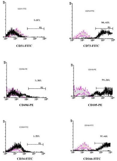
Characterization by flow cytometry. In the
cytomeric analysis, UC-MSC were positive for CD73 ( 5-terminal nucleotidase or SH4), CD105 (Endoglin or SH2) and also for the CD166 adhesion molecule (ALCAM or SH1) which together is considered as a marker for MSC [23]. In contrast, they were negative for CD49d (integrin α4), and for hematopoitic lineages markers CD31 (PECAM1) and CD34 (Fig. 2).
Differentiation results. Differentiation studies
performed on UC-MSC revealed their potential to differentiate into adipocytes, osteoblasts, chondro-cytes and neuronal cellsin vitro. UC-MSC cultures in adipogenic differentiation medium, after 21 days, led to the appearance of larger round cells presenting numerous fat vacuoles in the cytoplasm. These lipid droplets were Oil red-O-positive while untreated control cultures did not have lipid droplets in their cytoplasm. (Fig. 3a and b).
UC-MSC under osteogenic inductive medium, led to the appearance of refringent crystals in the cells after 2 weeks. By the end of the second week of stimulation, most of the UC-MSC also became ALP-positive. Untreated control cultures growing in regular medium without any osteogenic differen-tiation stimuli did not show spontaneous osteoblast formation even after 3 weeks of cultivation (Fig. 3c and d).
When UC-MSC cultured as a pellet in the bottom of the tube and in chondrogenic medium, after 21 days, they originate a mass of cells with chondrocyte or chondroblast features such as rounded shape with a large vacuolated cytoplasm. These cartilage structures can be distinguished specifically with toluidine blue-sodium borate staining structures like this, did not form in untreated control cultures after this period (Fig. 3e and f).
Neurogenic induction in UC-MSC resulted in an apparent morphological change in the cultures. The cells that were responsive to the induction displayed small, spherical, and refractive cell bodies.
(a) (c)
(b) (d)
Fig. 1. Morphology and immunocytochemical characteristics of cells isolated from the human umbilical vein endothelial/subendothelial layer. Primary cultures containing MSC and endothelial cells or EC are stained in (a)and (b)with anti-vWF and anti-PECAM1 (CD31) antibodies, respectively. Oobviously, clear DAB signal for EC (compact round cells) indicates that they are vWF and PECAM1 (CD31)-positive, while MSC (spindle shape cells) remained pinkish and so they are vWF and PECAM1 negative. Immunocytochemical staining with anit-MySM antibody (c) and anti-ASMA antibody (d) show that isolated MSC are MySM negative, while they were ASMA-positive (d). Final magnifications for (a)and (b)were 100 and for (c)and (d)were 200. MSC, mesenchymal stem cells; EC, endothelial cell; vWF, von Willebrand factor; PECAM1, platelet/EC adhesion molecule-1; DAB, diamino benzidine; MySM, anti-myosin smooth muscle; ASMA, anti-α smooth muscle actin.
They also developed long cellular processes, some of which formed secondary, tertiary, and higher-order branches. In contrast, the unresponsive cells maintained the spread-out morphology observed in cells without induction (Fig. 3g and h). The differentiation status also was verified based on the expression of the human NSE, an enzyme found predominantly in neurons and neuroendocrine tissues (Fig. 3i and j).
RT-PCR results. When total RNA from induced
and non-induced cells were isolated and analyzed by RT-PCR, expression of β-actin, used as an internal control, was the same in undifferentiated and differentiated cells (Fig. 4e), whereas indicated lineage-specific genes were not expressed in control
non-induced cultures but were expressed in cells under inductive media (Fig. 4a, b, c and d).
DISCUSSION
MSC can be obtained from BM and from other sites in the adults or the fetus.We report the isolation of a cell population derived from the endothelium and subendothelium layers of the UC vein with morphological, immunophenotypical and different-tiation properties similar to those of MSC obtained from BM, as originally described by Friedensteinet al.[24, 25]. Although MSC of both origins (BM and UC) are highly similar from immunophenotypical and immunocytochemical points of views [3, 13],
CD31-FITC CD73-FITC
CD49d-PE CD105-PE
CD34-FITC CD166-FITC
Fig. 2. Flow cytometry histograms show the immunophenotype of MSC isolated from the postnatal human umbilical vein. UC-MSC were positive for CD73, CD105 and CD166. These cells were negative for CD31, CD49d and CD34. UC-UC-MSC, Umbilical cord (UC)-derived mesenchymal stem cells (MSC).
(a) (b) (c)
(d) (e) (f)
(g)
(h)
(i) (j)
Fig. 3. Differentiation potential of umbilical vein-derived MSC. (a)and (b)show the results of Oil Red O staining in control untreated medium, and in cell cultures growing within 2 weeks in adipogenic medium, respectively. Results of osteogenic differentiation showing non-stimulated (c)and stimulated cells (d)stained with diagnostic alkaline phosphatase kit. (e)and (f)show the results of toluidine blue-sodium borate staining in control untreated MSC and in MSC cultures exposured to chondrogenic medium for 21 days, respectively. Neurogenic differentiation evidenced by morphological changes (h)and by immunocytochemical staining for NSE (j). (g)and (i) are negative untreated control cells for (h)and (j), respectively. Final magnifications for (i)and (j)are 400 and for (f)is 40. The other magnifications are 100. MSC, mesenchymal stem cells.
(a) (b)
(c) (d)
(e)
Fig. 4. Room temperature-PCR analysis for expression of adipogenic (a), osteogenic (b), chondrogenic (c), and neurogenic (d) marker genes. Total RNA was isolated from differentiated and non-induced (negative control) cells and used for synthesis of cDNA which then was amplified using specific primers for each marker gene. All experiments performed in triplicate, representative gels shown. (a), UC-MSC induced toward adipogenic lineage express Adipsin (lane 2, 271 bp), lipoprotein lipase (lane 4, 717 bp), and FABP4 (lane 6, 357 bp) after up to 21 days. Non-induced cells (lanes 3, 5, and 7) were analyzed as a negative control for each primer. Lane 1 is molecular weight marker and lane 8 shows expression of β-actin as an internal control in non-induced control cells; (b), UC-MSC exhibit osteogenic capacity in vitro. Lane 1, Molecular weight marker; OPN (lane 2, 500 bp), OPG (lane 4, 435 bp), and ALP (lane 6, 300 bp) were expressed in cells under osteogenic differentiation condition. Lane 3, 5 and 7 are negative non-induced control cells for OPN, OPG, and ALP, respectively. Loading of PCR product of β-actin in non-induced cells served as an internal control indicated in lane 8. (c), In chondrogenic differentiation condition, syndercan (lane 2, 409 bp), Col II (lane 4, 394 bp), and Perlecan (lane 6, 300 bp) were detected after over 3 weeks of induction. Lane 3, 5 and 7 are negative non-induced cells for Syndercan, Col II, and Perlecan, respectively. Lane 1, molecular weight marker; Lane 8, expression of β-actin as an internal control in non-induced control cells. (d), Neurogenic differentiation condition led to expression of Nestin (lane 2, 500 bp), Neuro D1 (lane 4, 450 bp), and GFAP (lane 6, 400 bp). Lane 3, 5, and 7 are negative non-induced control cells for Nestin, neurogenic differentiation factor 1, and glial fibrillary acidic protein, respectively. Lane 1, molecular weight marker; Lane 8, expression of β-actin as an internal control in non-induced control cells. (e), Loading of PCR products of β-actin served as internal control in adipogenic (lane 2), osteogenic (lane 3), chondrogenic (lane 4), neurogenic (lane 5), and in non-induced control cells (lane 6). Lane 1 is molecular weight marker. All bands are present at a molecular weight of 512 bp. UC-MSC, Umbilical cord (UC)-derived mesenchymal stem cells (MSC); OPN, osteopontin; osteoprotegerin (OPG); ALP, alkaline phosphatase.
it has been shown recently that some differences such as gene expression pattern are present between them. These differences could be functionally related to the origin of the MSC [14]. If confirmed, this would imply that MSC from a specific source may be more efficient for differentiating into particular target cells and thus more useful for a particular theraputic target [26]. So, the study of pluropotency and differentiation conditions of MSC isolated from various sources needs to be defined. In this way, we investigated multilineage activity of UC-MSC for further characterization of the pluripotency of these cells in accordance with the pluripotency of BM-MSC.
Recently, Romanov [3] and Covas [13] described cells isolated from the endothelium and subendothelium layers of the UC morphologically similar to those isolated here and also showed their ability for adipogenic and osteogenic differentiation, but this is one of the first studies that cells with these characteristics isolated from the endothelium/ subendothelium layers of the human umbilical vein have been extensively studied from differentiation capacity viewpoints. One of the reported methods used to isolate MSC from the endothelium/ subendothelium layers of the umbilical cord vein is with 1X endothelial growth factor (EGF) and vascular endothelial growth factor (VEGF) for enrichment of culture medium [13]. However, cells which have been isolated by this method can not used widely in the clinical trials because the effects of EGF and VEGF on the cells remain to be more defined [27].
We isolated and cultured MSC from the endothelium/subendothelium layers of umbilical cord vein without using these materials. After isolation, cells in primary cultures were evaluated by immunocytochemical staining. In this way, we tested cells in primary culture, with four monoclonal antibodies. The expression of vWF and PECAM-1 which are specific for EC, in some cells with small flat morphology, revealed that those were EC. ASMA which can be expressed in MCS types, was expressed in cells with fibroblastoid morphology and MySM which is specific for smooth muscle cells, was not expressed in any cell type, indicated that our primary culture was not infected with vein-derived smooth muscle cells. Results of flow cytometry studies on expanded UC-MSC completely were similar with the results of cytomeric analysis in BM-MSC [1]. In the differentiation analysis, our results indicate that adipogenic, osteogenic, chondrogenic and neurogenic potential of the
UC-MSC is very similar to BM-derived UC-MSC and mediums for induction of BM-MSC toward a specific lineage can be used for induction of UC-MSC in a same manner.
In addition to lineage specific staining, we used RT-PCR technique for studying the expression of each lineage related differentiation marker in molecular level. Like immunophenotypical properties, RT-PCR results of UC-MSC were in matched with the results of studies of BM-MSC studies [1, 14]. These results indicate that UC-MSC have a range of good properties such as high proliferation potency, high differentiation capacity, low risk for viral contamination, more accessible and easier to get in comparison with BM-MSC, and similar potential of differentiation in comparison with BM-MSC. Altogether we conclude that UC-MSC can be consider as a good and potent alternative for BM-MSC and the Umbilical cord/placenta vessels can serve as a rich source of MSC which may be used in experimental and clinical demands.
REFERENCES
1. Deans, R.J. and Moseley, A.B. (2000) Mesenchymal stem cells: biology and potential clinical use. Exp. Hematol. 28:875-884.
2. Forbes, S.J., Vig, P. and Poulsom, R. (2002) Adult stem cell plasticity: new pathways of tissue regeneration become visible. Clin.Sci.103:355-369.
3. Romanov, Y.A., Svintsitskya, V.A. and Smirnov, V.N. (2003) Searching for alternative sources of postnatal human mesenchymal stem cells: Candidate MSC-like cells from umbilical cord. Stem Cells 21:
105-110.
4. Anker, P.S. and Noort, W.A. (2003) Mesenchymal stem cells in human second-trimester bone marrow, liver, lung and spleen exhibit a similar immunophenotype but a heterogeneous multilineage differentiation potential. Hematologica 88:845-852.
5. Anker, P.S. and Scherjon, S.A. (2003) Amniotic fluid as a novel source of mesenchymal stem cells for theraputic transplantation. Blood 102:1548-1549.
6. Lee, O.K., Kuo, T.K. and Chen, W.M. (2004) Isolation of multipotent mesenchymal stem cells from umbilical cord blood. Blood 103: 1669-1675.
7. Campagnoli, C., Roberts, I.A. and Kumar, S. (2001) Identification of mesenchymal stem/progenitor cells in human first-trimester fetal blood, liver, and bone marrow. Blood 98: 2396-2402.
8. Goodwin, H.S. and Bicknese, A.R. (2001) Multilineage differentiation activity by cells isolated from umbilical cord blood: expression of bone, fat, and neural markers. Biol. Blood Marrow Transplant
7: 581-588.
9. Hou, L., Cao, H. and Wei, G. (2002) Study of in vitro
expansion and differentiation into neuron-like cells of human umbilical cord bloodmesenchymal stem cells.
Zhonghua. Xue. Ye. 23: 415-419.
10. Rosada, C., Justesen, J. and Melsvic, D. (2003) The human umbilical cord blood: a potential source for osteoblast progenitor cell. Calcif. Tissue Int. 72: 135-142.
11. Mareschi, K. and Biasin, E. (2001) Isolation of human meenchymal stem cells: bone marrow versus umbilical cord blood. Hematologica 86: 1099-1100.
12. Wexler, S.A., Donaldson, C. and Dening-Kendall, P. (2003) Adult bone marrow is a rich source of human mesenchymal stem cells but umbilical cord and mobilized adult blood are not. Br. J. Hematol. 121: 368-374.
13. Covas, D.T., Siufi, J.L.C. and Silva, A.R.L. (2003) Isolation and culture of umbilical vein mesenchymal stem cells. Braz. J. Med. Biol. Res. 36: 1179-1183.
14. Panepucci, R.A., Siufi, J.L.C. and Silva, W.A. (2004) Comparison of gene expression of umbilical cord vein and bone marrow-derived mesenchymal stem cells. Stem Cells 22: 1263-1278.
15. Janderova, L., McNeil, M. and Murrell, A.N. (2002) Human mesenchymal stem cells as an in vitromodel
for human adipogenesis. Obesity. Res. 11: 65-74.
16. Ramriez, J.L., Castro, F. and Kuri, W. (1992) Quantitation of adipose conversion and triglycerides by staining intracytoplasmic lipids with Oil Red O.
Histochem. 97: 493-497.
17. Johnstone, B., Hering, T.M. and Caplan, A.I. (1998)
In vitro chondrogenesis of bone marrow-derived
mesenchymal progenitor cells. Exp. Cell Res. 238: 265-272.
18. Ichiro, S., Benjamin, L.L. and Jason, R. (2002) Expansion of human adult stem cells from bone marrow stroma: Conditions that maximize the yields of early progenitors and evaluate their quality. Stem Cells 20: 530-541.
19. Woodbury, D., Schwars, E.J. and Black, I.B. (2000) Adult rat and human bone marrow sromal cells differentiate into neurons. J. Neurosci. Res. 61: 364-370.
20. Zuk, P.A., Zhu, M., Ashjin, P. and Hedrick, M. (2002) Human adipose tissue is a source of multipotent stem cells. Mol. Biol. Cell 13: 4279-4295.
21. Kim, J.W., Kim, S.Y., Park, S.Y. and Ryu, H.M. (2004) Mesenchymal progenitor cells in the human umbilical cord. Ann. Hematol.22: 1263-1278.
22. Zhang, Y.I., Li, C., Jiang, X. and Mao, N. (2004) Human placenta-derived mesenchymal progenitor cells support culture expansion of long term culture-initiating cells from cord blood CD34+ cells.
Exp. Hematol. 32: 657-664.
23. Haynesworth, S.E., Baber, M.A. and Caplan, A.I. (1992) Cell surface antigens on human marrow-derived mesenchymal stem cells are detected by monoclonal antibodies. Bone 13: 69-80.
24. Friedenstein, A.J., Deriglazoa, U.F. and Kulagina, N.N. (1974) Precursors for fibroblast in different populations of hematopoieticcells as detected by the
in vitro colony assay method. Exp. Hematol. 2: 83-92.
25. Friedenstein, A.J., Gorskaja, J.F. and Kulagina, N.N. (1976) Fibroblast precursors in normal and irradiated mouse hematopoietic organs. Trend. Mol. Med. 4: 267-274.
26. Davini, S., Marandin, A. and Mersin, N. (2003) Mesenchymal progenitor cells differentiate into an endothelial phenotype, enhance vascular density, and improve heart unction in a rat cellular cardiomyoplasty model. Circulation 108 (suppl 1): II253-II258.
27. Braker, J.N. and Wagner, J.E. (2003) Umbilical ord blood transplantations: current practice and future innovations. Crit. Rev. Oncol. Hematol. 48: 35-43.