M
AREKB
OLANOWSKI1, 2, W
OJCIECHP
LUSKIEWICZ3, A
NNAS
KRZEK2, J
ANUSZB
OLANOWSKI2,
P
IOTRA
DAMCZYK4Beneficial Effects of Tai Chi on Women’s Skeletal
Status Assessed by Quantitative Ultrasound
at the Hand Phalanges – One−Year Follow−up Study
Korzystny wpływ ćwiczeń Tai Chi na układ kostny kobiet oceniany
na podstawie ilościowej ultrasonografii paliczków – obserwacja roczna
1 Department of Endocrinology, Diabetology and Isotope Therapy, Silesian Piasts University of Medicine
in Wrocław, Poland
2 Department of Physiotherapy, University of Physical Education, Wrocław, Poland
3 Metabolic Bone Diseases Unit, Department of Internal Diseases, Diabetology, and Nephrology,
Silesian University of Medicine, Katowice, Poland
4 Department of Pediatric Nephrology and Endocrinology, Silesian University of Medicine, Katowice, Poland
Adv Clin Exp Med 2007, 16, 5, 675–681 ISSN 1230−025X
ORIGINAL PAPERS
© Copyright by Silesian Piasts University of Medicine in Wrocław
Abstract
Objectives. The advantageous effect of physical exercise on bones has been proven. Quantitative ultrasound (QUS) at different skeletal sites is an accepted method for indirect and noninvasive assessment of bone quality and fracture risk. Earlier, the present authors showed higher values of ultrasound transmission in the hand phalanges of a group of Polish women practicing Tai Chi. The aim of the present study was a prospective assessment of skele− tal status in women practicing Tai Chi.
Material and Methods. One−year follow−up of skeletal status was assessed using QUS measurements at the hand phalanges in a group of 46 exercising women (mean age: 59.3 ± 8.7 years).
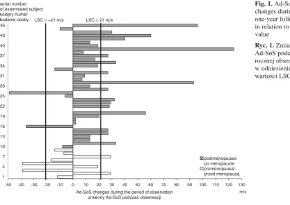
Results.At follow−up, increases in the mean amplitude−dependent speed of sound (Ad−SoS) from 1995 ± 80 to 2012 ± 75 m/s (p= 0.001) and mean Z−scorefrom 0.673 ± 1.054 to 1.053 ± 1.178 (p= 0.0005) were observed in the exercising subjects. These changes revealed statistically significant positive correlation with subject age (r= 0.39, p= 0.007 and r= 0.35, p= 0.018, respectively). The increase in Ad−SoS exceeded the value of the “least significant change” in 19 subjects (41%) of the total group and in 2 (22%) and 17 (46%) women in the pre− and postmenopausal subgroups, respectively.
Conclusions.A beneficial effect of regular Tai Chi exercise on skeletal status as assessed by phalangeal QUS is observed in older women. Tai Chi gymnastics may be recommended in the prevention of postmenopausal osteo− porosis, especially in older individuals (Adv Clin Exp Med 2007, 16, 5, 675–681).
Key words:osteoporosis, phalanges, quantitative ultrasound, Tai Chi exercise, women.
Streszczenie
Wprowadzenie. Znany jest korzystny wpływ ćwiczeń fizycznych na układ kostny. Ilościowa ultrasonografia (QUS) różnych części szkieletu jest uznaną metodą pośredniej i nieinwazyjnej oceny jakości kości i zagrożenia zła− maniami. We wcześniej przeprowadzonych badaniach własnych wykazano większą szybkość przechodzenia fali ultradźwięków w paliczkach rąk w grupie polskich kobiet ćwiczących Tai Chi.
Cel pracy.Prospektywna ocena za pomocą QUS układu kostnego kobiet ćwiczących Tai Chi.
Materiał i metody.Roczna obserwacja statusu kostnego została dokonana za pomocą metody QUS paliczków rę− ki w grupie 46 kobiet ćwiczących Tai Chi. Średnia wieku wynosiła 59.3 ± 8.7 lat.
Exercise has a positive impact on both muscle strength and the maintenance of postural balance. Physical activity in the elderly is a very important factor influencing general efficiency and it can pre− vent fractures as it reduces the likelihood of falls [1, 2]. The advantageous effect of physical exercise on bone mineral density (BMD) has been proven [3, 4]. Tai Chi is a system of low−weight−bearing gymnastics and is the major regular physical exer− cise practiced by elderly Chinese populations. There are some suggestions of a positive effect of Tai Chi exercise on BMD in postmenopausal women and osteoporotic fracture prevention [5–7]. The incidence of osteoporotic fractures is a major health problem in the elderly. The frac− tures are related to decreased bone density, impaired bone quality, and susceptibility to falls. Quantitative ultrasound (QUS) at different skeletal sites is an accepted method for indirect and nonin− vasive assessment of bone quality and fracture risk [8–11]. Phalangeal QUS was introduced about a decade ago and it has been validated in various clinical studies. By measuring the amplitude− −dependent speed of sound (Ad−SoS), this tech− nique may provide useful information not only on bone mass, but also on bone tissue architecture and elasticity. There are reports on good correlation between ultrasound parameters and age, osteo− porosis, and fracture risk. Several studies revealed changes in phalangeal ultrasound properties due to pregnancy, menopause, aging, and certain bone disorders [10, 12–15].
In a previous study, the present authors showed higher values of Ad−SoS, reflecting ultra− sound transmission in the hand phalanges of a group of Polish women practicing Tai Chi [16]. This could suggest the positive effect of this form of exercise on bone status and potential antifrac− ture impact. The aim of the present study was to assess the influence of one−year regular exercises on the QUS parameter studied in Polish Tai Chi women gymnasts.
Material and Methods
A follow−up examination after one year of reg− ular exercise was carried out on 46 women aged 27–75 years (mean: 59.3 ± 8.7 years) taking part in a Tai Chi program. They were involved in a pro−
gram of regularly exercising women at Tai Chi clubs in Wroclaw (Lower Silesia, Poland). Thirty− −seven of the women were postmenopausal. The data on the subjects are shown in Table 1. Subjects having factors of potential influence on bone metabolism (prolonged diseases of the thy− roid, liver, or kidney, gastrointestinal surgery, or using medications such as corticosteroids, anticon− vulsants, or thyroid hormone or being treated with drugs other than HRT for osteoporosis) were not included. All subjects gave their informed consent. The local ethics committee approved the study protocol.
Anthropometric Measurements
In all subjects, body weight, height, and body mass index (BMI as the weight in kilograms divid− ed by the squared height in meters, kg/m2) were
established.
Exercise Scores
The exercise duration and intensity scores for the Tai Chi exercising subjects were calculated as follows. The duration of exercise score was defi− ned as 0 (onset), 1 (< 6 months), 2 (6–12 months), 3 (12–24 months), and 4 (> 24 months). The inten− sity of exercise score was defined as: 0 (occasion− ally), 1 (once a week), 2 (twice a week), 3 (three times a week), and 4 (four or more times a week). The above scores were introduced by the authors themselves [16].
QUS Measurement
Skeletal status was assessed by QUS measure− ments at the proximal phalanges using the DBM Sonic 1200 device (IGEA, Carpi, Italy). This unit consists of two probes mounted on an electronic caliper, one emitter, and one receiver. The latter records the ultrasound energy after it has crossed the phalanx. The amplitude−dependent speed−of− −sound (Ad−SoS, m/s) in the distal metaphyses of the proximal phalanges of the second through the fifth fingers of the dominant hand was determined. SoS in bone tissue was calculated considering the first signal with an amplitude of at least 2 mV at the receiving probe; thus the measured SoS is amplitude−dependent. Acoustic coupling was
Wnioski.Korzystny wpływ regularnych ćwiczeń Tai Chi na układ kostny oceniany przez QUS paliczków obser− wuje się u starszych kobiet. Ćwiczenia Tai Chi można zalecać w profilaktyce osteoporozy pomenopauzalnej, zwłaszcza u starszych kobiet (Adv Clin Exp Med 2007, 16, 5, 675–681).
achieved using a standard ultrasound gel. All mea− surements were carried out by one experienced operator. The precision of the QUS measurements expressed as the root mean square percent confi− dence value (RMS CV%) was 0.37% and was established on the basis of 50 bone scans with repositioning of the device caliper (five measure− ments in ten subjects).
Statistics
All statistical analyses were carried out using STATISTICA for WINDOWS. The mean values and SD between the groups studied were com− pared using Student’st−test. Correlations between variables were calculated according to Pearson’s or Spearman’s tests where appropriate. The chi−square test was used to compare the “scores” of exercise duration and the coefficients of correlation between exercising women and con− trols were compared using Fisher’s test. To follow reliable changes of Ad−SoS in individual subjects, the least significant change (LSC) was calculated.
The LSC, or critical difference, denotes the mini− mum difference between two successive results in an individual that can be considered to reflect a real change. The LSC was calculated using the formula: CV% × 2 × 1.41, which would represent a statistical difference at the 95% confidence level [17]. Differences were statistically significant at apvalue of < 0.05.
Results
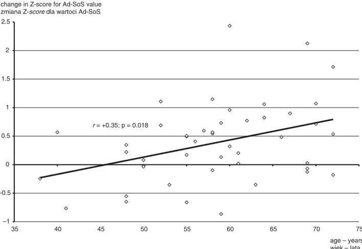
Repeated measurement after one year of reg− ular exercise showed increases in the mean Ad−SoS value from 1995 ± 80 to 2012 ± 75 m/s (p = 0.001) and Z−score from 0.673 ± 1.054 to 1.053 ± 1.178 (p= 0.0005) in the exercising subjects. The Ad−SoS changes in the individual gymnasts (LSC value for Ad−SoS: 21 m/s) are shown in Figure 1. Among all subjects the increase in Ad−SoS exceeded the value of LSC in 19 subjects (41%), and in pre− and postmenopausal women in 2 (22%) and 17 (46%), respectively. The follow−up sub− jects had both duration of exercise and intensity of exercise scores at the onset of follow−up greater than the entire group of the gymnasts (2.6 ± 1.4 vs. 2.22 ± 1.5 and 2.4 ± 1.1 vs. 1.87 ± 1.1, respec− tively) [16]. After one−year follow−up there were statistically significant negative correlations between Ad−SoS and age, BMI, and years since menopause (YSM), similar to those at the onset of the observation (Table 2). The changes in Ad−SoS and Z−score values after one−year follow−up revealed statistically significant positive correla− tions with the ages of the exercising subjects (r= 0.39, p= 0.007 and r= 0.35, p= 0.018, respec− tively). Figure 2 shows the correlation between changes in Z−score for the Ad−SoS value and age during the period of observation in the women exercising with Tai Chi.
Table 1. Clinical characteristics of the subjects practicing Tai Chi at the baseline and follow−up. Values presented as the mean ± SD
Tabela 1. Charakterystyka kliniczna osób ćwiczących Tai Chi na początku i po roku obserwacji. Wartości przedstawiono jako średnie ± SD
Variable Tai Chi baseline Tai Chi follow−up p
(Parametr) (Początek) (Obserwacja roczna)
n = 46 n = 46
Age – years 58.3 ± 8.7 59.3 ± 8.7 –
(Wiek – lata)
Body mass – kg 64.8 ± 10.8 64.6 ± 10.7 ns.
(Masa ciała – kg)
Height – cm 161.1 ± 5.3 161.1 ± 5.3 ns.
(Wzrost – cm)
BMI – kg/m2 25.0 ± 4.0 24.9 ± 3.9 ns.
(Współczynnik BMI – kg/m2)
YSM – years 11.5 ± 6.5 (n = 37) 12.5 ± 6.5 (n = 37) –
(YSM – lata)
Duration of exercise score 2.6 ± 1.4 3.4 ± 0.7 0.000
(Współczynnik czasu ćwiczeń)
Intensity of exercise score 2.4 ± 1.1 2.4 ± 1.1 ns.
(Współczynnik intensywności ćwiczeń) BMI – body mass index.
YSM – years since menopause.
Discussion
This study is the first showing skeletal status assessed prospectively by quantitative ultrasound in female gymnasts exercising with Tai Chi. Very promising data on the beneficial influence of this
kind of physical activity obtained at the baseline [16] were supported by a longitudinal observation and this finding seems to be the most important part of the study.
Osteoporosis, followed by fractures, is a major health problem of the aging population and the
Fig. 1.Ad−SoS changes during the one−year follow−up in relation to LSC value
Ryc. 1. Zmiany Ad−SoS podczas rocznej obserwacji w odniesieniu do wartości LSC
−50 −40 −30 −20 −10 0 10 20 30 40 50 60 70 80 90 100 110 120 130 1
4 7 10 13 16 19 22 25 28 31 34 37 40 43 46
Ad−SoS changes during the period of observation zmienny Ad−SoS podczas obserwacji
postmenopausal po menopauzie premenopausal przed menopauzą LSC = –21 m/s LSC = 21 m/s
serial number of examinated subject kolejny numer badanej osoby
m/s
Table 2. Correlation (Pearson or Spearman, accordingly) between Ad−SoS and other variables studied in the group of sub− jects practicing Tai Chi at baseline and follow−up (n = 46, except for YSM, where n = 37)
Tabela 2. Korelacje (według Pearsona lub Spearmana, odpowiednio) między Ad−SoS i innymi badanymi parametrami w grupie osób ćwiczącychTai Chi na początku i po roku obserwacji (n = 46, z wyjątkiem YSM, gdzie n = 37)
Variable Baseline p Follow−up p
(Parametr) Correlation coefficient Correlation coefficient (Współczynnik korelacji) (Obserwacja roczna
– współczynnik korelacji)
Age –0.72 0.000 –0.55 0.00007
(Wiek)
Body mass –0.22 ns. –0.23 ns.
(BMI)
Height +0.11 ns. +0.19 ns.
(Wzrost)
BMI –0.27 ns. –0.29 0.04
(Współczynnik BMI)
YSM –0.49 0.002 –0.39 0.01
Exercise duration +0.25 ns. +0.13 ns.
(???)
Exercise intensity +0.21 ns. +0.06 ns.
(???)
BMI – body mass index. YSM – years since menopause.
evaluation of procedures which are able to prevent them is of great importance. Physical activity is one of these, since it leads to better postural stabil− ity and less susceptibility to falling [1]. Moreover, there are some suggestions that physical activity on a regular basis in the elderly could influence bone strength and density [3, 4]. This might be due to some beneficial impact on bone turnover by skeletal muscle activity [18, 19]. Tai Chi is a pop− ular, low−weight−bearing exercise regularly prac− ticed by the elderly Chinese population and has recently became popular in Europe as well. There are limited data on the influence of these exercises on BMD. Studies carried out on Chinese popula− tions showed a significant 2.6− to 3.6−fold retarda− tion of bone loss in both the trabecular and cortical compartments of the distal tibia assessed by peripheral quantitative computed tomography (pQCT) in postmenopausal women exercising with Tai Chi [5]. Other studies, carried out using dual−energy X−ray absorptiometry (DXA) and pQCT techniques, showed significantly higher BMD (7.1% to 14.8%) in the lumbar spine, proxi− mal femur, and the ultradistal tibia in postmeno− pausal Tai Chi gymnasts than in controls [6, 7].
QUS at the hand phalanges allows indirect assessment of bone properties using bone and soft tissue ultrasound transmission expressed in Ad− SoS values. There are numerous reports on decreases in Ad−SoS in subjects with bone deteri− oration, as in menopause, pregnancy, immobiliza− tion, disease, and hormonal dysfunctions [10, 14, 20–22]. The most important data were provided by a multicenter European study in which a group more than 10,000 women was evaluated and frac−
ture prediction was proven [14]. In another small longitudinal study, QUS phalangeal measurements showed an ability to predict fracture risk at the hip [23]. A very recent study presented the value of pha− langeal ultrasonography in the assessment of 10−year probability for clinical vertebral fractures [12].
A previous study by the present authors showed higher values of Ad−SoS, reflecting ultra− sound transmission in the hand phalanges, in the entire group and in the subgroups older than 50 of Tai Chi exercising subjects compared with con− trols. The control group used at the baseline assessments (1030 non−exercising women) was matched for age, body mass, and number of years since menopause, which allowed making reliable comparisons. This suggested the positive effect of these exercises on bone status and potential anti− fracture impact. One of the most important clinical findings of the study is the efficacy of Tai Chi exercises to improve skeletal status only in post− menopausal females. Moreover, improvement expressed as differences between exercising women and controls increased with age, so Tai Chi should be recommended rather in older post− menopausal women [16].
In the present study, further increases in mean Ad−SoS and Z−score values were noted after one− year of follow−up. The increases were more often observed in older subjects. These changes corre− lated positively with the age of the exercising sub− jects, indicating the beneficial impact of exercise on older individuals. The subjects studied again after one−year had duration of exercise and inten− sity of exercise scores at the onset of the follow−up greater than the entire group of the gymnasts. This
Fig. 2. Correlation between changes in the Z−score for the Ad−SoS value and age during the period of observation
Ryc. 2. Zależność między zmianą Z−scoredla wartości Ad−SoS i wiekiem podczas obserwacji
–1 –0.5 0 0.5 1 1.5 2 2.5
35 40 45 50 55 60 65 70 75
age – years wiek – lata
r= +0.35; p = 0.018 change in Z−score for Ad−SoS value
could confirm the beneficial effects of both the intensity and the duration of the exercises on the QUS parameters assessed.
To the present authors’ knowledge, this is the first study describing prospectively the influence of Tai Chi exercises on ultrasound velocity at the hand phalanges, especially regarding premeno− pausal subjects. Moreover, there are no data in the literature on the skeletal advantages of Tai Chi exercise in premenopausal women. On the other hand, the present authors’ previous study on young female handball players did not show any deviation from the normal range in these women [24]. In another study, higher tibial, but not radial, QUS results were observed in young female swim− mers than in controls [25].
In the baseline study, the values of Ad−SoS correlated positively with exercise duration but not with exercise intensity in the entire group [16]. In the present follow−up, no such correlation was observed; the subjects reported similar intensity of exercises, but the duration of the gymnastics increased by 1 year. This study has shown negative correlation between Ad−SoS on the one hand and age, BMI, and the number of years since menopause on the other. The negative correlation between Ad−SoS and age and YSM reflect the nat−
ural age−dependent bone deterioration and the influence of menopause [13, 14, 26]. The subjects with greater body mass and BMI had lower values of Ad−SoS [26, 27].
The present study has several limitations. The one−year follow−up study was performed in a group of 46 out of 115 women. This is less than half of the baseline study population, but exercis− ing persons usually give up their physical activi− ties in such a proportion. This follow−up study was carried out in the subgroup of more advanced gymnasts; they had both duration of exercise and intensity of exercise scores at the onset of the fol− low−up greater than those of the entire group of gymnasts (115 women). The intensity of exercise score remained at the same level during the period of observation. Only one skeletal site was evaluat− ed and bone densitometry was not implemented. Despite these limitations, the study results are promising and further longitudinal observation is in progress.
In conclusion, a beneficial effect of regular Tai Chi exercise on skeletal status as assessed by pha− langeal QUS is observed in older women. Tai Chi gymnastics may be recommended in the preven− tion of postmenopausal osteoporosis, especially in older individuals.
References
[1] Sinaki M, Itoi E, Wahner HW, Wollan P, Gelzcer R, Mullan BP, Collins DA, Hodgson SF:Stronger back mus− cles reduce the incidence of vertebral fractures: A prospective 10 year follow−up of postmenopausal women. Bone 2002, 30, 836–841.
[2] Skrzek A, Bolanowski M:Strength of trunk and thigh musculature and bone mineral density in women aged 40–79 years. Isokinet Exerc Sci 2006, 14, 341–347.
[3] Chien MY, Wu YT, Hsu AT, Yang RS, Lai JS:Efficacy of a 24−week aerobic exercise program for osteopenic postmenopausal women. Calcif Tissue Int 2000, 67, 443–448.
[4] Wallace BA, Cumming RG:Systematic review of randomized trials of the effect of exercise on bone mass in pre− and postmenopausal women. Calcif Tissue Int 2000, 67, 10–18.
[5] Chan K, Qin L, Lau M, Woo J, Au S, Choy W, Lee K, Lee S:A randomized, prospective study of the effects of Tai Chi Chun exercise on bone mineral density in postmenopausal women. Arch Phys Med Rehabil 2004, 85, 717–722.
[6] Qin L, Au S, Choy W, Leung P, Neff M, Lee K, Lau M, Woo J, Chan K:Regular Tai Chi Chuan exercise may retard bone loss in postmenopausal women: A case−control study. Arch Phys Med Rehabil 2002, 83, 1355–1359.
[7] Qin L, Choy W, Leung K, Leung PC, Au S, Hung W, Dambacher M, Chan K:Beneficial effects of regular Tai Chi exercise on musculoskeletal system. J Bone Miner Metab 2005, 23, 186–190.
[8] Schott AM, Weill−Engerer S, Hans D, Duboeuf F, Delmas PD, Meunier PJ:Ultrasound discriminates patients with hip fracture equally well as dual energy X−ray absorptiometry and independently of bone mineral density. J Bone Miner Res 1995, 10, 243–249.
[9] Hans D, Dargent−Molina P, Schott AM, Sebert JL, Cornier C, Kotzki PD, Delmas PD, Pouilles JM, Breart G, Meunier PJ:Ultrasonographic heel measurements to predict hip fracture in elderly women: the EPIDOS prospective study. Lancet 1996, 348, 511–514.
[10] Hellmeyer L, Ossendorf A, Ziller V, Tekesin I, Schmidt S, Hadji P:Quantitative ultrasonometry of the pha− langes during pregnancy: a longitudinal study. Climacteric 2006, 9, 446–451.
[11] Glüer CC, Wu CY, Genant HK:Broadband ultrasound attenuation signals depend on trabecular orientation: an in vitro study. Osteoporos Int 1993, 3, 185–191.
[12] Kanis JA, Johnell O, Oden A, De Laet C, de Terlizzi F:Ten−year probabilities of clinical vertebral fractures according to phalangeal quantitative ultrasonography. Osteoporos Int 2005, 16, 1065–1070.
[13] Pluskiewicz W, Drozdzowska B.Quantitative ultrasound (QUS) at the calcaneus and hand phalanges in Polish healthy postmenopausal women. Ultrasound Med Biol 2001, 27, 373–377.
Reginster JY, de Terlizzi F, Cadossi R.Phalangeal osteosonogrammetry study: Age−related changes, diagnostic sensitivity, and discrimination power. J Bone Miner Res 2000, 15, 1603–1614.
[15] Bolanowski M, Pluskiewicz W, Adamczyk P, Daroszewski J:Quantitative ultrasound at hand phalanges in patients with acromegaly. Ultrasound Med Biol 2006, 32, 191–195.
[16] Bolanowski M, Pluskiewicz W, Skrzek A, Adamczyk P, Bolanowski J:Bone status assessed by quantitative ultrasound at hand phalanges in women exercising Tai Chi. Hum Movement 2006, 7, 162–167.
[17] Glüer CC, Blake G, Lu Y, Blunt BA, Jergas M, Genant HK:Accurate assessment of precision errors: how to measure the reproducibility of bone densitometry techniques. Osteoporos Int 1995, 4, 262–270.
[18] Langberg H, Skovgaard D, Asp S, Kjaer M: Time pattern of exercise−induced changes in type I collagen turnover after prolonged endurance exercise in humans. Calcif Tissue Int 2000, 67, 41–44.
[19] Rudberg A, Magnusson P, Larsson L, Joborn H:Serum isoforms of bone alkaline phosphatase increase during physical exercise in women. Calcif Tissue Int 2000, 66, 342–347.
[20] Alexandersen P, de Terlizzi F, Tanko LB, Bagger YZ, Christiansen C:Comparison of quantitative ultrasound of the phalanges with conventional bone densitometry in healthy postmenopausal women. Osteoporos Int 2005, 16, 1071–1078.
[21] Bolanowski M, Pluskiewicz W:Quantitative ultrasound of the hand phalanges and calcaneus revealed skeletal abnormalities due to primary hyperparathyroidism: a case report. Ultrasound Med Biol 2002, 28, 265–259.
[22] Pluskiewicz W, Drozdzowska B: Ultrasound measurements of proximal phalanges in Polish early post− menopausal women. Osteoporos Int 1998, 8, 578–583.
[23] Mele R, Masci G, Ventura V, de Aloysio D, Biocchi M, Cadossi R:Three−year longitudinal study with quanti− tative ultrasound at the hand phalanx in a female population. Osteoporos Int 1997, 7, 550–557.
[24] Bolanowski M, Pluskiewicz W, Jędrzejuk D, Zimmer K:Results of ultrasonic study of hand phalanx in young female handball players (in Polish). Medycyna Sportowa 1999, 15, 14–16.
[25] Falk B, Bronshtein Z, Zigel L, Constantini N, Eliakim A:Higher tibial ultrasound in young female swimmers. Br J Sports Med 2004, 38, 461–465.
[26] Rico H, Aguado F, Arribas I, Hernandez ER, Villa LF, Seco C, Gervas JJ:Behavior of phalangeal bone ultra− sound in normal women with relation to gonadal status and body mass index. Osteoporos Int 2001, 12, 450–455.
[27] Ventura V, Mauloni M, Mura M, Paltrinieri F, de Aloysio D:Ultrasound velocity changes at the proximal pha− langes of the hand in pre−, peri− and postmenopausal women. Osteoporos Int 1996, 6, 368–375.
Address for correspondence:
Marek Bolanowski
Department of Endocrinology, Diabetology, and Isotope Therapy Silesian Piasts University of Medicine
Pasteura 4 50−367 Wroclaw Poland
Tel.: +48 71 784 27 40
E−mail: [email protected] Conflict of interest: None declared Received: 10.07.2007