Joanna Kiśluk
1, A, C, D, Mariusz Gryko
1, B, C, Katarzyna Guzińska-Ustymowicz
2, D–F,
Andrzej Kemona
2, E, Bogusław Kędra
1, E, FImmunohistochemical Diagnosis of Gastrointestinal
Stromal Tumors – An Analysis of 80 Cases
from 2004 to 2010*
Diagnostyka immunohistochemiczna guzów stromalnych
przewodu pokarmowego – analiza 80 przypadków z lat 2004–2010
1 Second Department of General and Gastroenterological Surgery, Medical University of Białystok, Bialystok, Poland 2 Department of General Pathomorphology, Medical University of Bialystok, Białystok, Poland
A – research concept and design; B – collection and/or assembly of data; C – data analysis and interpretation;
D – writing the article; E – critical revision of the article; F – final approval of article; G – other
Abstract
Background. Gastrointestinal stromal tumors (GISTs) are the most common cancers of mesenchymal origin in the abdomen. An approximately 95% of GISTs show positive expression of the membrane receptor c-kit (CD117 antigen), which currently constitutes the basis for histopathological diagnosis if this type of tumor is suspected.
Objectives. The aim of this study was to perform and investigate wide parametric immunohistochemical and mor-phological analyses of stromal tumors diagnosed in the Podlasie province in the years 2004–2010.
Material and Methods. The study group consisted of 80 patients who had undergone surgical treatment for mes-enchymal tumors of the gastrointestinal tract. The immunostaining technique was performed using the surgically resected material that was formalin-fixed and embedded in paraffin blocks, then sliced into sections and stained with the monoclonal antibodies CD117, CD34, SMA, S-100 and Ki-67.
Results. CD117 was positively expressed in 77 cases, which confirmed the diagnosis of GIST. In 66 of the cases of CD117-positive stromal tumors, positive immunoreactivity for CD43 was observed. Nearly 49% (38 cases) of the GISTs were negative for SMA by immunohistochemistry. Most of the cases (57.5%) were reported in the stomach, while 17.5% were located in the intestines; 18.75% presented in different locations (the colon, ovary or gall blad-der).
Conclusions. The use of a highly sensitive IHC panel can increase the accuracy of GIST diagnoses. Detailed immu-nohistochemical studies are useful in successfully identifying any case of GIST, which is crucial to clinical treatment (Adv Clin Exp Med 2013, 22, 1, 33–39).
Key words: gastrointestinal stromal tumor, GIST.
Streszczenie
Wprowadzenie. Guzy stromalne przewodu pokarmowego (gastrointestinal stromal tumor – GIST) należą do naj-częstszych nowotworów w jamie brzusznej o pochodzeniu mezenchymalnym. Około 95% guzów GIST wykazuje pozytywną ekspresję receptora błonowego c-kit (antygen CD117), co obecnie stanowi podstawę rozpoznania histo-patologicznego przy podejrzeniu tego typu nowotworu.
Cel pracy. Ze względu na częste występowanie guzów GIST w przewodzie pokarmowym oraz różnorodność histo-logiczną celem pracy była szeroko parametrowa analiza immunohistochemiczna oraz morfologiczna guzów pod-ścieliskowych rozpoznanych na Podlasiu w latach 2004–2010.
Materiał i metody. Grupę badaną stanowiło 80 pacjentów z usuniętymi operacyjnie guzami mezenchymalny-mi przewodu pokarmowego. Do analizy immunohistochemezenchymalny-micznej wykorzystano utrwalony w formalinie materiał pooperacyjny, który został przeprowadzony do postaci kostek parafinowych, a następnie skrojony do barwień immunohistochemicznych z użyciem przeciwciał monoklonalnych: CD117, CD34, SMA, S-100 oraz Ki-67.
Adv Clin Exp Med 2013, 22, 1, 33–39 ISSN 1899–5276
OrIGINAl PAPErS
© Copyright by Wroclaw Medical University
Gastrointestinal stromal tumors are defined as a group of tumors derived from mesenchymal interstitial cells of Cajal precursor that serve as a positive control for evaluating KIT (CD117) expression immunohistochemically [1]. Stromal tumors are among the most common mesenchy-mal neoplasms of the gastrointestinal tract. This type of cancer has been also been reported in soft tissues of the abdominal cavity, where GIST metastases very often appear [2]. reports from many countries indicate that there are approxi-mately 20–40 cases of stromal tumors per million population [3–5]. Because of the long term de-velopment of clinical malignant stromal tumors (10–15 years), the incidence of cancer may be much greater. Most of the tumors are located in the stomach (70%) and the small intestine (15%). GISTs can be also found in the colorectum (7%), esophagus (7%) and rare cases have been reported in the retroperitoneum (1%) [6]. A detailed histo-pathological examination (specifying the type of cells that predominate in the site of H+E stain-ing) and immunohistochemical investigation are useful in identifying GIST tumors. Most stromal tumors are benign neoplasms, but approximately 20–30% of cases are malignant tumors. The de-gree of possible malignancy is based on the re-sults of diagnostic parameters such as tumor size, location and mitotic index (MI: the number of mitotic figures in 50 large fields of view). Micro-scopically, stromal tumors are composed of epi-theliolid cells, spindle type cells, pleomorphical cells and mixed type [7].
The diversity histological features and the specific immunophenotype allows GISTs to be di-stinguished from other neoplasms of the gastroin-testinal tract. For years, the many types of GISTs were diagnosed and classified as leiomyoblasto-ma, leiomyosarcoleiomyoblasto-ma, schwannoleiomyoblasto-ma, leiomyoma or neurofibroma because of the similarity of their microscopic features. Stromal tumors have been presented as a specific subtype of mesenchymal tissue tumors since 2004. Due to the high inciden-ce of GISTs in the gastrointestinal tract and their histological variety, the aim of this study was to carry out wide parametric immunohistochemical
and morphological analyses of stromal tumors diagnosed in the Podlasie province in the years 2004–2010.
Material and Methods
The study involved a group of 80 patients who had been diagnosed with mesenchymal tumors and surgically treated in the Second Department of General and Gastroenterological Surgery at the Medical University of Bialystok (Poland) from 2004 through 2010. All archival paraffin-embed-ded tissue blocks were from the Academic Center for Pathological Diagnosis in Bialystok.
The postoperative material was fixed in 10% formalin. Fragments of tumor were embedded in paraffin. Consecutive 5-µm sections were cut from the paraffin blocks and mounted on normal and adhesive slides. The prepared samples were dehy-drated by passing them through a series of graded alcohols (70%, 96% and 2 × absolute alcohol), and dewaxed with xylenes. The 5-µm paraffin sections were stained with hematoxylin and eosin. The his-tological type of the tumor and the type of prevail-ing cells in representative areas were determined and marked on the H&E slides. Tumors located in the stomach were characterized by spindle cells with palisade-like arrays of nuclei. The histo-logical characteristics of stromal tumors located within the small intestine include a predominance of spindle cells, numerous bands of eosinophilic collagen fibres (called skenoid fibers) and a prolif-eration of blood vessels. GISTs in other locations don’t have a typical histological pattern and most of them consist of a mixture of spindle and epi-thelioid cells. The number of mitotic figures was counted in 50 large fields of view (50 HPF) to de-termine the mitotic index.
Immunohistochemistry Methods
The 5-µm-thick sections were mounted on ad-hesive slides and placed in a water bath at 99.8°C for 60 minutes, then immersed in citric buffer at pH = 6.0. The retrieval of antigens was carried out accord-Wyniki. W 77 przypadkach wykazano dodatnią ekspresję CD117, co potwierdziło rozpoznanie GIST. W 66 przy-padkach guzów stromalnych CD117-dodatnich stwierdzono pozytywną ekspresję na CD34. W 38 przyprzy-padkach z 77 ekspresja SMA była ujemna (49%). Najwięcej przypadków odnotowano w obrębie żołądka (57,5%). 17,5% przypadków dotyczyło umiejscowienia w obrębie jelita cienkiego, zaś 18,75% w innych miejscach jamy brzusznej (jelito grube, jajnik, pęcherzyk żółciowy).Wnioski. Wyniki panelowych badań IHC z dużą czułością potwierdzają rozpoznanie nowotworu GIST. Dokładna analiza immunohistochemiczna umożliwia scharakteryzowanie każdego przypadku GIST, co jest niezbędne do podjęcia procedur leczniczych (Adv Clin Exp Med 2013, 22, 1, 33–39).
ing to immunohistochemical procedures. In this study, the following specific antibodies were used: CD117 (Monoclonal Mouse Anti-Human CD117 clone 104D2, DAKO), CD34 (Monoclonal Mouse anti-human CD34 clone QBEnd 10, DAKO), SMA (Monoclonal Mouse Anti-Human Smooth Muscle Actin clone 1A4, DAKO), S-100 (polyclonal rabbit anti-S-100, DAKO) and Ki-67 (Monoclonal Mouse anti-human Ki-67 clone MIB-1, DAKO). The slides were incubated with 0.5% hydrogen peroxide solu-tion in methanol to block endogenous peroxidase. After incubation with the primary antibodies, the sections were incubated with biotinylated antimouse antibody and followed by horseradish peroxidase-conjugated streptavidin (lSAB kit, DAKO). The col-ored peroxidase reaction product was visualized us-ing chromogen B-DAB (DAKO). Positive reactions were counted in at least 500 tumor cells from each slice and examined under a light microscope (×400).
Assessment of tumor aggressiveness was per-formed using such tumor parameters as location, the largest diameter and the number of mitotic fig-ures in 50 large fields of view (50 HPF) [6].
Results
Characteristics of the Study Group
Depending on the Result
of CD117 Expression
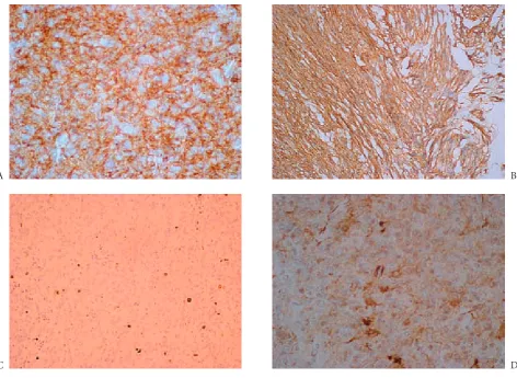
CD117 was positively expressed in 77 cases, which confirmed the diagnosis of GIST. Most of the tumors demonstrated a cytoplasmic and Golgi staining pattern (Figure 1A). In three cases a membranous staining pattern was ob-served (Figure 1B). Nearly 61.25% of the posi-tive CD-117 the tumor cells occurred in adults over 60 years. There was no gender predilection (48.75% of the CD-117-positive cases were males and 47.5% were females). Most of the GISTs were located in the stomach (57.5%), and 17.5% were in the small intestine. retroperitoneal tumors were found in 2.5% of the cases, while 18.75% were located in different areas (colon, ovary or gall bladder). In 52 of cases with confirmed diagnoses of GIST by immunohistochemistry methods, tumor size did not exceed 5 cm. In 20
Fig. 1. A) CD117-positive GIST tumor with cytoplasmic staining, B) CD117-positive GIST tumor with membranous staining pattern, C) Ki-67 expression in 1% of cells, D) Positive CD34 expression
Ryc. 1. A) Pozytywna ekspresja CD117, reakcja cytoplastyczna, B) pozytywna ekspresja CD117, reakcja błonowa, C) pozytywna ekspresja Ki-67 w 1% komórek, D) pozytywna ekspresja CD34
A
C
B
cases the tumor volume was from 5 cm to 10 cm; only 5 tumors had a size exceeding 10 cm. All the cases were characterized by low mitotic activity (in 72.5% of the cases fewer than 5 mitotic fig-ure were found in the 50 HPF, 16.25% of GISTS showed a mitotic index ranging from 5 to 10 / 50 HPF. In only 7.5% the mitotic index was > 10 / 50 HPF). A detailed analysis of the study group is shown in Table 1.
Expression of CD117, CD34,
SMA, S-100 and Ki-67
in the Study Group
In 66 of the CD117-positive stromal tumors, positive expression for CD34 was observed (Fig-ure 1D). Positive expression of smooth muscle ac-tin (SMA) was observed in only 39 cases. All of the CD117-positive tumors were characterized by a negative result of immunohistochemical reac-tion for S-100 compared with the CD117-negative GISTs, which clearly showed a positive reaction to this protein. In 24 of 77 cases, no expression of proliferative Ki-67 protein was observed. The oth-er GIST tumors showed mostly weak positive ex-pression: In only 4 cases the reaction for Ki-67 was seen in more than 10% of the tumor cells; in 12 cases Ki-67-positive tumor cells ranged from 5% to 10%; while the expression of this protein was seen in ≤ 5% of the cell in 37 patients. Expression of Ki-67 in GIST cells is shown in Figure 1C. De-tailed characteristics of individual protein expres-sion in the study group are presented in Table 2.
Discussion
Gastrointestinal stromal tumors have been identified and diagnosed for several years. Im-munophenotyping and histological observa-tions of the tumor stroma led to the distinction of GISTs from other gastrointestinal neoplasms without a glandular or epithelial formations, such
Table 1. Characteristics of the study group, sorted by CD117 expression
Tabela 1. Charakterystyka grupy badanej w zależności od wyniku ekspresji na CD117
CD117 (+) CD117 (–) (N = 77)
96% (N = 3)4%
Age (Wiek) < 60 (N = 29)
≥ 60 (N = 51)
28 (35%) 49 (61.25%) 1 (1.25%) 2 (2.5%) Sex (Płeć)
Male (N = 41) (Mężczyźni) Female (N = 39) (Kobiety) 39 (48.75%) 38 (47.50%) 2 (2.5%) 1 (1.25%) location (Umiejscowienie) stomach (N = 47)
intestinal (N = 18)
retroperitoneum (N = 2)
other (colon, gallblader, esophagus) (N = 15)
46 (57.5%) 14 (17.5%) 2 (2.5%) 15 (18.75%) 1 (1.25%) 2 (2.5%) 0 0 Size (rozmiar) ≤ 5 cm (N = 55)
5–10 cm (N = 20)
≥ 10 cm (N = 5)
52 (65%) 20 (25%) 5 (6.25%) 3 (3.75%) 0 0
Mitotic Index IM (Wskaźnik mitozy) ≤ 5 / 50 HPF (N = 59)
5–10 / 50 HPF (N = 15)
≥ 10 / 50 HPF (N = 6) 58 (72.5%) 13 (16.25%) 6 (7.5%) 1 (1.25%) 2 (2.5%) 0
Table 2. Expression of CD117, CD34, SMA, S-100 and Ki-67 in the study group
Tabela 2. Ekspresja CD117, CD34, SMA, S-100 oraz Ki-67 w grupie badanej
Total (N = 80)
(razem) CD117 (+) CD117 (–)
CD34 (+) 66 3
(–) 11 0
SMA (+) 39 3
(–) 38 0
S-100 (+) 0 3
(–) 77 0
Ki-67 (–) 24 0
≤ 5% 37 1
5–10 % 12 1
as leiomyomas, sarcomas, rhabdomyosarcomas, neurinomas, neurofibromas and meningiomas [1, 8, 9]. The mechanism leading to the malignant transformation of GIST has not been definitively elucidated. The results of immunohistochemical studies suggest that stromal tumor are derived from intramural ganglion cells (called Cajal cells), located in the Auerbach plexus area, or from their precursors [7]. Cajal cells are regarded as elements of pacemakers located in the tubular organs of the gastrointestinal tract that can differentiate toward both smooth muscle cells lining the walls of diges-tive organs and cells of the autonomic nervous sys-tem, coordinating peristalsis. Other theories sug-gest that stromal tumors arise from the neoplastic transformation of pluripotent precursors of stem cells to pacemaker cells.
According to data in the literature, the vast majority of stromal tumors (approximately 95%) show positive membrane expression of c-kit re-ceptor tyrosine kinase (CD117) [10, 11]. Because of the high sensitivity and specificity of KIT, it is a useful marker for differentiating GISTs from other mesenchymal tumors of the gastrointestinal tract [7]. In the current study, 77 tumors (96%) out of 80 cases expressed CD117-positive cells.
Population-based studies conducted world-wide among patients with stromal tumors have shown that expression of CD34 is also important in a GIST diagnosis. The data in the literature demon-strate that positive expression of CD34 is observed in 60–70% of GISTs and is associated with one of the probable mechanisms of carcinogenesis in the group of tumors that focus on neoplastic transfor-mation of pluripotent stem cells [4, 10, 11]. The authors of the current study found that 11 patients had a lack of CD34 expression and a simultane-ous positive reaction for CD117. Six of these cases were small intestinal GISTs and five were gastric GISTs. This observation concurred with the report by Miettinen et al., who noted a similar distribu-tion of CD34 expression in GISTs [11, 12].
Many authors emphasize difficulties interpreting the expression of smooth muscle markers, including smooth muscle actin (SMA) in stromal neoplasms. Entrapped smooth muscle cells from muscularis pro-pria or adjacent muscularis mucosae can cause a false impression of positive expression of SMA in a GIST. According to Fletcher et al [9], SMA expression in GISTs is found in 20–40% of the lesions located in the stomach and about 50% of those in the intestine, and is often linked with negative expression of CD34. In the current investigation positive SMA expression was observed in all the CD117-negative tumors and in 51% of the CD117-positive tumors. It was noted that positive SMA expression did not correlate with negative expression of CD34 staining.
Determining S-100 protein expression is con-sidered a way to distinguish GISTs from other mesenchymal tumors. Strongly positive expres-sion of S-100 protein was observed in tumors of nervous origin, especially in schwannomas [13]. The expression of S100 proteins is very rare in GISTs. reports have shown that only stromal tu-mors located in the small intestine are character-ized by a greater tendency to express positive reac-tion (about 10%), located in both the cytoplasm and the cell nucleus [14]. In the current study none of the CD117-positive tumors showed expression of S-100. S-100-positive cells were found in three GISTs with negative CD117 expression.
The location of GISTs in the gastrointestinal tract may influence the degree of malignancy. As noted by Patil et al [10], most malignant stromal tumors are localized in intestine. In the current study 57.5% of all CD117-positive GISTs were observed in gastric locations. The most common locations of stromal tumors noted in the litera-ture [1, 8, 12] are similar to those in the current study. Tumors located in the small intestine ac-counted for 20% of the cases, and 17.5% of them had a positive CD117 expression. Only three cases of KIT-postive tumors were observed in the colon. Determining the location is necessary to assess the risk of disease progression for each completely di-agnosed GIST [2]. In the current study, the study group had a low mitotic index (< 5 abnormal mi-totic figures counted in 50 high power fields of view in 72.5% of all the tumors examined) and a small tumor size (65% of the tumors were less than 5 cm in diameter, and 25% had a diameter of 5–10 cm), which allowed them to be classified as a group with a low risk of progression and low tumor aggressiveness.
Table 3. Differential diagnosis of Gastrointestinal Stromal Tumor – immunohistochemistry methods [10]
Tabela 3. Diagnostyka różnicowa GIST – metody immunohistochemiczne [10]
Diagnosis
(rozpoznanie) CD117 CD34 SMA S-100
GIST
(Guz stromalny przewodu pokarmowego) +++ +++ (70%) + (40%) –
leiomyoma
(Mięśniak gładki) – +/– +++ –
leiomyosarcoma
(Mięśniakomięsak gładkokomórkowy) – + (10%) +++ –
Schwannoma
(Nerwiak osłonkowy) – – – +++
The differential diagnosis of GISTs in the current study corresponded with the immuno-histochemistry methods used by other authors. Summaries of CD117, CD34, S-100 and SMA ex-pression in various mezenchymal tumors are pre-sented in Table 3.
In summary, the immunohistochemical analyses of the study group allowed GISTs to be differentiated from other mesenchymal tumors. The results of panel tests using antibodies against CD117, CD34, SMA and S-100, along with the as-sessment of the mitotic index in H&E staining, confirmed the diagnoses of GIST. In doubtful
cases with negative expression of CD117, a final diagnosis is not possible on the basis of deter-mining CD34, SMA, S-100 or Ki-67 expression. The coincidence of positive expression of CD34, SMA and S-100 in CD-117 negative cases in the current study is interesting but requires further study on a larger group of patients. It is important that a positive reaction for these individual anti-bodies specifically indicated a possible pathway of mutation toward stromal neoplasm in GISTs confirmed using molecular methods. A detailed immunostaining study is useful to successfully identify cases of GIST.
References
Miettinen M, Lasota J:
[1] Gastrointestinal stromal tumors (GISTs): definition, occurrence, pathology, differential diagnosis and molecular genetics. Pol J Pathol2003, 54, 3–24.
Miettinnen M, ElRifaj EL, Sobin LH, Lasota J:
[2] Evaluation of malignancy and prognosis of gastrointestinal stromal tumors: a review. Hum Pathol 2002, 33, 478–483.
Kondblom LG, Meis-Kindblom J, Bumming P et al.:
[3] Incidence, prevalence, phenotype and biologic spectrum of gastrointestinal stromal cell tumors (GIST) – a population-based study of 600 cases. Ann Oncol 2002, 13 (Suppl 5), 157, 16.
Nilsson B, Bumming P, Meis
[4] -Kindblom JM et al.: Gastrointestinal stromal tumors: the incidence, prevalence, clinical course, and prognostication in the preimatinib mesylate era – a population-based study in western Sweden. Cancer 2005, 103, 821–829.
Tran T, Davila JA, El-Serag HB:
[5] The epidemiology of malignant gastrointestinal stromal tumors: an analysis of 1,458 cases from 1992 to 2000. Am J Gastroenterol 2005, 100, 162–168.
Miettinen M, Lasota J:
[6] Gastrointestinal stromal tumors: pathology and prognosis at different sites. Semin Diagn Pathol 2006, 23, 70–83.
Miettinen M, Lasota J:
[7] Gastrointestinal Stromal Tumors review on Morphology, Molecular Pathology, Prognosis, and Differential Diagnosis. Arch Pathol lab Med 2006, 130, 1466–1478.
Miettinen M, Majidi M, Lasota J:
[8] Pathology and diagnostic criteria of gastrointestinal stromal tumors (GISTs): a review. Eur J Cancer 2002, 38 (Suppl 5), 39–51.
Fletcher CDM, Berman JJ, Corless C et al.:
[9] Diagnosis of gastrointestinal stromal tumors: a consensus approach. Hum Pathol 2002, 33, 459–465.
Patil DT, Rubin BP:
[10] Gastrointestinal Stromal Tumor: Advances in Diagnosis and Management. Arch Pathol lab Med 2011, 135, 1298–1310.
Miettinen M, Lasota J:
[11] KIT (CD117): a review on expression in normal and neoplastic tissues, and mutations and their clinicopathologic correlation. Appl Immunohistochem Mol Morphol 2005, 13, 205–220.
Miettinen M, Makhlouf H, Sobin LH, Lasota J:
Trabelsi A, Mestiri S, Stita W, Mokni M, Sriha B, Rammeh S, Yahyaoui S, Korbi S:
[13] Mesenchymal tumors of the
digestive tract, immunohistochemistry contribution Ann Biol Clin (Paris) 2007, 65, 365–368.
Liu FY, Qi JP, Xu FL, Wu AP:
[14] Clinicopathological and immunohistochemical analysis of gastrointestinal stromal tumor. World J Gastroenterol 2006, 12, 4161–4165.
Nishimura R, Osako T, Okumura Y, Tashima R, Toyozumi Y, Arima N:
[15] Changes in the Er, Pgr, HEr2, p53
and Ki-67 biological markers between primary and recurrent breast cancer: discordance rates and prognosis. World J Surg Oncol 2011, 9, 131–137.
Tolonen TT, Tammela TL, Kujala PM, Tuominen VJ, Isola JJ, Visakorpi T:
[16] Histopathological variables and
bio-markers enhancer of zeste homologue 2, Ki-67 and minichromosome maintenance protein 7 as prognosticators in primarily endocrine-treated prostate cancer BJU Int. 2011 May 18. doi: 10.1111/j.1464-410X.2011.10253.
Address for correspondence:
Joanna Kiśluk
Second Department of General and Gastroenterological Surgery Medical University of Bialystok
Skłodowskiej-Curie 24A 15-276 Białystok Poland
Tel.: +48 510 506 386 E-mail: [email protected]
Conflict of interest: None declared received: 5.12.2011
![Table 3. Differential diagnosis of Gastrointestinal Stromal Tumor – immunohistochemistry methods [10]Tabela 3](https://thumb-us.123doks.com/thumbv2/123dok_us/8769926.1756450/6.595.67.529.92.239/table-differential-diagnosis-gastrointestinal-stromal-immunohistochemistry-methods-tabela.webp)
