Omer E. Yapca
1, A, F, Bunyamin Borekci
2, A, F,
Mehmet I. Turan³
, A, D, F, Mine Gulapoglu
4, B, C, FThe Effect of Agomelatine on Oxidative Stress Induced
with Ischemia/Reperfusion in Rat Ovaries
¹ Obstetrics and Gynecology, Sorgun State Hospital, Yozgat, Turkey
² Department of Obstetrics and Gynecology, Ataturk University, Erzurum, Turkey ³ Department of Pediatrics, Ataturk University, Erzurum, Turkey
4 Department of Biochemistry, Ataturk University, Erzurum, Turkey
A – research concept and design; B – collection and/or assembly of data; C – data analysis and interpretation;
D – writing the article; E – critical revision of the article; F – final approval of article; G – other
Abstract
Background. Ovarian ischemia and reperfusion can lead to serious and irreversible health problems.
Objectives. The aim of this study is to investigate the protective effect of agomelatine against ovarian ischemia/ /reperfusion injury in rats using biochemical methods.
Material and Methods. Thirty female rats were divided into three groups (the number of animals in each group = 10), a control group in which ischemia/reperfusion was established (IRC), an ischemia/reperfusion + agomelatine (IRA) group and a healthy group given a sham operation (SG). Total glutathione (tGSH) and malondialdehyde (MDA) levels and glutathione peroxidase(GPx), superoxide dismutase (SOD) and myeloperoxidase (MPO) enzyme activ-ity were measured in ovarian tissue extracted at the end of the experiment.
Results. Biochemical results revealed MDA levels of 19.1 ± 2.03, 5.8 ± 1.5 and 5.5 ± 1.4 µmol/g protein in ovarian tissue in the IRC, IRA and SG groups, respectively. MPO activity in the IRC, IRA and SG groups was 7.87 ± 2.7, 4.0 ± 2.0 and 3.0 ± 1.0 U/g protein, respectively. tGSH levels were 1.87 ± 1.13, 4.37 ± 1.4 and 5.87 ± 1.64 nmol/g tein, respectively. GPx activity in the IRC, IRA and SG groups was 7.37 ± 1.68, 18.6 ± 3 and 17.75 ± 3.2 U/g pro-tein, and SOD activity 31.1 ± 2.9, 45.3 ± 3.7 and 54 ± 4.2 U/g propro-tein, respectively. The level of 8-OH/ /Gua, a product of DNA damage, was 2.18 ± 0.2 pmol/L in the IRC group, 1.28 ± 0.2 pmol/L in the IRA group and 0.93 ± 0.01 pmol/L in the SG group.
Conclusions. Agomelatine prevented ovarian ischemia/reperfusion injury (Adv Clin Exp Med 2014, 23, 5, 715–721).
Key words: agomelatine, rat, ovary, antioxidants, ischemia, reperfusion.
Adv Clin Exp Med 2014, 23, 5, 715–721 ISSN 1899–5276
ORIGINAL PAPERS
© Copyright by Wroclaw Medical University
Ischemia emerges as the result of subtotal or to-tal interruption of blood flow to tissue for various reasons. Ovarian ischemia is a pathological condi-tion seen as a result of the ovary twisting around its own vascular structures (torsion) [1]. Prolonged exposure to ischemia may lead to irreversible dam-age in ovarian tissue. In order to prevent irrevers-ible damage, the ovary exposed to torsion is reper-fused through detorsion [2]. However, reperfusion of ischemic tissue may lead to more severe injury than that caused by ischemia in the tissue [3]. High levels of molecular oxygen and overproduction of toxic oxygen radicals with reperfusion of ischemic
tissue are implicated as causes of reperfusion inju-ry. Overproduction of toxic oxygen radicals causes severe damage in cells by leading to lipid peroxida-tion and DNA damage [4]. The use of drugs with antioxidant activity is therefore being tested in the prevention and treatment of ischemia/reperfusion injury [5, 6]. The agomelatine used in our study is a synthetic analogue of melatonin. Like melatonin, agomelatine stimulates MT1 and MT2 membrane
to exhibit a powerful antioxidant effect through ML1 receptors. Membrane ML1 receptors are also known to be present in ovarian tissue [9]. The fact that agomelatine both stimulates melatonin recep-tors and antagonizes serotonin receprecep-tors indicates that the drug may be useful in the prevention of ischemia/reperfusion injury.
Scans of the literature revealed no data con-cerning the protective effect of agomelatine against oxidative damage induced with ischemia/reperfu-sion in rat ovaries. The purpose of this study was therefore to investigate whether agomelatine does have a protective effect against oxidative stress in-duced with ischemia/reperfusion in rat ovaries.
Material and Methods
Animals
The study was performed with 30 female al-bino Wistar rats weighing 225–235 g. These were obtained from the Atatürk University Medical Ex-perimental Practice and Research Center. In order to adapt to their environment, animals were kept and fed for a while at normal room temperature (22°C) in the Department of Pharmacology labo-ratory where the experiment was to be conducted.
Chemical Substances
Agomelatine 25 mg tablets were obtained from Les Laboratoires Servier, France. All other chemi-cal substances used for the experiments were pro-vided by IE Ulagay, Turkey.
General Procedure
The female rats to be used in the experi-ment were divided into 3 groups, a control group (N = 10) in which ischemia/reperfusion was to be established (IRC), an ischemia/reperfusion + + agomelatine (IRA) group (N = 10) and a healthy group (N = 10) given a sham operation (SG). Sur-gical procedures were performed with 25 mg/kg in-traperitoneal (i.p.) thiopental sodium anesthesia.
Surgical Procedures
Before rat ovaries were subjected to the isch-emia/reperfusion procedure, the IRA group was given a 25 mg/kg dose of agomelatine by tube. The IRC and SG group rats were given distilled water as solvent by the same route. Once the drugs had been administered, the inferior part of the abdo-men was accessed through a 2–2.5 cm vertical inci-sion. An arterial clip was later attached to the lower
part of the right ovary (the region where the ova-ry joins the uterus) of the rats in the IRC and IRA groups, and ischemia was established for 3 h. No ischemia was established in the SG ovaries. At the end of that period the arterial clip was removed and 2-h reperfusion was established. Once reper-fusion had been established all animals were sac-rificed with high-dose anesthesia, and biochemical analyses were performed. The biochemical findings from the IRA group were analyzed by comparing them with those from the IRC and SG groups.
Biochemical Analysis
of Ovarian Tissue
Specimen Preparation
At this stage, 0.2 g were weighed from each ovary extracted. The ovaries were homogenized in 0.5% HDTMAB (0.5% hexadecyltrimethylam-monium bromide) containing pH = 6 potassi-um phosphate buffer for MPO assay, 1.15% po-tassium chloride solution for MDA assay and pH = 7.5 phosphate buffer for other measure-ments, all made up to 2 mL in an iced environ-ment. They were subsequently centrifuged at 10,000 rpm for 15 min at +4°C. The supernatant part was used as a specimen for analysis.
Chemical Parameter
Screening
Malondialdehyde (MDA) Assay
Based on spectrophotometric measurement at an emission wavelength of 532 nm of the absor-bance of the pink complex formed at high tem-perature (95°C) by thiobarbituric acid (TBA) and MDA [10].
Myeloperoxidase (MPO)
Activity Assay
Oxidation reaction performed with MPO-me-diated H2O2 containing
4-aminoantipyrene/phe-nol solution as substrate was used to determine MPO enzyme activity [11].
Total Glutathione (tGSH) Assay
Glutathione Peroxidase
(GPx) Assay
The enzyme GPx reduces H2O2 to water in the
presence of H2O2 and catalyzes the oxidation of
re-duced glutathione (GSH) to oxidized glutathione (GSSG). The GSSG that forms reduces one reductase again to GSH through the glutathi-one reductase reaction in which it is used as reduc-ing substrate of NADPH. The absorbance decrease resulting from the oxidation of NADPH to NADP is measured spectrophotometrically at 340 nm and GPx enzyme activity calculated from this [13].
Superoxide Dismutase (SOD)
Activity Assay
This is based on measurements at a wave-length of 560 nm of the absorbance of formazan, a purple compound forming with the reduction of nitroblue tetrazolium (NBT) in the reaction envi-ronment by O2¯radicals produced by the addition
of the enzyme xanthine oxidase to the reaction en-vironment. The intensity of the reduction reac-tion depends on the activity of the Cu/Zn SOD en-zyme activity in the specimen. The more enen-zyme in the environment, the less O2¯radical there is to
react with NBT. The intensity of the purple color emerging with the presence of the formazan thus decreases in proportion [14].
Isolation of DNA
from Ovarian Tissue
Ovarian tissue was drawn and DNA isolat-ed using Shigenaga et al.’s modifiisolat-ed method [15]. Samples (50–200 mg) were homogenized at 4°C in 1 mL of homogenization buffer (0.1 mol/L NaCl, 30 mmol/L, Tris, pH 8.0, 10 mmol/L EDTA, 10 mmol/L 2-mercaptoethanol, 0.5% (vol/vol) Triton X-100) with 6 passes of a Teflon-glass ho-mogenizer at 200 rpm. The samples were centri-fuged at 4°C for 10 min at 1000 g to pellet nuclei. The supernatant was discarded, and the crude nu-clear pellet re-suspended and re-homogenized in 1 mL of extraction buffer (0.1 mol/L Tris, pH 8.0, 0.1 mol/L NaCl, 20 mmol/L EDTA) and re-centri-fuged as above for 2 min. The washed pellet was resuspended in 300 µL of extraction buffer with a wide-orifice 200 μL Pipetman tip (Eppendorf, Germany). The resuspended pellet was subse-quently incubated at 65°C for 1 h with the presence of 0.1 mL of 10% sodium dodecyl sulfate, 40 μL proteinase K, and 1.9-mL leukocyte lysis buffer. Afterward, ammonium acetate was added to the crude DNA sample to give a final concentration of 2.5 mol/L, and centrifuged in a microcentrifuge for
5 min. The supernatant was removed and mixed with 2 volumes of ethanol to precipitate the DNA fraction. After centrifugation, the pellet was dried under reduced pressure and dissolved in sterile water. The absorbance of this fraction was mea-sured at 260 and 280 nm. Purification of DNA was determined as A260/280 ratio 1.8.
DNA Hydrolysis
with Formic Acid
Approximately 50 mg of DNA was hydrolyzed with 0.5 mL of formic acid (60%, v/v) for 45 min at 150°C [16]. The tubes were allowed to cool. The contents were then transferred to Pierce micro-vi-als, covered with Kleenex tissues cut to size (se-cured in place using a rubber band), and cooled in liquid nitrogen. Formic acid was then removed by freeze-drying. Before analysis by HPLC, they were re-dissolved in the eluent (final volume 200 µL).
Measurement
of 8-Hydroxy-2 Deoxyguanine
(8-OH Gua) with a High
Performance Liquid
Chromatography (HPLC) System
The amount of 8-OH gua and guanine (Gua) was measuredby using a HPLC system equipped with an electrochemicaldetector (HP Agilent 1100 module series, E.C.D. HP 1049 A), as described previously [16].
Statistical Analysis
The results obtained from the experiments are expressed as “mean ± standard deviation” (x ± SD). One-way ANOVA was used to deter-mine degree of significance in differences between groups. Fisher’s post hoc least significant differ-ences (LSD) test was then performed. All statisti-cal procedures were performed on SPSS 18.0. Sig-nificance was set at p < 0.05.
Results
levels (p < 0.0001) but no significant different with the SG group (p > 0.05). MPO activity in the IRC, IRA and SG groups’ ovarian tissues was 7.87 ± 2.7, 4.0 ± 2.0 and 3.0 ± 1.0 U/g protein, re-spectively (Fig. 2). tGSH levels were 1.87 ± 1.13, 4.37 ± 1.4 and 5.87 ± 1.64 nmol/g protein, again respectively (Fig. 3). There was a statistically sig-nificant difference between the IRA group and the IRC group in terms of MPO activity (p < 0.001) but none with the SG group (p > 0.05). There was a statistically significant difference in terms of tGSH levels between the IRA group and the IRC group (p < 0.001) and also the SG group (p < 0.05). GPx activity in the IRC, IRA and SG groups was 7.37 ± 1.68, 18.6 ± 3 and 17.75 ± 3.2 U/g pro-tein, respectively (Fig. 4), and SOD activity 31.1 ± 2.9, 45.3 ± 3.7 and 54 ± 4.2 U/g protein,
Fig. 1. Comparison of malondialdehyde (MDA) levels of groups. MDA levels defined in µmol/g protein. Bars are means ± standard deviation. IRC, control group in which ischemia/reperfusion would be established; IRA, ischemia/reperfusion + agomelatine treated group; SG, healthy group given a sham operation
0 5 10 15 20 25
**
*
IRC IRA SG
M
D
A
(
µmol/g protein)
* p < 0.0001 ** p > 0.05
Fig. 2. Comparison of myeloperoxidase (MPO) enzyme activities of groups. MPO enzyme activities defined in U/g protein. Bars are means ± standard deviation. IRC, control group in which ischemia/reper-fusion would be established; IRA, ischemia/reperischemia/reper-fusion + agomelatine treated group; SG, healthy group given a sham operation
0 5 10 15
**
*
IRC IRA SG
MPO (U/g protein)
* p < 0.001 ** p > 0.05
Fig. 3. Comparison of total glutathione (tGSH) levels of groups. tGSH levels defined in nmol/g protein. Bars are means ± standard deviation. IRC, control group in which ischemia/reperfusion would be established; IRA, ischemia/reperfusion + agomelatine treated group; SG, healthy group given a sham operation
0 2 4 6
8
**
*
IRC IRA SG
tGSH (nmol/g protein)
* p < 0.001 ** p < 0.05
Fig. 4. Comparison of glutathione peroxidase(GPx) enzyme activities of groups. GPx enzyme activities defined in U/g protein. Bars are means ± standard deviation. IRC, control group in which ischemia/reper-fusion would be established; IRA, ischemia/reperischemia/reper-fusion + agomelatine treated group; SG, healthy group given a sham operation
0 5 10 15 20 25
**
*
IRC IRA SG
GPx (U/g protein)
* p < 0.0001 ** p > 0.05
Fig. 5. Comparison of superoxide dismutase (SOD) enzyme activities of groups. SOD enzyme activities defined in U/g protein. Bars are means ± standard deviation. IRC, control group in which ischemia/reper-fusion would be established; IRA, ischemia/reperischemia/reper-fusion + agomelatine treated group; SG, healthy group given a sham operation
0 20 40 60 80
*
*
IRC IRA SG
SOD (U/g protein)
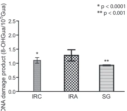
respectively (Fig. 5). The difference in GPx ac-tivity was statistically significant between the IRA and IRC groups (p < 0.0001), but there was no statistically significant difference with the SG group (p > 0.05). There was a statistically signif-icant difference in SOD activity between the IRA group and the IRC (p < 0.0001) and SG groups (p < 0.0001). Levels of 8-OH/Gua, a product of DNA damage, were 2.18 ± 0.2 pmol/L in the IRC group, 1.28 ± 0.2 pmol/L in the IRA group and 0.93 ± 0.01 pmol/L in the SG group (Fig. 6). There was a significant difference in terms of DNA dam-age product between the IRA group and the IRC (p < 0.0001) and SG (p < 0.001) groups.
Discussion
This study investigated the protective effect of agomelatine against oxidative stress induced with ischemia/reperfusion in rat ovaries. Ovarian isch-emia emerges as the result of torsion of the ovaries for various reasons. The detorsion procedure per-formed in order to reperfuse the ischemic ovaries leads to reperfusion injury. Organ-protective sur-gery is most appropriate in young girls, since ovar-ian functions need to be protected [17]. Therefore, detorsion with a conservative treatment option is increasingly recommended [18]. The fact that ox-idative injury is more severe in ischemic ovaries not receiving antioxidant treatment and in which post-ischemic reperfusion was established shows the importance of conservative therapy before surgery [5, 6]. As our experimental results show,
agomelatine therapy significantly prevented oxi-dative injury arising in the right ovaries of rats in which ischemia/reperfusion was established. We elected to use the right-side ovaries because in lit-erature the pathology is in the right ovary in 2/3 of patients [19]. Agomelatine significantly reduced the ischemia/reperfusion-associated rise in MDA in ovarian tissue compared with the IRC group. MDA is the final product of lipid peroxidation and one of the important parameters in the evaluation of ischemia/reperfusion associated oxidative inju-ry [20]. Lipid peroxidation leads to impaired cell membrane permeability, decreased membrane po-tential and cell damage. Cell damage further inten-sifies with MDA formation [21].
Another parameter known as an oxidant in the cell is the enzyme MPO. In the presence of chlo-ride ions, MPO reduces hydrogen peroxide to hy-pochlorous acid. Hyhy-pochlorous acid is a powerful oxidant and leads to tissue damage as it can easi-ly enter into reaction with several biological mol-ecules [22]. The fact that MPO activity in ovari-an tissue receiving agomelatine was lower thovari-an in the IRC group reveals that it protects ovarian tis-sue against ischemia/reperfusion injury. Kumbasar et al. showed that MPO activity in ovarian tissues in which ischemia/reperfusion was established was lower compared to that in healthy tissues [23]. This information from the literature agrees with our own experimental results and indicates that agomelatine is effective in the prevention of ovar-ian ischemia/reperfusion injury.
The level of GSH in the IRA group receiving agomelatine was significantly higher than that in the IRC group. There are known to be various an-tioxidant defense mechanisms that eliminate the harmful effects of toxic oxidants in tissues. The idant/antioxidant balance altering in favor of ox-idants leads to oxidative tissue damage. The im-portant effects of antioxidants protect against oxidation in target molecules such as proteins, nu-cleic acids and carbohydrates, as well as cell mem-brane lipids [24]. GSH is an important antioxidant and reducing agent. It protects cells against oxi-dant damage by entering into reactions with glu-tathione, free radicals and peroxides. It prevents protein oxidation by holding SH groups in an un-reduced form [25]. Drugs that protect against isch-emia/reperfusion injury in ovarian tissue have been shown to prevent a decrease in GSH in ovarian tis-sue [26]. The mechanisms of injury established by ischemia/reperfusion in many organs, such as the brain, heart, lungs, liver, stomach, ovaries and in-testines, have been investigated and free radicals have been reported to be one of the major compo-nents of ischemia/reperfusion injury [27, 28]. This information from the literature is also compatible
Fig. 6. Comparison of DNA damage product levels of groups. DNA damage product levels defined in 8-OHGua/105Gua. Bars are means ± standard
devia-tion. IRC, control group in which ischemia/reperfu-sion would be established; IRA, ischemia/reperfuischemia/reperfu-sion + agomelatine treated group; SG, healthy group given a sham operation
0.0 0.5 1.0 1.5 2.0 2.5
**
*
IRC IRA SG
DNA
damage product (8-OHGua/1
0
5 G
ua
) * p < 0.0001
with our study findings. GPx and SOD activity were significantly higher in ovarian tissue in which ischemia/reperfusion injury was established and receiving agomelatine compared to the IRC group. GPx is one of the important enzymatic antioxidant parameters establishing reduction of peroxide and organic hyperoxides [29].
Agomelatine significantly prevented DNA oxi-dation in ovarian tissue in which ischemia/reperfu-sion injury was established. The product reflecting oxidation of DNA in tissue is 8-hydroxyguanidine
(8-OHGua) [30]. Studies have shown that 8-OH -Gua levels in damaged tissue rise in parallel to increases in oxidant parameters [3]. These facts from the literature are also in agreement with our results. In conclusion, ischemia/reperfusion leads to oxidative damage in ovarian tissue. Agomela-tine significantly prevented the oxidative damage caused by ischemia/reperfusion in ovarian tissue. This in turn shows that agomelatine may be useful in the prevention of damage associated with ovar-ian torsion and detorsion.
References
Beaunoyer M, Chapdelaine J, Bouchard S, Ouimet A:
[1] Asynchronous bilateral ovarian torsion. J Pediatr Surg
2004, 39, 746–749.
Celik A, Ergun O, Aldemir H, Ozcan C, Ozok G, Erdener A, Balyk E:
[2] Long-term results of conservative
manage-ment of adnexal torsion in children. J Pediatr Surg 2005, 40, 704–708.
Ingec M, Isaoglu U, Yilmaz M, Calik M, Polat B, Alp HH, Kurt A, Gundogdu C, Suleyman H:
[3] Prevention of
ischemia-reperfusion injury in rat ovarian tissue with the on-off method. J Physiol Pharmacol 2011, 62, 575–582.
Etensel B, Ozkisacik S, Ozkara E, Karul A, Oztan O, Yazici M, Gursoy H:
[4] Dexpanthenol attenuates lipid
peroxida-tion and testicular damage at experimental ischemia and reperfusion injury. Pediatr Surg Int 2007, 23, 177–181.
Isaoglu U, Yilmaz M, Calik M, Polat B, Bakan E, Kurt A, Albayrak Y, Suleyman H:
[5] Biochemical and
histopatho-logical investigation of the protective effect of disulfiram in ischemia-induced ovary damage. Gynecol Endocrinol 2012, 28, 143–147.
Ingec M, Calik M, Gundogdu C, Kurt A, Yilmaz M, Isaoglu U, Salman S, Akcay F, Suleyman H:
[6] Biological and
Histopathological Investigations of Moclobemide on Injured Ovarian Tissue Following Induction of Ischemia-Reperfusion in Rats. Int J Fertil & Steril 2012, 6, 19–26.
Zlotos DP:
[7] Recent advances in melatonin receptor ligands. Arch Pharm (Weinheim) 2005, 338, 229–247.
Turkoz Y, Celik O, Hascalik S, Cigremis Y, Hascalik M, Mizrak B, Yologlu S:
[8] Melatonin reduces
torsion-detor-sion injury in rat ovary: biochemical and histopathologic evaluation. J Pineal Res 2004, 37, 137–141.
Song Y, Chan CW, Brown GM, Pang SF, Silverman M:
[9] Studies of the renal action of melatonin: evidence that the
effects are mediated by 37 kDa receptors of the Mel1a subtype localized primarily to the basolateral membrane of the proximal tubule. Faseb J 1997, 11, 93–100.
Ohkawa H, Ohishi N, Yagi K:
[10] Assay for lipid peroxides in animal tissues by thiobarbituric acid reaction. Anal Biochem 1979, 95, 351–358.
Wei H, Frenkel K:
[11] In vivoformation of oxidized DNA bases in tumor promoter-treated mouse skin. Cancer Res 1991, 51, 4443–4449.
Sedlak J, Lindsay RH:
[12] Estimation of total, protein-bound, and nonprotein sulfhydryl groups in tissue with Ellman’s reagent. Anal Biochem 1968, 25, 192–205.
Paglia DE, Valentine WN:
[13] Studies on the quantitative and qualitative characterization of erythrocyte glutathione peroxidase. J Lab Clin Med 1967, 70, 158–169.
Sun Y, Oberley LW, Li Y:
[14] A simple method for clinical assay of superoxide dismutase. Clinical Chemist 1988, 34, 497–500.
Shigenaga MK, Aboujaoude EN, Chen Q, Ames BN:
[15] Assays of oxidative DNA damage biomarkers 8-oxo-2’-
-deoxyguanosine and 8-oxoguanine in nuclear DNA and biological fluids by high-performance liquid chromatog-raphy with electrochemical detection. Meth Enzymol 1994, 234, 16–33.
Kaur H, Halliwell B:
[16] Measurement of oxidized and methylated DNA bases by HPLC with electrochemical detec-tion. Biochem J 1996, 318 (Pt 1), 21–23.
Cohen SB, Oelsner G, Seidman DS, Admon D, Mashiach S, Goldenberg M:
[17] Laparoscopic detorsion allows
spar-ing of the twisted ischemic adnexa. J Am Assoc Gynecol Laparosc 1999, 6, 139–143.
Eckler K, Laufer MR, Perlman SE:
[18] Conservative management of bilateral asynchronous adnexal torsion with necrosis in a prepubescent girl. J Pediatr Surg 2000, 35, 1248–1251.
Duntan C:
[19] Torsion of the ovary. J. B. Lippincott Company, Philadelphia 1994.
Gutteridge JM:
[20] Lipid peroxidation and antioxidants as biomarkers of tissue damage. Clin Chem 1995, 41, 1819– –1828.
Girotti AW:
[21] Lipid hydroperoxide generation, turnover, and effector action in biological systems. J Lipid Res 1998, 39, 1529–1542.
Lavelli V, Peri C, Rizzolo A:
[22] Antioxidant activity of tomato products as studied by model reactions using xanthine oxidase, myeloperoxidase, and copper-induced lipid peroxidation. J Agric Food Chem 2000, 48, 1442–1448.
Kumbasar S, Yapca O, Bilen H, Suleyman B, Ozgeris F, Borekci B, Suleyman H:
[23] The Effect of Lacidipine on
Rangan U, Bulkley GB:
[24] Prospects for treatment of free radical-mediated tissue injury. Br Med Bull 1993, 49, 700–718.
Di Mascio P, Murphy ME, Sies H:
[25] Antioxidant defense systems: the role of carotenoids, tocopherols, and thiols. Am J Clin Nutr 1991, 53, Suppl 1, 194–200.
Kurt A, Ingec M, Isaoglu U, Yilmaz M, Cetin N, Calik M, Polat B, Akcay F, Gundogdu C, Suleyman H:
[26] An
investigation about the inhibition of acute ischemia/reperfusion damage by dexmedetomidine in rat ovarian tissue. Gynecol Endocrinol 2012.
Coskun KA GA, Halici Z, Oral A, Seyrek M, Bayir Y, Kilic C, Yigit T, Ozer T, Uzar AI:
[27] The Effects of Amlodipine
on the Biochemical and Histopathological Changes in the Rabbit Ileum Subjected to Ischemia-Reperfusion. EAJM 2011, 43, 33–38.
Ceretta LB, Reus GZ, Abelaira HM, Ribeiro KF, Zappellini G, Felisbino FF, Steckert AV, Dal-Pizzol F, Quevedo [28]
J: Increased oxidative stress and imbalance in antioxidant enzymes in the brains of alloxan-induced diabetic rats. Exp Diabetes Res 2012, 2012, 302682.
Drevet JR:
[29] The antioxidant glutathione peroxidase family and spermatozoa: a complex story. Mol Cell Endocrinol 2006, 250, 70–79.
Grollman AP, Moriya M:
[30] Mutagenesis by 8-oxoguanine: an enemy within. Trends Genet 1993, 9, 246–249.
Address for correspondence:
Mehmet I. Turan
Department of Pediatrics, Faculty of Medicine Ataturk University
25240 Erzurum Turkey
E-mail: [email protected] Tel.: +90 505 26 04 621
Conflict of interest: None declared