Anna Choromańska
1, Jolanta Saczko
1, Julita Kulbacka
1,
Nina Skolucka
1, Michał Majkowski
2The Potential Role of Photodynamic Therapy in the
Treatment of Malignant Melanoma – an
in vitro
Study*
Możliwości zastosowania terapii fotodynamicznej
w leczeniu czerniaka – badania
in vitro
1 Department of Medical Biochemistry, Wroclaw Medical University, Poland
2 Laboratory of Cytobiochemistry, Faculty of Biotechnology, University of Wroclaw, Poland
Abstract
Background. Melanoma is the most severe of skin neoplasms as it may grow rapidly and metastasize. The applica-tion of photodynamic therapy (PDT) opens up new prospects in the treatment of this tumor. Numerous studies suggest that the exposure of tumor cells to PDT can lead to cellular and molecular mechanisms which mediate oxidative stress in cells.
Objectives. The aim of this study was to evaluate in vitro the influence of photodynamic therapy on the human melanoma Me45 cell line.
Material and Methods. Photofrin® (Ph) was used as a photosensitizer.
Results. Viability studies have shown that there are significant differences between cells after PDT and cells with-out irradiation. After 24 hours of incubation with a 20 µg/ml concentration of Ph and with irradiation, less than 20% of the cells survived. In the control (without PDT), 65% of the cells survived.
Conclusions. The mitochondrial localization of Ph is significant, as it may lead to disturbances of mitochondrial transmembrane potential and finally to apoptotic cell death. The expressions of manganese superoxide dismutase and heme oxygenase and the level of carbonyl and thiol groups are indicating factors for oxidative stress in Me45 cells (Adv Clin Exp Med 2012, 21, 2, 179–186).
Key words: photodynamic therapy, Photofrin, melanoma, apoptosis.
Streszczenie
Wprowadzenie. Czerniak należy do grupy silnie złośliwych nowotworów skóry, ponieważ cechuje go szybki wzrost oraz wczesne tworzenie przerzutów. Zastosowanie terapii fotodynamicznej (PDT) otwiera nowe perspektywy w leczeniu tego nowotworu. Liczne badania sugerują, że ekspozycja komórek nowotworowych na PDT może prowadzić do uruchamia-nia komórkowych i molekularnych mechanizmów, które prowadzą do stresu oksydacyjnego w komórkach guza.
Cel pracy. Ocena wpływu PDT na ludzką linię komórkową czerniaka (Me45).
Materiał i metody. Jako fotouczulacz zastosowano Photofrin® (Ph).
Wyniki. Badanie przeżywalności komórek wykazało znaczące różnice między żywotnością komórek po zastosowa-nej PDT a komórkami bez naświetlania. Po 24-godzinzastosowa-nej inkubacji komórek po PDT z 20 mg/ml Ph przeżyło mniej niż 20% komórek, podczas gdy w próbie z Ph, ale bez naświetlania, przeżyło 65% komórek.
Wnioski. Zaobserwowano mitochondrialne umiejscowienie fotouczulacza, co może prowadzić do zaburzenia potencjału transbłonowego mitochondriów i ostatecznie do śmierci komórek na drodze apoptozy. Ekspresja mito-chondrialnej dysmutazy ponadtlenkowej oraz hemooksygenazy-1, a także zwiększone stężenie markerów uszko-dzenia białek świadczą o zaistniałym stresie oksydacyjnym w komórkach czerniaka linii Me45 (Adv Clin Exp Med 2012, 21, 2, 179–186).
Słowa kluczowe: terapia fotodynamiczna, Photofrin, czerniak, apoptoza.
Adv Clin Exp Med 2012, 21, 2, 179–186 ISSN 1899–5276
OrIgINAL PAPErS
© Copyright by Wroclaw Medical University
Photodynamic therapy (PDT) is an approved, minimally invasive and highly selective therapeu-tic approach to a variety of tumors [1]. It is based on the accumulation of specific photosensitizers in tumor tissue, followed by irradiation with visible light. The hydrophobic photosensitizer tends to lo-calize in the plasma and intracellular membranes, making these structures especially sensitive to pho-tooxidative damage. It concentrates specifically within the malignant tissue. The photochemical in-teractions of the photosensitizer, light and molecu-lar oxygen produce singlet oxygen and other forms of active oxygen [2, 3]. They induce the disintegra-tion of cellular structures and moduladisintegra-tion of ge-netic information [4]. Cell death in PDT may occur by apoptosis or by necrosis [5, 6]. The reactions of free radicals with lipids and proteins in membranes may cause alterations of membrane function [7]. During oxidative stress, we observed high levels of protein carbonyls, a reduction in the concentration of protein thiol groups, and high levels of lipid per-oxides [8]. Antioxidant enzyme activity increases considerably to eliminate the initial radicals and also the more toxic products of spontaneous free-radical reaction. Two of the main antioxidant en-zymes are superoxide dismutase (SOD) and heme oxygenase (HO-1). SOD can even partially prevent the photodestruction caused by PDT [9, 10].
Melanoma is the most serious type of skin cancer because of its ability to grow rapidly and to metastasize. There is no fully effective treat-ment and the application of photodynamic ther-apy opens up new perspectives in the therther-apy of this cancer, especially if tumor cells are killed via apoptosis. The definitive treatment for primary cu-taneous melanoma is wide surgical excision [11]. Advanced melanoma may also be treated with ra-diation therapy, chemotherapy, immunotherapy or a combination of these methods. In comparison to these treatments, PDT is a method of destroying cancer without damaging healthy tissue. Lim et al. [6] maintain that many kinds of malignant tumors have been effectively treated by hematoporphyrin derivative (e.g. Photofrin®) PDT, but an exception is pigmented melanoma. The reason is the com-petition between melanoma melanin absorption and the absorption of porphyrins. For many years this neoplasm, mainly due to the presence of mela-notic biopolymers, has been included in the group of neoplasms resistant to irradiation. However, the research conducted in recent years has indicated a high degree of differentiation radiosensitivity in melanoma cells [12].
The current study presents the potential role of photodynamic therapy in the treatment of ma-lignant melanoma. The aim of this study was to investigate if Photofrin®-PDT induces oxidative
stress in the human pigmented melanoma Me45 cell line. The authors examined the cell viability, level of oxidative stress markers and the photosen-sitizer localization, which is a very important fac-tor for the mode of cell death.
Material and Methods
Cell Culture
The human pigmented malignant melanoma (Me45) cell line (derived from a lymph node me-tastasis of skin melanoma in a 35-year-old male) was established in 1997 at the radiobiology De-partment of the Center of Oncology in gliwice, Poland. The cells were grown in DMEM (medium, Sigma) with the addition of 10% fetal bovine se-rum (Biowhittaker) and supplemented by antibiot-ics (Antibiotic-Antimycotic Stabilized, Sigma). For experiments, the cells were removed by trypsin-ization (Trypsin-EDTA, Sigma) and washed with PBS. The cells were maintained in a humidified atmosphere at 37
˚
C and 5% CO2.Photodynamic Therapy
Photofrin®(Ph, QLT Phototherapeutics, Inc. Vancouver, Canada), a photosensitizer that has been accepted for clinical studies, was used for the photodynamic therapy. The cells were incubated for 18 h in the dark with 5, 10 or 20 µg/ml of Ph in DMEM (13, 14). Then they were irradiated for 10 minutes with a light intensity of 10 mW/cm2
using a lamp (OPTEL, Opole, Poland) with polar-ized light and a red filter (632.8 nm) and incubat-ed again for 3, 6 or 24 h at 37
˚
C and 5% CO2 inDMEM. Cell irradiation was performed in a me-dium without FBS. After irradiation, the meme-dium without FBS was replaced by a full medium.
Cell Viability
The cell viability was determined by MTT as-say (Sigma, In Vitro Toxicology Asas-say), which determines mitochondria metabolic function. For the experiment, cells were seeded into 96-well mi-croculture plates at 1 × 10⁴ cells/well and allowed
vi-able treated cells relative to untreated control cells. All experiments were performed four times.
The Localization of Ph
and Colocalization
with Mitochondria
From the culture dishes, the cells were trypsinized and conducted on cover glasses (24x24 mm, Thermo Scientific). The cells were incubated with the photosensitizer (20 µg/ml) for 18 hours. After washing with PBS, the cells were fixed for 10 minutes in 4% buffered formalin, washed in PBS and incubated with 100 nM Mito-Tracker green (Molecular Probes) for 15 minutes. After that, the cells were examined under a confocal scanning laser microscope (Carl Zeiss gmbH, Jena, ger-many). Using a UV excitation filter (300–400 nm) we observed Ph emission in the red region of the spectrum (400–800 nm). Mito-Tracker green was excited using an argon-ion laser 488 nm line.
Expression of Mitochondrial
Superoxide Dismutase (MnSOD)
and Heme Oxygenase-1 (HO-1)
The cells were plated into eight-dip glass (Nunc). For immunocytochemical detection of MnSOD and HO-1, polyclonal antibodies were used (1:100, Santa Cruz, USA). Formalin-fixed cells were immunostained with a DAKO LSAB 2 Kit. Stained cells were determined by counting 100 cells in three randomly selected fields. Cells were judged as positive if the stained reaction was ob-served in more than 5% of cells. The intensity of immunocytochemical staining was evaluated as: (-) negative, (+) weak, (++) moderate and (+++) strong. Positive and negative controls were per-formed. All experiments were repeated twice.
Determination of Markers
of Oxidative Stress
The Level of Carbonyl Groups
Oxidative damage to proteins was assessed by the determination of carbonyl groups based on the reaction with dinitrophenylhydrazine (DNPH) (Sigma). Briefly, proteins were precipi-tated by the addition of 20% trichloroacetic (Sig-ma) acid and redissolved in 10 mM DNPH, and the absorbance was read at 370 nm. The results were calculated using an extinction coefficient of ε = 21.01 mmol-1/cm-1 for aliphatic hydrazone.
Ex-periments were repeated three times.
The Level of Thiol Groups
After treatment with drugs in vitro, the cells were removed by trypsinization and washed twice in PBS. The cells (about 5 × 10⁶) were used for
assay according to Ellman’s method [15]. Experi-ments were repeated three times.
Statistical Analysis
Statistical analysis was performed by a Stu-dent’s t-test with significance level P < 0.05.
Results
Cell Viability
The viability studies have shown that there are significant differences between the samples after PDT and samples without irradiation. This experi-ment showed that Ph is an efficient photosensitizer, which may be applied successfully for photodynam-ic therapy of melanoma. In samples without irradia-tion, the viability was much higher. After 24 hours of incubation with a 20 µg/ml concentration of Ph and with irradiation, less than 20% of the cells survived (p = 0.00002). In the same combination but without irradiation, 65% of the cells survived (Fig. 1). Data concerning the concentrations of 5 and 10 µg/ml were not presented because these concentrations did not significantly influence cell proliferation. Only the highest concentration (20 µg/ml) of Ph decreased cell viability and this concentration was selected for further experiments.
*
Fig. 1. The viability relationship of Me45 cells on time after irradiation and without irradiation. results expressed as a mean ± SD. * p < 0.05
Localization of Ph and
Mitochondria Identification
The intracellular distribution of Ph in the Me45 cell line was monitored after 18 hours of incuba-tion. Mito-Tracker green was used as a molecule marker. Ph was mainly localized in the mitochon-drial membranes and cytoplasm (Fig. 2).
Expression of Mitochondrial
Superoxide Dismutase (MnSOD)
and Heme Oxygenase-1 (HO-1)
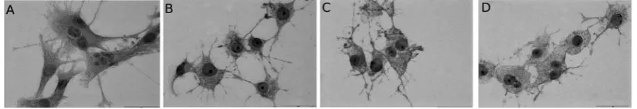
After each period of incubation followed ir-radiation, a strong immunocytochemical reaction was observed, which indicated the expression of MnSOD (Fig. 3). The authors also observed mor-phological changes in the cells after all doses of PDT as compared with control cells. The same results for the morphological changes and the im-munocytochemical reaction were also observed for HO-1 (Fig. 4). The control cells did not display any morphological changes. In all cells, a strong immu-nocytochemical reaction was observed (Tab. 1, 2).
Determination of Markers
of Oxidative Stress
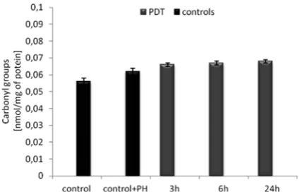
The level of carbonyl groups did not significant-ly increase with the time of incubation (Fig. 5). The results obtained are nearly the same as for the con-trol group. For the concon-trol cells it was 0.056 nmol/ mg of protein, while at 3, 6 and 24 hours after in-cubation, the level of carbonyl groups was 0.066, 0.067 and 0.068 nmol/mg of protein, respectively.
The level of thiol groups decreased regularly with the time of incubation (Fig. 6). For the control cells it was 18.44 nmol/mg of protein. After 3, 6 and 24 hours this level decreased from 13.9 to 12.1 to 9.4 nmol/mg of protein, respectively.
Discussion
For more than twenty years, scientists have maintained that PDT is not effective for pigmented melanomas [16]. However in this paper the authors demonstrate that Ph-based photodynamic therapy in-duces an increased response of antioxidant enzymes (MnSOD, HO-1) and protein damage in Me45 cells.
Fig. 3. The MnSOD expression in Me45 A) control cells; B) cells after 3 h incubation after Ph-PDT (cPh = 20 μg/ml
and light dose 6 J/cm2); C) cells after 6 h incubation after Ph-PDT (cPh = 20 μg/ml and light dose 6 J/cm2); D) cells
after 24 h incubation after Ph-PDT (cPh = 20 μg/ml and light dose 6 J/cm2)
Ryc. 3. Immunocytochemiczna ocena ekspresji białka MnSOD w komórkach Me45 A) komórki z grupy kontrolnej; B) komórki po 3 godzinach inkubacji po Ph-PDT (cPh = 20 μg/ml, dawka promieniowania: 6 J/cm2); C) komórki po
6 godzinach inkubacji po Ph-PDT (cPh = 20 μg/ml, dawka promieniowania: 6 J/cm2); D) komórki po 24 godzinach
inkubacji po Ph-PDT (cPh = 20 μg/ml, dawka promieniowania: 6 J/cm2)
Fig. 2. The intracellular distribution of: A) Ph in Me45 cell line after 18 hours of incubation; B) Mito-Tracker green in Me45 cell line after 15 minutes incubation; C) Co-localization of Ph and Mito-Tracker green in Me45 cell line
Other researchers have also shown that PDT of melanoma with porphyrin and porphyrin deriv-atives [17, 18] or chlorine [6, 19] is effective. Some authors have applied PDT with the water-soluble photosensitizer PCI-0123 on heavily pigmented and metastatic B16F10 melanoma. Their results indicated significant tumor growth delay and in-creased longevity of the treated animals [20]. In parallel to the increase in protein carbonylation, the authors showed a dependent loss of free thi-ols. Protein oxidation is a useful marker to evalu-ate oxidative stress. The level of carbonyl groups is the most general and well-used biomarker of severe oxidative protein damage. Carbonyls are relatively difficult to induce, compared with me-thionine sulphoxide and cysteinyl derivatives, and might therefore indicate more severe oxidative stress [21].
Strong expression of mitochondrial superox-ide dismutase and heme oxygenase-1 are direct indicators of oxidative stress. MnSOD is the cell’s primary defense against free radical mediated
damage [22]. golab at el. also demonstrated that PDT with Ph increases the level of MnSOD pro-tein, which was confirmed in current results [23]. HO-1 has a number of potential protective effects against oxidative stress. Its expression is closely related to oxidative stress levels in the cells [24]. The authors observed a strong immunocy-tochemical reaction which indicated expression of MnSOD and HO-1 after just 3 hours of incubation after PDT. An increase in the expression of HO-1 following photodynamic therapy was observed by gomer et al. [25] and Nowis et al [26]. They dem-onstrated that overexpression of HO-1 protects cancer cells against PDT with Photofrin. Other authors also indicated that PDT with 5-ALA was seen to result in an increased formation of rOS and a further enhancement of HO-1 induction [27]. Chen et al. revealed that PDT with methyl-ene blue (MB) causes oxidative stress, which plays an essential role in initiating apoptotic cell death. Their data has implicated mitochondria in the process of MB-PDT-induced tumor regression,
Table 1. Theintensity of immunocytochemical reaction of MnSOD and the percentage of cells positively stained Tabela 1. Intensywność reakcji immunocytochemicznej MnSOD oraz procent komórek pozytywnie wybarwionych
Intensity of reaction (Intensywność reakcji)
The percentage of cells positively stained
(Odsetek komórek pozytywnie wybar-wionych)
Control − 100
Control + Ph − 100
3h after PDT +++ 95
6h after PDT +++ 95
24h after PDT ++/+++ 90
Table 2. Theintensity of immunocytochemical reaction of HO-1 and the percentage of cells positively stained Tabela 2. Intensywność reakcji immunocytochemicznej HO-1 oraz procent komórek pozytywnie wybarwionych
Intensity of reaction (Intensywność reakcji)
The percentage of cells positively stained
(Odsetek komórek pozytywnie wybar-wionych)
Control − 100
Control + Ph − 100
3h after PDT +++ 95
6h after PDT +++ 90
24h after PDT ++/+++ 85
Fig. 4. The HO-1 expression in Me45: A) control cells; B) cells after 3 h incubation after Ph-PDT (cPh = 20μg/ml and
light dose 6 J/cm2); C) cells after 6 h incubation after Ph-PDT (c
Ph = 20 μg/ml and light dose 6 J/cm2); D) cells after
24 h incubation after Ph-PDT (cPh = 20 μg/ml and light dose 6 J/cm2)
Ryc. 4. Immunocytochemiczna ocena ekspresji białka HO-1 w komórkach Me45: A) komórki z grupy kontrolnej; B) komórki po 3 godzinach inkubacji po Ph-PDT (cPh = 20 μg/ml, dawka promieniowania: 6 J/cm2); C) komórki po
6 godzinach inkubacji po Ph-PDT (cPh = 20 μg/ml, dawka promieniowania: 6 J/cm2); D) komórki po 24 godzinach
because MB binds to the environment of the mito-chondrial matrix [28]. In the current study, the au-thors demonstrated that the viability of Me45 cells is dependent on the duration of incubation after irradiation. Moreover, Ph accumulates mainly in the mitochondrial membranes. Mitochondrial damage leads to excessive cytochrome c release, which in turn triggers apoptosis [29]. Present re-sults strongly suggest the influence of PDT with the usage of Ph on rOS generation, which is a
sig-nal for the development of morphological chang-es, as presented on images from a fluorescent mi-croscope. The authors have shown that Ph-PDT can provoke the death of melanoma cells. The next step of present research will be the determination of cell death after photodynamic therapy.
PDT with the application of Photofrin® ap-pears to be a promising technique, which could be combined with chemotherapy or radiotherapy, particularly in the early phase of melanoma.
Fig. 5. The level of carbonyl groups in Me45 cells after Ph-PDT in comparison to control cells without treat-ment. results expressed as a mean ± SD
Ryc. 5. Stężenie grup karbonylowych w komórkach Me45 po Ph-PDT w porównaniu do komórek z grupy kontrolnej bez terapii. Wyniki wyrażone jako średnia ± SD
*
Fig. 6. The level of thiol groups in Me45 cells after Ph-PDT in comparison to control cells without treat-ment. results expressed as a mean ± SD.* p < 0.05
Ryc. 6. Stężenie grup tiolowych w komórkach Me45 po Ph-PDT w porównaniu do komórek z grupy kontrol-nej bez terapii. Wyniki wyrażone jako średnia ± SD. * p < 0,05
References
[1] Brown SB, Brown EA, Walker I: The present and future role of photodynamic therapy in cancer treatment. Lancet Oncol 2004,5, 497–508.
[2] Castano AP, Demidova TN, Hamblin MR: Mechanisms in photodynamic therapy: part two – cellular signaling, cell metabolism and modes of cell death. Photodiag Photodyn Ther 2005,2, 1–23.
[3] Wawrzuta A, Saczko J, Kulbacka J, Chwiłkowska A: Can photodynamic therapy be an alternative method in melanoma treatment? Przegl Dermatol 2009, 96, 240–243.
[4] Ramiro DA, Bruno JM, Arselio PC, Carlos BD: Intracellular signaling mechanisms in photodynamic therapy. Biochim Biophys Acta 2004, 1704, 59–86.
[5] Robertson CA, Hawkins Evans D, Abrahamse H: Photodynamic therapy (PDT): A short review on cellular mechanisms and cancer research applications for PDT. J Photochem Photobiol B 2009, 96, 1–8.
[6] Lim DS, Ko SH, Lee WY: Silkworm-pheophorbide a mediated photodynamic therapy against B16F10 pigmented melanoma. J Photochem Photobiol B 2004,74, 1–6.
[7] Saczko J, Skrzypek W, Chwilkowska A, Choromanska A, Pola A, Gamian A, Kulbacka J: Photo-oxidative action in cervix carcinoma cells induced by HpD-mediated photodynamic therapy. Exp Oncol 2009, 31, 195–199.
[8] Kolagal V, Karanam SA, Dharmavarapu PK, D’Souza R, Upadhya S, Kumar V, Kedage V, Muttigi MS, Shetty JK, Prakash M: Determination of oxidative stress markers and their importance in early diagnosis of uremia-related complications. Indian J Nephrol 2009, 19, 8–12.
[9] Dolgachev V, Oberley LW, Huang TT, Kraniak JM, Tainsky MA, Hanada K, Separovic D: A role for manganase superoxide dismutase in apoptosis after photosensitization. Biochem Biophys res Commun 2005, 332, 411–417.
[10] Vile GF, Basu-Modak S, Waltner C, Tyrrell RM: Heme oxygenase 1 mediates an adaptive response to oxidative stress in human skin fibroblasts. Proc Natl Acad Sci USA 1994, 91, 2607–2610.
[11] Moncieff MD, Thompson JF, Quinn M, Stretch JR: reconstruction after wide excision of primary cutaneous melanomas: part II – the extremities. Lancet Oncol 2009,10, 810–815.
[12] Kusmierz D, Latocha M, Zielinska A, Nawrocka-Musial D, Sliupkas-Dyrda E: The Expression of the Melanogenesis Pathway genes TYR, TYRP−1, and TYRP−2 and the Synthesis of Melanin in SH−4 Melanoma Cells After Photodynamic Therapy with Photolon. Adv Clin Exp Med 2009, 18, 449–459.
[14] Saczko J, Chwilkowska A, Kulbacka J, Berdowska I, Zielinski B, Drag-Zalesinska M, Wysocka T, Lugowski M, Banas T: Photooxidative action in cancer and normal cells induced by the use of photofrin in photodynamic therapy. Folia Biol 2008, 54, 24–29.
[15] Ellman GL: Tissue sulfhydryl groups. Arch Biochem Biophys 1959, 82, 70–77.
[16] Ambrosone CB, Ahn J, Singh KK, Rezaishiraz H, Furberg H, Sweeney C, Coles B, Trovato A: Polymorphisms in genes related to Oxidative Stress (MPO, MnSOD, CAT) and Survival After Treatment for Breast Cancer. Cancer res 2005, 65, 1105–1111.
[17] Kolarova H, Macecek J, Nevrelova P, Huf M, Tomecka M, Bajgar R, Mosinger J, Strnad M: Photodynamic therapy with zinc-tetra (p-sulfophenyl) porphyrin bound to cyclodextrin induces single stand breaks of cellular DNA in g361 melanoma cells. Toxicol in Vitro 2005, 19, 971–974.
[18] Nowak-Sliwinska P, Karocki A, Elas M, Pawlak A, Stochel G, Urbanska K: Verteporfin, Photofrin II and mero-cyanine 540 as PDT photosensitizer against melanoma cells. Biochem Biophys res Commun 2006, 349, 549– 555.
[19] Radestock A, Elsner P, Gitter B, Hipler UC: Induction of Apoptosis in HaCaT Cells by Photodynamic Therapy with Chlorin e6 or Pheophorbide A. Skin Pharmacol Physiol 2007, 20, 3–9.
[20] Woodburn KW, Fan Q, Kessel D, Luo Y, Young SW: Photodynamic Therapy of B16F10 Murine Melanoma with Lutetium Texaphyrin. J Invest Dermatol 1998,110, 746–751.
[21] Yagc R, Ersoz I, Erdurmu M, Gurel A, Duman S: Protein carbonyl levels in the aqueous humour and serum of patients with pseudoexfoliation syndrome. Eye2008, 22, 128–131.
[22] Saczko J, Kulbacka J, Chwilkowska A, Pola A, Lugowski M, Marcinkowska A, Malarska A, Banas T: Cytosolic superoxide dismutase activity after photodynamic therapy, intracellular distribution of Photofrin II and hypericin, and P-glycoprotein localization in human colon adenocarcinoma. Folia Histochem Cytobiol 2007, 45, 93–97.
[23] Golab J, Nowis D, Skrzycki M, Czeczot H, Baranczyk-Kuzma A, Wilczynski GM, Makowski M, Mroz P, Kozar K, Kaminski R, Jalili A, Kopec M, Grzela T, Jakobisiak M: Antitumor effects of photodynamic therapy are potentiated by 2-methoxyestradiol. A superoxide dismutase inhibitor. J Biol Chem 2003,278, 407–414.
[24] Hoshidaa S, Nishidab M, Yamashitaa N, Igarashib J, Aokib K, Horia M, Kuzuyab T, Tada M: Heme Oxygenase-1 Expression and Its relation to Oxidative Stress During Primary Culture of Cardiomyocytes. J Mol Cell Cardiol 1996, 28, 1845–1855.
[25] Gomer CJ, Luna M, Ferrario A, Rucker N: Increased transcription and translation of heme oxygenase in Chinese hamster fibroblasts following photodynamic stress or Photofrin II incubation. Photochem Photobiol 1991, 53, 275–279.
[26] Nowis D, Legat M, Grzela T, Niderla J, Wilczek E, Wilczynski GM, Glodkowska E, Mrowka P, Issat T, Dulak J, Jozkowicz A, Was H, Adamek M, Wrzosek A, Nazarewski S, Makowski M, Stoklosa T, Jakobisiak M, Golab J:
Heme oxygenase-1 protects tumor cells against photodynamic therapy-mediated cytotoxicity. Oncogene 2006, 25, 3365–3374.
[27] Frank J, Lornejad-Schafer MR, Schoffl H, Flaccus A, Lambert Ch, Biesalski HK: Inhibition of heme oxyge-nase-1 increases responsiveness of melanoma cells to ALA-based photodynamic therapy. Int J Oncol 2007, 31, 1539–1545.
[28] Chen Y, Zheng W, Li Y, Zhong J, Ji J, Shen P: Apoptosis induced by methylene-blue-mediated photodynamic therapy in melanomas and the involvement of mitochondrial dysfunction revealed by proteomics. Cancer Sci 2008,99, 2019–2027.
[29] Das N, Gupta S, Mazumdar S: Direct Observation of release of Cytochrome cfrom Lipid-Encapsulated Protein by Peroxide and Superoxide: A Possible Mechanism for Drug-Induced Apoptosis. Biochem Biophys res Commun 2001, 286, 311–314.
Address for correspondence:
Anna Choromańska
Department of Medical Biochemistry Wroclaw Medical University Chałubińskiego 10
50-368 Wrocław Poland
E-mail address: [email protected] Tel.: 0048 71 784 13 75, 500 777 527
Conflict of interest: None declared