M
ONIKAK
OSACKA, R
ENATAJ
ANKOWSKAThe nm−23 Protein Expression in Non−Small−Cell
Lung Cancer*
Ekspresja proteiny nm−23 w niedrobnokomórkowym raku płuca
Chair and Department of Pulmonology and Lung Cancer, Silesian Piasts University od Medicine in Wrocław, Poland
Adv Clin Exp Med 2008, 17, 3, 307–312 ISSN 1230−025X
ORIGINAL PAPERS
© Copyright by Silesian Piasts University of Medicine in Wrocław
Abstract
Background. NM−23 gene is considered a metastasis suppressor gene. Expression of its product is reported to cor− relate inversely with metastatic potential and prognosis in some cancers, but its significance in lung cancer is unclear. Lung cancer is currently the most frequently diagnosed neoplasm in males and the second most frequent in females in Poland and most developed countries. The frequency and unfavorable prognosis in this malignancy justifies studies on prognostic factors.
Objectives. The aim of this study was to evaluate the prognostic value of nm−23 protein expression in non−small− cell lung cancer and to compare nm−23 protein expression with some clinico−pathological findings.
Material and Methods.nm−23 protein expression was examined in 95 patients (mean age: 59.48 ± 7.86 years) with non−small−cell lung cancer who had had surgical treatment. The histopathological diagnoses were squamous cell carcinoma in 59 patients, adenocarcinoma in 26, large cell carcinoma in 5, and non−small−cell lung cancer of unspecified type in 5. nm−23 expression was determined by immunohistochemistry on paraffin−embedded tissue using a polyclonal antibody to nm−23. All patients were observed for at least 24 months, at which time 49 patients were alive and 46 had died.
Results.Positive cytoplasmatic staining was observed in 69 of the tumor tissues (72.63%). No relationship was found between nm−23 expression and survival. No correlation was found between nm−23 expression and clinico− pathological parameters, including gender, histological type, tumor differentiation stage, and nodal involvement. Higher expression of nm−23 protein was found in patients with squamous carcinoma without tumor cell emboli in blood vessels (p = 0.0161).
Conclusions. These data suggest that the nm−23 protein expression probably has no prognostic value in lung can−
cer and limited significance in squamous cell lung cancer (Adv Clin Exp Med 2008, 17, 3, 307–312).
Key words: nm−23 protein, non−small−cell lung cancer, prognostic factors.
Streszczenie
Wprowadzenie.Gen NM−23jest zaliczany do genów antymetastatycznych. W części nowotworów stwierdzono odwrotną korelację między ekspresją jego produktu a zdolnością do tworzenia przerzutów i rokowaniem. Zależ−
ność ta i znaczenie genu NM−23w raku płuca pozostaje jednak niejasne. Rak płuca jest najczęściej występującym
nowotworem złośliwym u mężczyzn i drugim u kobiet w Polsce i w większości rozwiniętych krajów świata. Roz− powszechnienie raka płuca i niekorzystne rokowanie skłania do badań nad czynnikami prognostycznymi.
Cel pracy.Ocena znaczenia rokowniczego ekspresji proteiny nm−23 w raku niedrobnokomórkowym płuca oraz jej porównanie z wybranymi wskaźnikami klinicznymi.
Materiał i metody.Ekspresję proteiny nm−23 oznaczono u 95 chorych na niedrobnokomórkowego raka płuca, u których przeprowadzono zabieg operacyjny. Średni wiek w badanej grupie 59,48 ± 7,86 lat. U 59 chorych roz− poznano raka płaskonabłonkowego, u 26 gruczolakoraka, u 5 raka wielkokomórkowego, a u 5 raka niedrobnoko− mórkowego bez określenia podtypu histopatologicznego. Ekspresję proteiny nm−23 oznaczono w materiale guza metodą immunohistochemiczną. Zastosowano przeciwciało poliklonalne Rabbit Anti Human nm−23 Protein DA− KO A 0096. Pacjenci zostali poddani 2−letniej obserwacji. 2 lata przeżyło 49 chorych. 46 osób zmarło.
Tumor metastasis, defined as the formation of progressively growing secondary tumor foci at sites other than the primary lesion, is responsible for a high degree of mortality in cancer patients [1, 2]. Metastasis suppressor genes, including NM−23,
KAI−1, KiSS−1, BrMS1, MKK4, RhoGDI2, CRSP3, and VDUP−1, inhibit the formation of metastases without affecting the growth rate of the primary tumor [2, 3]. NM−23 was identified in the murine K 1735 melanoma cell line in 1988 by P. Steeg and coworkers. They found an inverse correlation between NM−23 gene expression and metastatic potential [4]. These observations were confirmed by other studies. Transfection of NM−23H1 cDNA into highly metastatic murine melanoma, rat mam− mary adenocarcinoma, human breast cancer, and melanoma cells reduced their invasiveness and metastatic potential in vivo[2]. Nm−23 is located on chromosome 17q21[5]. Eight human homo− logues (NM−23 H1−H8) have been described so far, but only H1 and H2 have been tested for their role in tumor spread.
The mechanism of metastasis suppression by
NM−23 gene is still unknown. Its gene products have been identified as nucleoside diphosphate kinases (NDPKs). The isoforms NM−23 H1 and
NM−23 H2 encode 20.5−kDa and 18.5−kDa pro− teins. They have a highly homologous (88%) amino−acid sequence and are sometimes called kinase−A and kinase−B [6]. Kinase B was previ− ously identified as a transcription factor of the c− Myc oncogene [7]. Roymans and coworkers iden− tified rat nm−23R1/NDPK beta, a homologue of the human metastasis suppressor nm−23−H1, as a component of the centrosomal complex. They demonstrated that nm−23R1 is located in the cen− trosome of dividing and non−dividing cells. In addition, human nm−23−H1 was also shown to be present in the centrosome of different human cell types [8]. Kantor et al. reported that murine melanoma cell lines and human breast carcinoma cells that were stably transfected with nm−23−H1 lost their ability to migrate in response to a variety of chemoattractants, including serum platelet− derived growth factor and insulin−like growth fac− tor 1 [9]. The protective activity on metastatic behavior was confirmed in some human neo− plasms, mostly in breast cancer and melanoma. However, in other types of neoplasms (e.g. col−
orectal, gastric, renal, and lung cancer) the role of
NM−23is still disputable [10,11]. The aim of this study was to evaluate the prognostic value of nm− 23 protein expression in non−small−cell lung can− cer and to compare the expression of nm−23 with some clinico−pathological findings.
Material and Methods
Ninety−five patients with pathologically con− firmed non−small−cell lung cancer (65 men and 30 women, mean age: 59.48 ± 7.86 years) were eval− uated. They had undergone surgery, i.e. lobectomy, bilobectomy, pneumonectomy, or exploratory tho− racotomy. The histopathological diagnoses were squamous cell carcinoma in 59 patients, adenocar− cinoma in 26 patients, large−cell carcinoma in 5 patients, and non−small−cell lung cancer of unspecified type in 5 patients. According to the TNM staging system, 9 patients were in stage IA, 20 in IB, 2 in IIA, 17 in IIB, 31 in IIIA, 7 in IIIB, and 9 in stage IV. Forty−two patients received chemotherapy, 24 of whom received neoadjuvant chemotherapy. Twenty−five patients had radiother− apy after surgical treatment. All patients were observed for at least 24 months at which time 49 (52%) of the patients were alive and 46 (48%) had died. The study was approved by the appropriate ethics committees related to the institution and all the patients gave informed consent to participate in the study.
Immunohistochemistry
Formalin−fixed well−preserved tumor−tissue blocks from surgically resected lung cancer speci− mens were used for immunohistochemical study. The 4−µm sections of formalin−fixed tissues were mounted on silanized slides, deparaffinized in xylene, and rehydrated through serial baths of alcohol to water. The hydrated sections were treat− ed in 3% hydrogen peroxide for 10 minutes to eliminate endogenous peroxidase activity and washed in phosphate−buffered saline (PBS). The primary antibody used in this study was a poly− clonal antibody to nm−23 gene product (Polyclonal Rabbit Anti Human nm−23 Protein DAKO A 0096). The antibody recognizes human nm−23−H1 and nm−
Wyniki. Ekspresję proteiny nm−23 stwierdzono w 69 preparatach (72,63%). Nie wykazano związku proteiny nm−23 z przeżyciem ani z wybranymi wskaźnikami, takimi jak płeć, typ histopatologiczny, zajęcie węzłów chłonnych czy stopień zróżnicowania. Wykazano natomiast, że wyższą ekspresję nm−23 u chorych na raka płaskonabłonkowego bez zatorów z komórek nowotworowych w naczyniach.
Wnioski.Powyższe wyniki sugerują, że ekspresja białka nm−23 nie ma wartości prognostycznej w raku płuca. Ma
ona tylko ograniczone znaczenie w raku płaskonabłonkowym płuca (Adv Clin Exp Med 2008, 17, 3, 307–312).
23−H2. The polyclonal antibody−treated slides were rinsed in PBS solution and incubated with a biotiny− lated secondary antibody. The slides were then washed in PBS and incubated with an avidin−biotin− peroxidase complex for 15 minutes. After washing with PBS, a chromogenic reaction was developed by incubating with 3,3−diaminobenzidine tetrahy− drochloride (DAB+, Liquid K 3486 DAKO). Positive staining appeared as brown cell plasma. nm−23 protein accumulation was described as posi− tive if more than 10% of the cells were stained.
Statistical Method
Statistical analysis was performed using CSS Statistica for Windows (version 5.0). The chi− squared test was used for two or multiple groups.
Differences between samples were considered sig− nificant at p < 0.05. Survival curves were con− structed using the Kaplan−Meier method.
Results
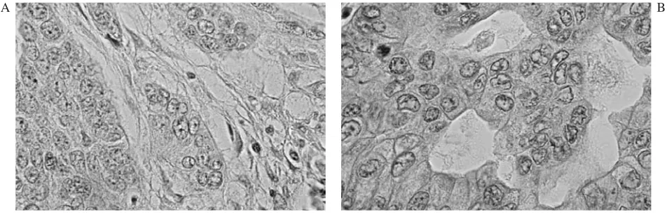
Sixty−nine of the 95 (72.63%) tumor tissue specimens were positive for nm−23 protein and 26 (27.36%) were negative. Figure 1 shows pic− tures of immunohistochemical staining. No sta− tistical difference was found in nm−23 protein expression regarding gender, histopathological type, tumor staging, degree of cancer cell differ− entiation, or nodal involvement (Table 1). However, nodal involvement and degree of can− cer cell differentiation were not determined in all
Ryc. 1. A. Brak ekspresji proteiny nm−23. B. Obecna ekspresja proteiny nm−23
Fig. 1. A. Negative immunostaining of nm−23 protein. B. Positive immunostaining of nm−23 protein
Table 1.Clinico−pathological findings and nm−23 protein expression Tabela 1. Wybrane wskaźniki i ekspresja proteiny nm−23
A. Gender nm−23 negative nm−23 positive Chi2 p
men 20 45
1.2 0.27
women 6 24
B. Histological type
squamous cell 19 40
1.5 0.22
adenocarcinoma 5 21
C. Stage
Stage I+II 11 37
0.97 0.33
Stage III+IV 15 32
D. Degree of cancer cell differentiation (G)
G 1−2 15 35
0.15 0.70
G 3 4 12
G 1 0 3
0.95 0.33
G 3 4 12
E. Metastases in lymph nodes (N)
N 0 11 28 0.25 0.61
N 1−3 12 39
N 0−1 14 42 0.02 0.88
N 2−3 9 25
patients. The prognostic value of nm−23 protein expression in all the patients with non−small−cell lung cancer and separately in patients with squa− mous cell carcinoma and adenocarcinoma was
analyzed, but there was no correlation between nm−23 protein expression and survival in any group (Table 2 and Figure 2). Higher expression of nm−23 protein was found in patients with
Table 2. Comparison of 24−month survival in all patients and in patients with squamous cell lung cancer and adenocarcinoma
Tabela 2. Porównanie 24−miesięcznego przeżycia z ekspresją proteiny nm−23 u wszystkich chorych oraz u chorych na raka płaskonabłonkowego i gruczolakoraka
Survival nm−23 negative nm−23 positive Chi2 p Cox
n (%) n (%) Mantel
A. All patients with NSCLC
< 24 months 13 (50%) 33 (48%) 0.04 0.85 0.62
> 24 months 13 (50%) 36 (52%) B. Patients with squamous cell carcinoma
< 24 months 10 (53%) 20 (50%) 0.04 0.85 0.68
> 24 months 9 (47%) 20 (50%)
F. Patients with adenocarcinoma
< 24 months 11 (52%) 2 (40%) 0.25 0.62 0.85
> 24 months 10 (48%) 3 (60%)
A.
0 10 20 30 40 50 60 70
Time (months) 0.0
0.1 0.2 0.3 0.4 0.5 0.6 0.7 0.8 0.9 1.0
C
umulativ
e
proportion
of
su
rv
iv
al
p=0.62 (Cox-Mantel Test)
nm-23 negative(n=26) nm-23 positive (n=69)
B.
0 10 20 30 40 50 60 70
Time (months) 0.0
0.1 0.2 0.3 0.4 0.5 0.6 0.7 0.8 0.9 1.0
C
umulativ
e
proportion
of
su
rv
iv
al
nm-23 negative (n=19) nm-23 positive (n=40) p=0.68 (Cox-Mantel Test)
C.
0 5 10 15 20 25 30 35
Time (months) 0.0
0.1 0.2 0.3 0.4 0.5 0.6 0.7 0.8 0.9 1.0
C
umulativ
e
proportion
of
su
rv
iv
al
nm-23 positive (n=5) nm-23 negative (n=21) p=0.85 (Cox-Mantel Test)
Fig. 2. Cumulative proportion of Kaplan−Meier survival according to nm−23 protein expression: A. In all patients with non−small−cell lung cancer, B. In patients with squamous cell lung cancer, C. In patients with adenocarcinoma
Ryc. 2.Skumulowana proporcja przeżywających Kaplana−Meiera w zależności od ekspresji proteiny nm−23:
squamous cell carcinoma without tumor cell emboli in the blood vessels (81.25% vs. 51.85%,
p= 0.0161) (Table 3).
Discussion
Lung cancer is currently the most frequently diagnosed cancer and the most common cause of cancer mortality in males. In women, only breast cancer occurs more frequently, but lung cancer remains the most common cause of cancer mortal− ity in the majority of developed countries. Despite many studies, improved diagnostics and therapeu− tics, as well as supportive care options, the prog− nosis remains unfavorable and long−term survival has practically not changed in the last 50 years [12]. This situation justifies a search for prognos− tic factors in lung cancer.
The expression of the NM−23 gene product has been reported to correlate inversely with prognosis and metastatic potential in some, but not all, tumors. Despite many efforts, the antimetastatic significance of NM−23 gene in non−small−cell lung cancer remains to be established. Kawakubo et al. reported that reduced expression of nm−23 protein was associated with lymph node metastasis and poor patient survival in pulmonary adenocarcino− ma, but correlation between nm−23 protein expres− sion and other clinical findings, such as histologi− cal grade, presence of distant metastases, or pri− mary tumor size, was not found [13]. Katakura et al. analyzed five−year survival in 117 patients with stage I non−small−cell lung cancer and showed that positive expression of nm−23 is a favorable prog− nostic factor [14]. Some data indicate that nm−23− H1 and −H2 isoform levels correlate with histolog− ical differentiation, but not with metastatic poten− tial or stage of pulmonary adenocarcinoma [15].
Many other studies confirm the observations of the present study. Bosnar et al. found no corre− lation between nm−23 protein expression and TNM stage, grade, primary tumor size, or patient survival in squamous cell lung cancer [16]. Mac Kinnon et al. evaluated a group of 162 patients with adenocarcinoma and, based on immunohisto− chemical assessment of nm−23 protein, did not find any prognostic value [17]. Mirejovsky et al. found no relationship between NM−23 gene prod− uct expression and histological type, grade of dif− ferentiation, or metastasis [18].
The present study demonstrated higher expres− sion of nm−23 protein in patients with squamous car− cinoma without tumor cell emboli in the blood ves− sels (p= 0.0161). This suggests that nm−23 protein may have limited significance only in this type of lung cancer. Observations in thyroid carcinoma indi− cate that the biological significance of NM−23gene expression may be quite different not only in differ− ent tissues, but also in different neoplasms of the same organ. Zafon et al. confirmed a significant inverse relationship between NM−23 gene product immunoreactivity and metastatic disease and poor prognosis in follicular carcinoma, but in papillary carcinoma he observed no relationship between nm− 23 protein immunoreactivity and survival or clinico− pathological parameters [19]. Lei et al. suggest that in lung cancer, NM−23 gene may play different roles in the pathogenesis and metastasis of squamous cell carcinoma and adenocarcinoma. He reported inverse correlation between positive staining and involve− ment of hilar or mediastinal lymph nodes only in patients with squamous cell carcinoma [20].
The association between NM−23 and tumor cell emboli in blood vessels may be explained by the mechanism of metastasis suppression. Some studies indicate that NM−23gene may play a role in cell migration [9] and other observations show Table 3. Tumor cell emboli in blood vessels according to nm−23 protein expression
and histological type
Tabela 3. Porównanie występowania zatorów z komórek nowotworowych z eks− presją proteiny nm−23 i typem histopatologicznym
Tumor cell emboli in blood vessels Chi2 p
present not present A. Patients with NSCLC
nm−23 positive 14 26 2.02 0.15
nm−23 negative 12 43
B. Patients with squamous cell carcinoma
nm−23 positive 14 26 5.80 0.0161
nm−23 negative 13 6
C. Patients with adenocarcinoma
nm−23 positive 1 10 1.26 0.26
that decreased NM−23 gene product expression could predict a risk of systemic micrometastasis in non−small−cell lung cancer [21]. The present study suggests that nm−23 protein expression has no prognostic value or antimetastatic role in lung can− cer, but the present authors realize that the group of patients was heterogeneous (different histologi−
cal types and stages), which decreases the value of these observations.
The authors concluded that nm−23 protein expression probably has no prognostic value in lung cancer. nm−23 protein expression has only limited significance in squamous cell lung cancer.
References
[1] Roymans D, Willems R, Van Blockstaele DR et al.:Nucleoside diphosphate kinase (NDPK/NM23) and the waltz with multiple partners: possible consequences in tumor metastasis. Clin Exp Metastasis 2002, 19, 465–476.
[2] Yoshida BA, Sokoloff MM, Welch DR et al.:Metastasis−Suppressor Genes: a review and perspective on an emerging field. J Natl Cancer Inst 2000, 92, 1717–1730.
[3] Outas T, Halverson D, Steeg P:Dexamethasone and Medroxyprogesterone Acetate elevate nm23−H1 metastasis suppressor gene expression in metastatic human breast carcinoma cells. Clin Cancer Res 2003, 9, 3763–3772.
[4] Steeg PS, Bevilacqua G, Kopper L et al.:Evidence for a novel gene associated with low tumor metastatic poten− tial. J Natl Cancer Inst 1998, 80, 200–204.
[5] Ma SH, Xu K, Huang T et al.:Relation of nm23 gene expression to CT sign and prognosis in peripheral nons− mall cell lung cancer. Chin Med J 2005, 118, 497–501.
[6] Otsuki Y, Tanaka M, Yoshii S et al.:Tumor metastasis suppressor nm23H1 regulates Rac GTPase by interaction with Tiam 1. Proc Natl Acad Sci USA 2001, 98, 4385–4390.
[7] Raveh S, Vinh J, Rossier J et al.:Peptidic determinants and structural model of human NDP kinase B(nm23−H2) bound to single−stranded DNA. Biochemistry 2001, 40, 5882–5893.
[8] Roymans D, Vissenberg K, De Jonghe C et al.: Identification of the tumor metastasis. Exp Cell Res 2001, 262, 145–153.
[9] Kantor JD, Mc Cormick B, Steeg PS et al.:Inhibition of cell motility after nm23 transfection of human and murine tumor cells. Cancer Res 1993, 971–973.
[10] Zhu J, Tseng Y−H, Kantor J et al.:Interaction of the Ras−related protein associated with diabetes Rad and the putative tumor metastasis suppressor NM23 provides a novel mechanism of GTPase regulation. Cell Biol 1999, 6, 14911–14918.
[11] Caligo MA, Grammatico P, Cipollini G et al.: A low nm23H1 gene expression identifying high malignancy human melanomas. Melanoma Res 1994, 4, 179–184.
[12] Skuladottir H, Olsen JH:Epidemiology of lung cancer. In: Lung cancer, ed.: Spiro S.G, ERS Journals Ltd, Sheffield 2001, 1–12.
[13] Kawakubo Y, Sato Y, Koh T et al.:Expression of nm23 protein in pulmonary adenocarcinomas: inverse corre− lation to tumor progression. Lung Cancer 1997, 17, 103–113.
[14] Katakura H, Tanaka F, Oyanagi T et al.:Clinical significance of nm23 expression in resected pathologic stage I, non small cell lung cancer. Ann Thorac Surg 2002, 73, 1060–1064.
[15] Sato Y, Tsuchiya B, Urao T et al.:Semiquantitative immunoblot analysis of nm23−H1 and H2 isoforms in ade− nocarcinomas of the lung: prognostic significance. Pathol Int 2000, 50, 200–205.
[16] Bosnar MH, Pavelic K, Krizanac S et al.:Squamous cell lung carcinomas: the role of nm−23−H1 gene. J Mol Med 1997, 75, 609–613.
[17] Mac Kinnon M, Kerr KM, King G et al.:p53,c−erbB−2 and nm23 expression have no prognostic significance in primary pulmonary adenocarcinoma. Eur J Cardiothorac Surg 1997, 11, 838–842.
[18] Mirejovsky T, Nhung NV, Mirejovsky P et al.:Expression of the nm−23 gene in pulmonary carcinomas. Cesk Patol 1998, 34, 136–138.
[19] Zafon C, Obiols G, Castellvi J et al.:Nm−23−H1 immunoreactivity as prognostic factor in differentiated thyroid carcinoma. J Clin Endocrinol Metab 2001, 86, 3975–3980.
[20] Lei WD, Zhang RG, Yan SZ:NDPK/nm23 expression and its correlation with lymph node metastasis in human lung cancer. Zhonghua Zhong Liu Za Zhi 1994, 16, 277–279.
[21] Ohta Y, Nozaki Z, Nozawa H et al.:The predictive value of vascular endothelial growth factor and nm23 for the diagnosis of occult metastasis in non−small cell lung cancer. Jpn J Cancer Res 2001, 92, 361–366.
Address for correspondence:
Monika Kosacka
Chair and Department of Pulmonology and Lung Cancer Silesian Piasts University od Medicine
Grabiszyńska 105 53−439 Wrocław Poland
Tel.: +48 71 334 95 59, E−mail: [email protected]
Conflict of interest: None declared