Bacteria Associated with Subcutaneous Abscesses of Cattle Caused by
Hypoderma
spp Larvae in North of Iran
Mosa Tavassoli1* Abbas Imani1
Mohammad Yousefnia Pasha2 Amir Tukmechi3
Hossein Tajik4
1
Department of Pathobiology, Faculty of Veterinary Medicine, Urmia University, Urmia, Iran
2
Private practitioner, Babol, Iran 3
Department of Pathobiology & Quality Control, Artemia & Aquatic Animal Research Institute, Urmia University, Urmia, Iran
4
Department of Food Hygiene and Quality Control, Faculty of Veterinary Medicine, Urmia University, Urmia, Iran.
Received: 5 May 2010, Accepted: 25 July 2010
Abstract
This study was performed from February to April 2006; several visits were made to abattoirs in the north of Iran for Hypoderma spp infestation. Necropsy inspection of slaughtered and skinned animals were carried out by examination of the inner skin surface and subcutaneous tissues. Warbles were isolated by squeezing nodules from subcutaneous tissues. In the case of abscess presence, aseptic sample were taken from abscesses. The parasitological and bacteriological examinations were performed on the samples. The results indicated that 104 out of 958 of slaughtered animals were infested to Hypoderma spp in which 48 (46.15 %), 34 (32.69 %) and 22 (21.15 %) were infested to Hypoderma bovis, Hypoderma lineatum and both species, respectively. Following bacterial analysis, the following bacteria were isolated: Escherichia coli, Streptococcus pyogenes, Staphylococcus aureus, Staphylococcus epidermidis and Klebsiella pneumonia.
Key words: Cattle, Subcutaneous abscess, Hypoderma spp, Micro-organism *
Corresponding author: Mosa Tavassoli, DVSc
Department of Pathobiology, Faculty of Veterinary Medicine, Urmia University, Urmia, Iran E-mail address: [email protected]
Veterinary Research Forum
Introduction
Cattle hypodermosis is a myiasis caused by the larvae of Hypoderma bovis and
Hypoderma lineatum (Diptera, Oestridae) and characterized by the presence of subcutaneous warbles in the dorsal parts of thorax and lumbar of the infested animals.1 This myiasis is present worldwide in tropical and subtropical areas and causes economic losses by reducing milk and meat production accompanied with hide damage.2 These two species of Hypoderma
infest mainly cattle, horses and occasionally humans.3
For the past 50 years, cattle hypodermosis has represented one of the most significant parasitic diseases in many countries of the northern hemisphere. Hypodermosis greatly impairs livestock production not only by inducing mechanical damage to the internal organs and skin but also by suppressing the host’s immune responses.2,4,5 As subcutaneous abscesses are mostly infected with different species of zoonotic bacteria, hence they may be ruptured during the skinning process, and their bacterial load can contaminate the surface of the carcass. Therefore, consumption of this infected meat may have public hygienic importance.
At the best of our knowledge, there has not been any study on the bacterial contamination of subcutaneous abscesses related to the Hypoderma spp larvae infestation in cattle. The aim of this study was to assess the Hypoderma spp in indigenous cattle and the analysis of bacterial contamination associated with the formation of abscesses.
Materials and Methods
This study was performed from February to April 2006 in north of Iran. Study area has a humid and temperate climate, with mean rainfall of about 1200 mm, relative humidity between 40 and 100 % and mean temperature of 17.5 oC.6
Necropsy inspection of slaughtered and skinned animals were carried out by examination of the inner skin surface and subcutaneous tissues. Warbles (L3) were
isolated by squeezing nodules from subcutaneous tissues. The larvae isolated from each infested animal were preserved in 70% alcohol for later identification. Animal age group was estimated on the basis of the dentition formula.7 On this basis, the animals were divided into two groups namely 2 and > 2 years old. Subcutaneous abscesses caused by myiasis were dissected with sterile surgery blade and sampled by the sterile swabs. The swabs were placed into sterile tubes containing Peptone Water Medium. Then, the samples were cultured on Brain Heart Infusion Agar (BHIA) and Blood Agar (BA) plates. Finally, the media were incubated at 37 oC for 24h in aerobic and anaerobic conditions. After incubation the appeared colonies were identified by routine bacteriological techniques such as gram staining and biochemical tests (Catalase, Coagulase, Simmon citrate, Urease, Gelatin melting, Nitrate reduction, TSI, OF and Carbohydrate fermentation).
Hypoderma spp larvae were identified according to Zumpt based on the peritreme structure. In H. bovis posterior spiracular plates surrounding the button has a narrow funnel- like channel whereas, in H. lineatum, it has a broad channel.8
Results
M.Tavassoli et al / Veterinary Research Forum. 2(Sept., 2010) 123-127
Table 1. The frequency of Hypoderma larvae on the infested cattle, in North of Iran
Months Number of
animals
Infested animals
Age and sex of infested animals
Male Female
≥ 2 years > 2 years ≥ 2 years > 2 years
February 294 27 3 16 2 6
March 511 61 9 33 4 15
April 153 16 3 8 3 2
Total 958 104 15 57 9 23
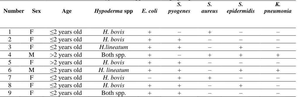
Table 2.The results of bacteriological study of subcutaneous abscesses, divided according to the
Hypoderma spp, sex and age groups
Number Sex Age Hypoderma spp E. coli
S. pyogenes
S. aureus
S. epidermidis
K. pneumonia
1 F ≥2 years old H. bovis + – + – –
2 F ≥2 years old H. bovis + + – – –
3 F ≥2 years old H.lineatum + + – + –
4 M >2 years old Both spp. + – + + +
5 F >2 years old H. bovis + + – – –
6 M ≥2 years old H. lineatum + + – + +
7 F ≥2 years old H. bovis – + + – –
8 F ≥2 years old H. bovis + + – + –
9 F ≥2 years old Both spp. + + – – –
seen in 48 (46.15 %), 34 (32.69 %) and 22 (21.15 %) samples, respectively. The bacteriological results indicated that, from 23 sampled abscesses 9 of them had bacterial contamination. The following bacteria were isolated: Escherichia coli,
Streptococcus pyogenes, Staphylococcus aureus, Staphylococcus epidermicus and
Klebsiella pneumonia (Table 2).
Discussion
In the past years, no detailed research has been conducted on the bovine hypodermosis in Iran. Besdies, the information related to basic epidemiology of the disease in Iran is scanty. Up to now, the only published work on cattle hypodermosis was showed the annual
isolate from subcutaneous abscesses. However, based on to previous reports, anaerobic bacteria were isolated more frequently from liver abscesses and their isolation was associated with colonic and biliary origins.12 The isolation of E. coli, S. pyogenes, S. aureus and S. epidermidis
from subcutaneous abscesses of cattle are in agreement with the results of Tadayon
et al, who showed the most frequented bacteria that isolated from subcutaneous abscesses of sheep and goat belong to the following genera: Corynebacterium,
Staphylococcus, Streptococcus,
Pasteurella, E.coli.13
In this work male animals showed higher mixed bacterial contamination than females. We did not find the plausible explanation for the less sensitivity of female animals than males to mixed bacterial contamination.
The third stage larva (L3) of both
Hypoderma species after migration reaches to the dorsal region of thorax and lumbar, finally it penetrates into the skin for releasing from animal. It should be pointed out that this migration is occurred in the last of winter and at this time of the year animals are kept in closed stalls with high density. On the other hand, the high prevalence of E. coli in these conditions is a good explanation for more isolation than the other bacteria from subcutaneous abscesses in cattle. Finally, based on our knowledge and the review of literatures, there are not enough studies about subcutaneous abscesses in cattle. On the other hand, because of economic lost and contamination of the abscesses by micro-organisms, more studies should be done to learn about incidence and antibiotic resistance profile in the other parts of the world.
Acknowledgements
The authors would like to appreciate the cooperation of Mazandaran Veterinary Organization, Iran.
References
1. Otranto D, Zalla P, Testini G, et al. Cattle grub infestation by Hypoderma sp. in Albania and risks for European countries. Vet Parasitol 2005; 128: 157–162.
2. Boulard C. Durably controlling bovine hypodermosis. Vet Res 2002; 33: 455– 464.
3. Boulard C, Villejoubert C, Moire N, et al. Serosurveillance of hypodermosis in a herd under therapeutic control. Effect of a low level of infestation. Vet Parasitol 1996 ;66: 109–117.
4. Scholl PJ. Biology and control of cattle grub. Annal Rev Entomol 1993; 15: 360–365.
5. Tarry DW. Biology, economic effects and early efforts to eradicate Hypoderma. In: Boulard C, Sol J, Pithan K, O’Brien D, Webster K, Sampimon OC ,eds. Improvement in the Control Methods for Warble Fly in Livestock, Commission of the European Communities, Brussels, 1998; 13–17. 6. Skerman KD, Shahlapour AA, Eslami
AH, et al. Observation on the incidence, epidemiology, control and economic importance of gastrointestinal parasites of sheep and goat in Iran. In: Soulsby EJL, ed. Reaction of the host to parasitism proceeding 3 International Conference World Association for the advancement of veterinary parasitology. Veterinary and Medical Review NGElwert Marburg-Iahn, 1967; 141-152.
7. Nickel R, Schummar A, Seiferld E. The viscera of the domestic mammals. Verlag Peul Parey, 1979; 88-93.
8. Zumpt F. Myiasis in Man and Animals in the Old World. Butterworths, London, 1965; 267.
9. Berkenkamp SD, Drummond RO. Hypodermosis. part I, the compendium. 1990; 12: 740-746.
M.Tavassoli et al / Veterinary Research Forum. 2(Sept., 2010) 123-127
Kazeroon abattoir. Pajouhesh-va-Sazandegi 2000; 4: 92–93.
11. Simsek S, Utuk AE, Koroglu E, et al. Seroprevalence of hypodermosis in cattle in some provinces of Turkey. Res Vet Sci 2008; 84: 246–249.
12. Brook I, Frazier EH. Microbiology of liver and spleen abscesses. J Med Microb 1998; 47: 1075-1080.