The Genomic Standards Consortium
Complete genome sequence of Syntrophobotulus glycolicus type
strain (FlGlyR
T)
Cliff Han1,2, Romano Mwirichia3, Olga Chertkov1,2, Brittany Held1,2, Alla Lapidus1, Matt Nolan1, Susan Lucas1, Nancy Hammon1, Shweta Deshpande1, Jan-Fang Cheng1, Roxanne Tapia1,2, Lynne Goodwin1,3, Sam Pitluck1, Marcel Huntemann1, Konstantinos Liolios1, Natalia Ivanova1, Ioanna Pagani1, Konstantinos Mavromatis1, Galina Ovchinikova1, Amrita Pati1, Amy Chen4, Krishna Palaniappan4, Miriam Land1,5, Loren Hauser1,5, Evelyne-Marie Brambilla6, Manfred Rohde7, Stefan Spring6, Johannes Sikorski6, Markus Göker6, Tanja Woyke1, James Bristow1, Jonathan A. Eisen1,8, Victor Markowitz4, Philip Hugenholtz1,9, Nikos C. Kyrpides1, Hans-Peter Klenk6*, and John C. Detter1,2
1 DOE Joint Genome Institute, Walnut Creek, California, USA
2 Los Alamos National Laboratory, Bioscience Division, Los Alamos, New Mexico, USA 3 Jomo Kenyatta University of Agriculture and Technology, Kenya
4 Biological Data Management and Technology Center, Lawrence Berkeley National Laboratory, Berkeley, California, USA
5 Oak Ridge National Laboratory, Oak Ridge, Tennessee, USA
6 DSMZ - German Collection of Microorganisms and Cell Cultures GmbH, Braunschweig, Germany
7 HZI – Helmholtz Centre for Infection Research, Braunschweig, Germany 8 University of California Davis Genome Center, Davis, California, USA
9 Australian Centre for Ecogenomics, School of Chemistry and Molecular Biosciences, The University of Queensland, Brisbane, Australia
*Corresponding author: Hans-Peter Klenk
Keywords: glycolate-oxidizing, Gram-negative with Gram-positive cell wall structure, strictly anaerobic, chemotrophic, mesophilic, non-motile, rod-shaped, spore-forming, Peptococca-ceae, Clostridiales, GEBA
Syntrophobotulus glycolicus Friedrich et al. 1996 is currently the only member of the genus
Syntrophobotulus within the family Peptococcaceae. The species is of interest because of its isolated phylogenetic location in the genome-sequenced fraction of tree of life. When grown in pure culture with glyoxylate as carbon source the organism utilizes glyoxylate through fermentative oxidation, whereas, when grown in syntrophic co-culture with homoacetogenic or methanogenic bacteria, it is able to oxidize glycolate to carbon dioxide and hydrogen. No other organic or inorganic carbon source is utilized by S. glycolicus. The subdivision of the family Peptococcaceae into genera does not reflect the natural relationships, particularly re-garding the genera most closely related to Syntrophobotulus. Both Desulfotomaculum and
Pelotomaculum are paraphyletic assemblages, and the taxonomic classification is in signifi-cant conflict with the 16S rRNA data. S. glycolicus is already the ninth member of the family
Peptococcaceae with a completely sequenced and publicly available genome. The 3,406,739 bp long genome with its 3,370 protein-coding and 69 RNA genes is a part of the
GenomicEncyclopedia ofBacteria andArchaea project.
Introduction
Strain FlGlyR
T(= DSM 8271) is the type strain of
Syntrophobotulus glycolicus
within the monotypic
genus
Syntrophobotulus
[1], which is affiliated to the
family
Peptococcaceae
within the order
Clostridiales
[2]. The genus name is derived from the latinized
Greek
syntrophos
meaning
having grown up with
one
, and the Latin
botulus
, sausage, a syntrophic
sau-sage-like item [3]. The species epithet is derived
from the Neo-Latin
acidum glycolicum
meaning
na-ture is excretion by algae and other photoautotrophs
and chemoautotrophs [4-7]. Strain FlGlyR
Twas
iso-lated from anoxic sewage sludge in Konstanz,
Ger-many [3], but was also mentioned in earlier reports
[8]. No further isolates have been reported until
now. Here we present a summary classification and
a set of features for
S. glycolicus
FlGlyR
T, together
with the description of the complete genomic
se-quencing and annotation.
Classification and features
A representative genomic 16S rRNA sequence of
strain FlGlyR
Twas compared using NCBI BLAST [9]
under default settings (
e.g
., considering only the
high-scoring segment pairs (HSPs) from the best
250 hits) with the most recent release of the
Green-genes database [10] and the relative frequencies of
taxa and keywords (reduced to their stem [11])
were determined, weighted by BLAST scores. The
most frequently occurring genera were
Desulfitobac-terium
(45.4%),
Desulfosporosinus
(19.3%),
Dehalo-bacter
(18.0%),
Heliobacterium
(13.8%) and
Syntro-phobotulus
(2.6%) (85 hits in total). Regarding the
single hit to sequences from members of the species,
the average identity within HSPs was 99.7%,
whe-reas the average coverage by HSPs was 99.7%.
Among all other species, the one yielding the highest
score was
Dehalobacter restrictus
(Y10164), which
corresponded to an identity of 95.0% and an HSP
coverage of 85.2%. (Note that the Greengenes
data-base uses the INSDC (= EMBL/NCBI/DDBJ)
annota-tion, which is not an authoritative source for
nomen-clature or classification.) The highest-scoring
envi-ronmental sequence was AJ278164 (‘
Dehalobacter
sp. clone SHD-11' [12]), which showed an identity of
95.3% and an HSP coverage of 86.8%. The most
fre-quently occurring keywords within the labels of
en-vironmental samples which yielded hits were 'soil'
(6.5%), 'microbi' (6.4%), 'respons' (4.8%), 'paddi,
rice' (4.6%) and 'condit' (4.5%) (165 hits in total).
The most frequently occurring keyword within the
labels of environmental samples which yielded hits
of a higher score than the highest scoring species
was 'dehalobact' (100.0%) (1 hit in total). The
BLAST analysis results concur with earlier reports
on the ecology and the physiology of the isolate
whereby it was isolated from a co-culture with other
sulfate-reducing bacteria [3,8].
Figure 1 shows the phylogenetic neighborhood of
S.
glycolicus
in a 16S rRNA based tree. The sequences
of the four 16S rRNA gene copies in the genome
differ from each other by up to eight nucleotides,
and differ by up to 14 nucleotides from the
pre-viously published 16S rRNA sequence X99706,
which contains two ambiguous base calls.
As two of the genera selected for Figure 1,
Desulfo-tomaculum
and
Pelotomaculum
, appeared as
para-phyletic in the tree, we conducted both
uncon-strained heuristic searches for the best tree under
the maximum likelihood (ML) [15] and maximum
parsimony (MP) criterion [17] as well as searches
constrained for the monophyly of all genera (for
details of the data matrix see the figure caption).
The bestknown ML tree had a log likelihood of
-12,054.61, whereas the best trees found under the
constraint had a log likelihood of -12,209.39 and
were significantly worse in the SH test as
imple-mented in RAxML [15] (p < 0.01). The best-known
MP trees had a score of 2,018 whereas the best
trees found under the constraint had a score of
2,076 and were significantly worse in the KH test as
implemented in PAUP* [17] (p < 0.0001).
Accor-dingly, the current classification of the group is in
significant conflict with the 16S rRNA data and
ap-parently does not reflect its natural relationships.
The classification could be improved if
combina-tions of phenotypic character states were found
which characterize a set of appropriately
rear-ranged, then monophyletic genera. However, it
might also be that the goal to 'define' each genus in
terms of unique combinations of few, potentially
arbitrarily selected character states is
over-ambitious, if not misleading in this group of
organ-isms. Apparently a taxonomic revision of the family
appears to be necessary which focuses more
strongly on the genealogy of the organisms than
previous treatments.
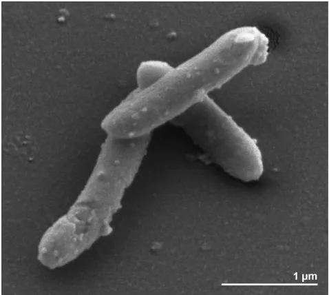
Cells of strain FlGlyR
Tare Gram-positive, spore
forming and slightly curved rods of 2.5-3.5 by 0.5 µm
in size [3] (Figure 2). Though the organism is
re-ported to be non-motile, numerous genes associated
with flagellar motility are present in the genome
(see below). Growth occurs between 15°C and 37°C
with an optimum at 28°C, and in a pH range of 6.7 to
8.3, with an optimum at pH 7.3 [3] (Table 1). The
reported habitat for this strain is sewage sludge and
anoxic freshwater sediments [3]. Initial isolation
condition was from defined co-cultures of
ferment-ing bacteria with homoacetogenic or methanogenic
bacteria which converted glycolate completely to
CO
2and H
2, with concomitant reduction of CO
2to
either acetate or methane [3,8]. Later strain FlGlyR
Twas identified as the primary fermenting partner in
these co-cultures and glyoxylate was the substrate
[3]. Strain FlGlyR
Tgrows optimally in freshwater
medium although growth also occurred in
brackish-water medium with 110 mM NaC1 and 5 mM MgCl
[3]. Strain FlGlyR
Tis strictly anaerobic, growing
chemotrophically in pure culture by fermentative
oxidation of glyoxylate [3]. In pure culture, glyoxylic
acid is fermented to carbon dioxide, hydrogen, and
glycolic acid [3]. However, in syntrophic co-culture
with, e.g.,
Methanospirillum hungatei
or
Acetobacte-rium woodii
as a partner, glycolic acid is converted to
carbon dioxide and hydrogen [3]. Glycolate
oxida-tion to glyoxylate and vice versa is coupled to a
membrane-bound electron transport system that
catalyzes either a proton potential-driven reversed
electron transport from glycolate to hydrogen or a
hydrogen-dependent glyoxylate reduction coupled
to ATP synthesis by electron transport
phosphoryla-tion [34,35]. Due to the oxygenase activity of the
D-ribulose-1,5-bisphosphate carboxylase at low CO
2and high O
2concentrations, the phosphoglycolate
formed in these organisms is subsequently
dephos-phorylated to glycolate [8]. It is reported that no
other organic or inorganic substrates are used [3],
even though a total of 78 carbohydrate transport
and metabolism genes are found the genome of this
organism (COGS table). Neither sulfate, sulfite,
thi-osulfate, elemental sulfur, nor nitrate are reduced
[3].
Figure 2. Scanning electron micrograph of S. glycolicus
Chemotaxonomy
Strain FlGlyR
Thas no cytochromes and the cells
contain menaquinone-7-10, with MK-9 as major
fraction [3]. Although the cells stain
Gram-negative, the ultrastructural analysis shows a
Gram-positive cell wall architecture [3].
Genome sequencing and annotation
Genome project history
This organism was selected for sequencing on the
basis of its phylogenetic position [36], and is part
of the
Genomic
Encyclopedia of
Bacteria and
Arc-haea
project [37]. The genome project is deposited
in the Genome On Line Database [18] and the
complete genome sequence is deposited in
Gen-Bank. Sequencing, finishing and annotation were
performed by the DOE Joint Genome Institute
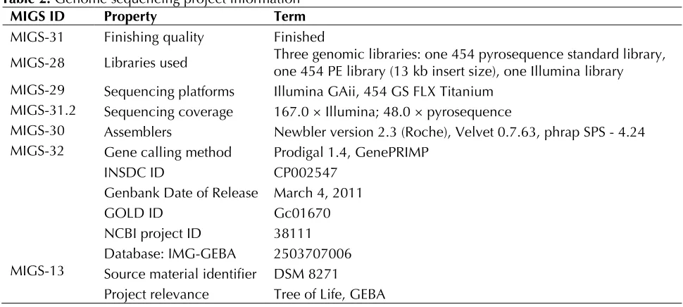
(JGI). A summary of the project information is
shown in Table 2.
Table 1. Classification and general features of S. glycolicus FlGlyRT according to the MIGS recommendations [22] and the NamesforLife database [23].
MIGS ID Property Term Evidence code
Current classification
Domain Bacteria TAS [24]
Phylum Firmicutes TAS [25,26]
Class Clostridia TAS [27,28]
Order Clostridiales TAS [29,30]
Family Peptococcaceae TAS [29,31]
Genus Syntrophobotulus TAS [3]
Species Syntrophobotulus glycolicus TAS [3]
Type strain FlGlyR TAS [3]
Gram stain negative TAS [3]
Cell shape rod shaped, slightly curved TAS [3]
Motility non-motile TAS [3]
Sporulation sporulating TAS [3]
Temperature range 15°C-37°C TAS [3]
Optimum temperature 28°C TAS [3]
Salinity tolerates ~6% NaCl TAS [3]
MIGS-22 Oxygen requirement strictly anaerobic TAS [3]
Carbon source glyoxylate TAS [3]
Energy metabolism chemotrophic TAS [3]
MIGS-6 Habitat marine, sludge, fresh water TAS [3]
MIGS-15 Biotic relationship free-living TAS [3]
MIGS-14 Pathogenicity not reported
Biosafety level 1 TAS [32]
Isolation anoxic sludge from municipal sewage treatment plant TAS [3,8]
MIGS-4 Geographic location Konstanz, Germany TAS [3,8]
MIGS-5 Sample collection time 1991 or before TAS [3,8]
MIGS-4.1 Latitude 47.67 NAS
MIGS-4.2 Longitude 9.16 NAS
MIGS-4.3 Depth unknown
MIGS-4.4 Altitude about 420 m NAS
Table 2. Genome sequencing project information
MIGS ID Property Term
MIGS-31 Finishing quality Finished
MIGS-28 Libraries used Three genomic libraries: one 454 pyrosequence standard library, one 454 PE library (13 kb insert size), one Illumina library MIGS-29 Sequencing platforms Illumina GAii, 454 GS FLX Titanium
MIGS-31.2 Sequencing coverage 167.0 × Illumina; 48.0 × pyrosequence
MIGS-30 Assemblers Newbler version 2.3 (Roche), Velvet 0.7.63, phrap SPS - 4.24 MIGS-32 Gene calling method Prodigal 1.4, GenePRIMP
INSDC ID CP002547
Genbank Date of Release March 4, 2011
GOLD ID Gc01670
NCBI project ID 38111 Database: IMG-GEBA 2503707006 MIGS-13 Source material identifier DSM 8271
Project relevance Tree of Life, GEBA
Growth conditions and DNA isolation
S. glycolicus
FlGlyR
T, DSM 8271, was grown
anae-robically in DSMZ medium 298b (FlGlyM-medium)
[38] at 28°C. DNA was isolated from 0.5-1 g of cell
paste using Jetflex Genomic DNA Purification kit
(GENOMED 600100) following the standard
pro-tocol as recommended by the manufacturer,
add-ing 10 µL proteinase K to the standard lysis
solu-tion for 50 minutes at 58°C. DNA is available
through the DNA Bank Network [39].
Genome sequencing and assembly
The genome was sequenced using a combination
of Illumina and 454 sequencing platforms. All
general aspects of library construction and
se-quencing can be found at the JGI website [40].
Py-rosequencing reads were assembled using the
Newbler assembler. The initial Newbler assembly
consisting of 38 contigs in two scaffolds was
con-verted into a phrap [41] assembly by making fake
reads from the consensus, to collect the read pairs
in the 454 paired end library. Illumina sequencing
data (602.6 Mb) was assembled with Velvet [42]
and the consensus sequences were shredded into
1.5 kb overlapped fake reads and assembled
to-gether with the 454 data. 454 draft assembly was
based on 163.5 Mb 454 draft data and all of the
454 paired end data. Newbler parameters are
-consed -a 50 -l 350 -g -m -ml 20. The
Phred/Phrap/Consed software package [41] was
used for sequence assembly and quality
assess-ment in the subsequent finishing process. After
the shotgun stage, reads were assembled with
pa-rallel phrap (High Performance Software, LLC).
Possible mis-assemblies were corrected with
ga-pResolution [40], Dupfinisher, or sequencing
cloned bridging PCR fragments with subcloning or
transposon bombing (Epicentre Biotechnologies,
Madison, WI) [43]. Gaps between contigs were
closed by editing in Consed, by PCR and by Bubble
PCR primer walks (J.-F. Chang, unpublished). A
total of 331 additional reactions were necessary to
close gaps and to raise the quality of the finished
sequence. Illumina reads were also used to correct
potential base errors and increase consensus
quality using a software Polisher developed at JGI
[44]. The error rate of the completed genome
se-quence is less than 1 in 100,000. Together, the
combination of the Illumina and 454 sequencing
platforms provided 215.0 × coverage of the
ge-nome. The final assembly contained 327,738
py-rosequence and 15,336,223 Illumina reads.
Genome annotation
Genome properties
The genome consists of a 3,406,739bp long
chro-mosome with a GC content of 46.4% (Figure 3 and
Table 3). Of the 3,439 genes predicted, 3,370 were
protein-coding genes, and 69 RNAs; 119
pseudo-genes were also identified. The majority of the
protein-coding genes (68.7%) were assigned a
putative function while the remaining ones were
annotated as hypothetical proteins. The
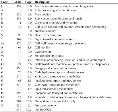
distribu-tion of genes into COGs funcdistribu-tional categories is
presented in Table 4.
Table 3. Genome Statistics
Attribute Value % of Total
Genome size (bp) 3,406,739 100.00%
DNA coding region (bp) 2,989,609 87.76% DNA G+C content (bp) 1,579,030 46.35%
Number of replicons 1
Extrachromosomal elements 0
Total genes 3,439 100.00%
RNA genes 69 2.01%
rRNA operons 4
Protein-coding genes 3,370 97.99%
Pseudo genes 119 3.46%
Genes with function prediction 2364 68.74%
Genes in paralog clusters 710 20.65%
Genes assigned to COGs 2,399 69.76%
Genes assigned Pfam domains 2,561 74.47% Genes with signal peptides 463 13.46% Genes with transmembrane helices 848 24.66%
CRISPR repeats 2
Table 4: Number of genes associated with the general COG functional categories Code value %age Description
J 152 5.8 Translation, ribosomal structure and biogenesis A 0 0.0 RNA processing and modification
K 230 8.8 Transcription
L 156 6.0 Replication, recombination and repair B 1 0.0 Chromatin structure and dynamics
D 33 1.3 Cell cycle control, cell division, chromosome partitioning Y 0 0.0 Nuclear structure
V 98 3.8 Defense mechanisms
T 162 6.2 Signal transduction mechanisms
M 170 6.5 Cell wall/membrane/envelope biogenesis N 68 2.6 Cell motility
Z 0 0.0 Cytoskeleton
W 0 0.0 Extracellular structures
U 43 1.7 Intracellular trafficking, secretion, and vesicular transport O 74 2.8 Posttranslational modification, protein turnover, chaperones C 156 6.0 Energy production and conversion
G 78 3.0 Carbohydrate transport and metabolism E 214 8.2 Amino acid transport and metabolism F 65 2.5 Nucleotide transport and metabolism H 140 5.4 Coenzyme transport and metabolism I 49 1.9 Lipid transport and metabolism
P 195 7.5 Inorganic ion transport and metabolism
Q 27 1.0 Secondary metabolites biosynthesis, transport and catabolism R 282 10.8 General function prediction only
Acknowledgements
We would like to gratefully acknowledge the help of
Maren Schröder (DSMZ) for growing S. glycolicus
cul-tures. This work was performed under the auspices of the US Department of Energy Office of Science, Biologi-cal and Environmental Research Program, and by the University of California, Lawrence Berkeley National Laboratory under contract No. DE-AC02-05CH11231,
Lawrence Livermore National Laboratory under Con-tract No. DE-AC52-07NA27344, and Los Alamos Na-tional Laboratory under contract No. DE-AC02-06NA25396, UT-Battelle and Oak Ridge National La-boratory under contract DE-AC05-00OR22725, as well as German Research Foundation (DFG) INST 599/1-2.
References
1. Euzéby JP. List of bacterial names with standing in nomenclature: A folder available on the internet.
Int J Syst Bacteriol 1997; 47:590-592
2. Boone DR, Castenholz RW, Garrity GM, eds. 2001. Bergey’s Manual of Systematic Bacteriolo-gy, 2nd ed. Springer-Verlag. New York, NY. 3. Friedrich M, Springer N, Ludwig W, Schink B.
Phylogenetic position of Desulfofustis glycolicus
gen. nov. sp. nov. and Syntrophobotulus glycoli-cus gen. nov. sp. nov., two new strict anaerobes growing with glycolic acid. Int J Syst Bacteriol
1996; 46:1065-1069
4. Whittingham CP, Pritchard GG. The production of glycolate during photosynthesis in Chlorella. Proc R Soc Lond 1963; 157:366-382.
5. Codd GA, Smith BM. Glycolate formation and excretion by the purple photosynthetic bacterium
Rhodospirillum rubrum.FEBS Lett 1974; 48
:105-108
6. Codd GA, Bowien B, Schlegel HG. Glycolate production and excretion by Alcaligenes eutro-phus.Arch Microbiol 1976; 110:167-171
7. Beudeker RF, Kuenen JG, Codd GA. Glycolate metabolism in the obligate chemolithotroph Thi-obacillus neapolitanus grown in continuous cul-ture. J Gen Microbiol 1981; 126:337-346. 8. Friedrich M, Laderer U, Schink B. Fermentative
degradation of glycolic acid by defined syntroph-ic cocultures. Arch Microbiol 1991; 156:398-404.
9. Altschul SF, Gish W, Miller W, Myers EW, Lip-man DJ. Basic local alignment search tool. J Mol Biol 1990; 215:403-410
10. DeSantis TZ, Hugenholtz P, Larsen N, Rojas M, Brodie EL, Keller K, Huber T, Dalevi D, Hu P, Andersen GL. Greengenes, a chimera-checked
16S rRNA gene database and workbench compat-ible with ARB. Appl Environ Microbiol 2006; 72:5069-5072
11. Porter MF. An algorithm for suffix stripping. Pro-gram: electronic library and information systems
1980; 14:130-137.
12. Schlötelburg C, von Wintzingerode C, Hauck R, von Wintzingerode F, Hegemann W, Göbel UB. Microbial structure of an anaerobic bioreactor population that continuously dechlorinates 1,2-dichloropropane. FEMS Microbiol Ecol 2002;
39:229-237
13. Lee C, Grasso C, Sharlow MF. Multiple sequence alignment using partial order graphs. Bioinformat-ics 2002; 18:452-464
14. Castresana J. Selection of conserved blocks from multiple alignments for their use in phylogenetic analysis. Mol Biol Evol 2000; 17:540-552
15. Stamatakis A, Hoover P, Rougemont J. A rapid bootstrap algorithm for the RAxML web-servers.
Syst Biol 2008; 57:758-771
16. Pattengale ND, Alipour M, Bininda-Emonds ORP, Moret BME, Stamatakis A. How many bootstrap replicates are necessary? Lect Notes Comput Sci
2009; 5541:184-200
17. Swofford DL. PAUP*: Phylogenetic Analysis Us-ing Parsimony (*and Other Methods), Version 4.0 b10. Sinauer Associates, Sunderland, 2002. 18. Liolios K, Chen IM, Mavromatis K, Tavernarakis
N, Hugenholtz P, Markowitz VM, Kyrpides NC. The Genomes On Line Database (GOLD) in 2009: status of genomic and metagenomic projects and their associated metadata. Nucleic Acids Res 2010; 38:D346-D354
19. Spring S, Lapidus A, Schröder M, Gleim D, Sims D, Meincke L, Glavina Del Rio T, Tice H, Copel-and A, Cheng JF, et al. Complete genome se-quence of Desulfotomaculum acetoxidans type strain (5575T). Stand Genomic Sci 2009; 1
:242-253
20. Kosaka T, Kato S, Shimoyama T, Ishii S, Abe T, Watanabe K. The genome of Pelotomaculum thermopropionicum reveals niche-associated evo-lution in anaerobic microbiota. Genome Res
2008; 18:442-448
21. Nonaka H, Keresztes G, Shinoda Y, Ikenaga Y, Abe M, Naito K, Inatomi K, Furukawa K, Inui M, Yukawa H. Complete genome sequence of the dehalorespiring bacterium Desulfitobacterium hafniense Y51 and comparison with Dehalococ-coides ethenogenes 195. J Bacteriol 2006; 188:2262-2274
22. Field D, Garrity G, Gray T, Morrison N, Selengut J, Sterk P, Tatusova T, Thomson N, Allen MJ, An-giuoli SV, et al. The minimum information about a genome sequence (MIGS) specification. Nat Biotechnol 2008; 26:541-547
23. Garrity G. NamesforLife. BrowserTool takes ex-pertise out of the database and puts it right in the browser. Microbiol Today 2010; 37:9.
24. Woese CR, Kandler O, Wheelis ML. Towards a natural system of organisms: proposal for the do-mains Archaea, Bacteria, and Eucarya.Proc Natl Acad Sci USA 1990; 87:4576-4579
25. Gibbons NE, Murray RGE. Proposals Concerning the Higher Taxa of Bacteria. Int J Syst Bacteriol
1978; 28:1-6
26. Garrity GM, Holt JG. The Road Map to the Ma-nual. In: Garrity GM, Boone DR, Castenholz RW (eds), Bergey's Manual of Systematic Bacteriolo-gy, Second Edition, Volume 1, Springer, New York, 2001, p. 119-169.
27. List Editor. List of new names and new combina-tions previously effectively, but not validly, pub-lished. List no. 132. Int J Syst Evol Microbiol
2010; 60:469-472
28. Rainey FA. Class II. Clostridia class nov. In: De Vos P, Garrity G, Jones D, Krieg NR, Ludwig W, Rainey FA, Schleifer KH, Whitman WB (eds), Ber-gey's Manual of Systematic Bacteriology, Second Edition, Volume 3, Springer-Verlag, New York, 2009, p. 736.
29. Skerman VBD, McGowan V, Sneath PHA. Ap-proved Lists of Bacterial Names. Int J Syst
Bacte-riol 1980; 30:225-420
30-1-225
30. Prévot AR. In: Hauderoy P, Ehringer G, Guillot G, Magrou. J., Prévot AR, Rosset D, Urbain A (eds), Dictionnaire des Bactéries Pathogènes, Second Edition, Masson et Cie, Paris, 1953, p. 1-692. 31. Rogosa M. Peptococcaceae, a new family to
in-clude the Gram-positive, anaerobic cocci of the genera Peptococcus, Peptostreptococcus and
Ruminococcus.Int J Syst Bacteriol 1971; 21
:234-237
32. BAuA 2005. Classification of bacteria and arc-haea in risk groups. TRBA 466 p. 336;
33. Ashburner M, Ball CA, Blake JA, Botstein D, But-ler H, Cherry JM, Davis AP, Dolinski K, Dwight SS, Eppig JT, et al. Gene Ontology: tool for the unification of biology. Nat Genet 2000; 25:25-29
34. Friedrich M, Schink B. Hydrogen formation from glycolate driven by reversed electron transport in membrane vesicles of a syntrophic glycolate-oxidizing bacterium. Eur J Biochem 1993;
217:233-240
35. Friedrich M, Schink B. Electron transport phos-phorylation driven by glyoxylate respiration with hydrogen as electron donor in membrane vesicles of a glyoxylate-fermenting bacterium. Arch Mi-crobiol 1995; 163:268-275
36. Klenk HP, Göker M. En route to a genome-based classification of Archaea and Bacteria? Syst Appl Microbiol 2010; 33:175-182
37. Wu D, Hugenholtz P, Mavromatis K, Pukall R, Dalin E, Ivanova NN, Kunin V, Goodwin L, Wu M, Tindall BJ, et al. A phylogeny-driven genomic encyclopaedia of Bacteria and Archaea. Nature
2009; 462:1056-1060
38. List of growth media used at DSMZ:
Biobanking 2011; 9:51-55.
40. The DOE Joint Genome Institute.
41. Phrap and Phred for Windows, MacOS, Linux,
and Unix
42. Zerbino DR, Birney E. Velvet: algorithms for de novo short read assembly using de Bruijn graphs.
Genome Res 2008; 18:821-829
43. Sims D, Brettin T, Detter JC, Han C, Lapidus A, Copeland A, Glavina Del Rio T, Nolan M, Chen F, Lucas S, et al. Complete genome sequence of
Kytococcus sedentarius type strain (541T). Stand
Genomic Sci 2009; 1:12-20
44. Lapidus A, LaButti K, Foster B, Lowry S, Trong S, Goltsman E. POLISHER: An effective tool for
us-ing ultra short reads in microbial genome assem-bly and finishing. AGBT, Marco Island, FL, 2008. 45. Hyatt D, Chen GL, LoCascio PF, Land ML,
Lari-mer FW, Hauser LJ. Prodigal: prokaryotic gene recognition and translation initiation site identifi-cation. BMC Bioinformatics 2010; 11:119
46. Pati A, Ivanova NN, Mikhailova N, Ovchinnikova G, Hooper SD, Lykidis A, Kyrpides NC. Gene-PRIMP: a gene prediction improvement pipeline for prokaryotic genomes. Nat Methods 2010;
7:455-457
47. Markowitz VM, Ivanova NN, Chen IMA, Chu K, Kyrpides NC. IMG ER: a system for microbial ge-nome annotation expert review and curation. Bio-informatics 2009; 25:2271-2278
![Figure 1. Phylogenetic tree highlighting the position of branches are support values from 700 ML bootstrap replicates [16] (left) and from 1,000 maximum parsimony boot-16S rRNA gene sequence under the maximum likelihood (ML) criterion [15] and rooted with](https://thumb-us.123doks.com/thumbv2/123dok_us/682761.2066654/2.612.74.532.376.649/phylogenetic-highlighting-position-bootstrap-replicates-parsimony-likelihood-criterion.webp)
![Table 1. Classification and general features of S. glycolicus FlGlyRT according to the MIGS recommendations [22] and the NamesforLife database [23]](https://thumb-us.123doks.com/thumbv2/123dok_us/682761.2066654/4.612.62.555.243.677/classification-features-glycolicus-flglyrt-according-recommendations-namesforlife-database.webp)