R E S E A R C H
Open Access
Notch1 signaling in melanoma cells
promoted tumor-induced
immunosuppression via upregulation of
TGF-
β
1
Zike Yang
1, Yanxia Qi
3, Nan Lai
1, Jiahe Zhang
1, Zehong Chen
1, Mingyu Liu
1, Wan Zhang
1, Rongcheng Luo
1*and Shijun Kang
2*Abstract
Background:The receptors of Notch family play an important role in controlling the development, differentiation, and function of multiple cell types. The aim of this study is to investigate the role of Notch1 signaling upon immune suppression induced by melanoma cells.
Methods:Melanoma cell line B16 cells were transfected by lentivirus containing mouse Notch1 gene or Notch1 shRNA to generate B16 cell line that highly or lowly expressed Notch1. Notch1 in anti-tumor immune response was comprehensively appraised in murine B16 melanoma tumor model in immunocompetent and immunodeficient mice. The ratios of CD3+CD8+cytotoxic T cells, CD49b+NK cells, CD4+CD25+FoxP3+Tregs and Gr1+CD11b+MDSCs in tumor-DLN or spleen were examined by flow cytometry. After the co-culture of B16 cells and CD8+T cells, the effects of Notch1 on the proliferation and activation of T cells were assessed by CCK8 assay, CFSE dilution and Chromium-release test. The mRNA expression and supernatant secretion of immunosuppressive cytokines, TGF-β1, VEGF, IL-10 and IFN-γwere measured by RT-PCR and ELISA, respectively.
Results:Downregulation or overexpression of Notch1 in B16 melanoma cells inhibited or promoted tumor growth in immunocompetent mice, respectively. Notch1 expression in B16 melanoma cells inhibited the infiltration of CD8 + cytotoxic T lymphocytes and NK cells and reduced IFN-γrelease in tumor tissue. It could also enhance B16 cell-mediated inhibition of T cell proliferation and activation, and upregulate PD-1 expression on CD4+and CD8+T cells. The percentage of CD4+CD25+FoxP3+Tregs and Gr1+CD11b+MDSCs were significantly increased in tumor
microenvironment, and all these were attributed to the upregulation of TGF-β1.
Conclusion:These findings suggested that Notch1 signaling in B16 melanoma cells might inhibit antitumor immunity by upregulation of TGF-β1.
Keywords:Malignant melanoma, Immunotherapy, Immunosuppression, Notch1, TGF-β1, Notch
* Correspondence:luorc02@vip.163.com;kangshijunlb@163.com 1Cancer Center, Integrated Hospital of Traditional Chinese Medicine,
Southern Medical University, No.13, Shiliugang Road, Haizhu District, Guangzhou 510315, Guangdong Province, People’s Republic of China
2Oncology Department, Nanfang Hospital, Southern Medical University,
No.1838, North of Guangzhou Avenue, Baiyun District, Guangzhou, Guangdong Province 510515, People’s Republic of China Full list of author information is available at the end of the article
Background
Malignant melanoma, one of the most highly aggressive tumors, resists to conventional chemotherapy and radio-therapy and has fatal outcomes. There are compelling evidences to show that melanoma cells escape the host’s immunity by actively developing multiple suppressive mechanisms within the cancer microenvironment [1]. For instance, melanoma cells evade T cell surveillance by creating an immunosuppressive environment via the production of cytokines such as transforming growth factor (TGF)-β1, vascular endothelial growth factor (VEGF) and IL-10, which recruit myeloid-derived sup-pressor cells (MDSCs) and T regulatory cells (Tregs). The promotion and recruition of MDSCs and Tregs by melanoma cells play a crucial role in tumor immune escape [2].
The Notch signaling is a highly conserved pathway that controls the differentiation, development and function of multiple cell types, such as stem cells [3]. Mammals have four Notch receptors (Notch1, Notch2, Notch3, and Notch4) that are bound by five ligands (Jagged-1, Jagged-2, DLL1, DLL3, and DLL4) families [4]. Aberrant Notch signaling has been identified in malignant melanoma to play an important role in the malignant biological behavior of melanoma [5]. Our previous study has shown that interference of both Notch co-activation factor MAML1 blocks the activation of Notch pathway in both human and mouse melanoma cells, suggesting a potential new treatment strategy [6]. Among the 4 receptors, Notch2-4 have been identified in multiple cell types, such as stem cells, hematopoietic cells, macrophage or nerve cells, and controlled their differentiation, development and function [7, 8]. The role of Notch1 has been proved to be closely related to melanoma progression and become a research hotspot recently [9]. Previous studies have demonstrated that Notch1 signaling promoted primary melanoma progres-sion by activating mitogen-activated protein kinase/ phosphatidylinositol 3-kinase-Akt pathways and up-regulating N-cadherin expression [10]. Moreover, Notch1 and NRG1 expression in melanoma promoted cell growth by activating PI3Kinase/Akt signaling pathway and facilitating the accumulation of p27 [11]. Additionally, activated Notch1 receptors in endothelial cells promoted neutrophil infiltration, tumor cell adhesion to the endo-thelium, intravasation, lung colonization and facilitated melanoma metastasis by generating a senescent, pro-inflammatory endothelium [12].
Although Notch signaling is known to be important for the malignant biological behavior of melanoma cells, little is known about the effects of aberrant activation of this pathway in melanoma on tumor-induced immuno-suppressive microenvironment. Our primary study has shown that siRNA-mediated Notch1 knockdown might
potentially enhance the effect of IL-2 immunotherapy in malignant melanoma [13]. In the present study, we further evaluated the role of Notch1 expression in mel-anoma cells on tumor-induced immunosuppression. This study was not only important for elucidating the mechanism of tumor-induced immune escape, but also provided a scientific basis for developing novel immuno-therapeutic strategies to target Notch1 in B16 melanoma cells to induce innate and adaptive immune responses against tumors.
Methods
Cells and animals
Murine malignant melanoma cell line B16 was purchased from China Center for Type Culture Collection. B16 cells were cultured in DMEM-high glucose (Thermo Fisher Scientific, Waltham, MA, USA) supplemented with 10% fetal bovine serum (Thermo Fisher Scientific, Waltham, MA, USA) at 37 °C in an atmosphere of 5% CO2.
In vivo study
Female C57BL/6 and BALB/c Nude mice were pur-chased from Laboratory Animal Center of Southern Medical University (Guangzhou, China). All mice were 6- to 8 weeks of age at the time of experiment, and at least 6 mice per group were used in each experiment. For tumor challenge experiments, 5 × 10⁵ B16, B16-shNotch1 or B16-Notch1 melanoma cells were subcuta-neously inoculated. Mice were observed carefully while tumor volume in mice was measured twice a week. Tumor volume was calculated by (length × width2)/2. All mice were sacrificed humanely after the experiments. Melanoma tissues in mice were surgically resected to perform further assay. Animal care and treatment were in accordance with institutional guidelines. All animal study protocols were reviewed and approved by the Animal Care and Use Committee of Southern Medical University, China.
Generation of stable Notch1 overexpression and knockdown melanoma cells
ELISpot assays
Mouse IFN-γ ELISpot kits (BD Biosciences, USA) were used according to the manufacturer’s instructions. Briefly, immune cells from spleens or tumor-DLNs (1 × 10⁵) were co-cultured with 30 Gy-irradiated B16 (5 × 10⁴) cells in 96-well plates precoated with mouse IFN-γ for 20 h at 37 °C in complete DMEM medium in tripli-cate. The cell suspension was aspirated and washed with deionized water. Then Biotinylated anti-mouse IFN-γ antibody was added and incubated for 2 h at room temperature. After washing with deionized water, a streptavidin-horseradish peroxidase solution was added and incubated for 1 h at room temperature. Then an aminoethyl carbozole substrate solution was added and incubated for 15 min. Spots in plate were counted using a stereomicroscope after washing.
Cell surface marker and intracellular cytokine staining At the indicated time points, tumor draining lymph nodes(tumor-DLNs) and spleens were harvested from the mice, and minced into small fragments and mechan-ically dispersed in 3-5 ml cold PBS. After filtering with 70 μm cell strainer (BD Falcon, USA), Single-cell suspensions were adjusted to 1 × 10⁶ cells in 100 μl of PBS. After this, single-cell suspensions of tumor cells were stained for 30 min on ice with 1 μg of antibodies labeled with fluorochromes CD45, CD3, CD4, CD8, CD49b, CD25, CD11b, Gr-1 or matched isotypic control antibodies(BD Biosciences, USA) and then fixed and permeabilized with a permeabilization buffer (BD Biosci-ences, USA). Cells were finally stained with antibody to IFN-γ or FoxP3 for 50 min at 4 °C, washed again, and analyzed by FACSCalibur (BD Biosciences, USA). Irrelevant IgG mAbs were used as a negative control. Ten thousands live events were acquired for analysis.
In vivo depletion of T and NK cells
To deplete CD8+T, CD4+T and NK cells before and dur-ing treatment with lentivirus-shNotch1, the transplanted mice received intravenous injections of 0.3 mg mAb from anti-CD8+ hybridoma (Bioxcell, USA) the anti-CD4+ hybridoma (Bioxcell, USA) or 0.5 mg of antiasialo-GM1 antibody (Wako Pure Chemical Indus-tries, Ltd). Antibody injection was started on the day of tumor innoculation, and the treatment was repeated every 5 days throughout the entire experimental period to ensure the depletion of the target immune cell subset.
Splenic T lymphocyte isolation
Splenic lymphocytes were isolated from splenic cell suspension using density gradient centrifugation (Ficoll-Hypaque, TBD Science, Tianjin, China). CD8+ T lym-phocytes were purified using microbead isolation kits followed by magnetic-activated cell sorting (MACS)
according to the manufacturer’s instructions (Miltenyi Biotec, Germany). Isolated cells were resuspended at a concentration of 1 × 10⁶cells/mL in RPMI-1640 medium (Thermo Fisher Scientific, USA) containing L-glutamine (2 mM), penicillin/streptomycin (100 U/mL), and 10% fetal calf serum (Thermo Fisher Scientific, Waltham, MA, USA).
Lymphocyte proliferation assay and apoptosis analysis 3.0μm Transwell chamber(Corning, USA) were used for co-culture of CD8+T cells and B16 cells. B16 cells were plated in lower chamber with 104cells per well. CD8+T lymphocytes were then added to the upper chamber in 1:1 ratio. For CFSE analysis, CD8+T cells were labelled with CFSE staining before co-cultured with B16 cells. After co-cultured for 48 h, CD8+cells in upper chamber were harvested for CCK8 assay or analysis of CFSE dilution and apoptosis.
In vitro cytotoxicity assay
CD8+T cells isolated from B16 tumor-bearing mice were cultured for 3 days with irradiated B16 stimulators in a complete 1640 RPMI medium. The responder cells were then collected and used as effector cells in a 8 h chro-mium release assay against indicated B16 target cells. B16 target cells were labeled by combining 5 × 10⁶cells with 50 μCi 51Cr (Perkin-Elmer Japan Co.) in a total volume of 0.2 ml complete RPMI for 1 h at 37 °C, followed by washing thrice with plain RPMI. For the chromium release assay, CD8+ effector cells were mixed with B16 target cells at the different ratio of 1:1, 5:1 and 25:1 in a 96-well round-bottom plate (BD Biosciences).
Enzyme-linked immunosorbent assay (ELISA)
B16 cells were seeded into 6-well plates at a density of 2 × 10⁵ cells/ml. The culture supernatants were har-vested after B16 cells had adhered to the bottom of the culture plate for 18h. Then, the supernatants were centrifuged to remove melanoma cells. In accordance with the manufacturer’s instructions, Culture -supernatants of B16 cells were measured by ELISA for the secretion of TGF-β1, VEGF and IL-10 (eBioscience, San Diego, CA, USA). Freshly excised tumor tissues were minced and homogenized for 1 min in tissue tearor. Then, supernatants were collected after centrifu-gation and subjected to ELISA for examining the secre-tion of TGF-β1, VEGF, IL-10 and IFN-γ using mouse ELISA kits according to the manufacturer’s protocol (eBioscience, San Diego, CA, USA).
Statistical analysis
cytotoxicity were assessed using a student’s t-test. Differ-ences were considered to be statistically significant when
P< 0.05. All statistical analyses were performed using SPSS 20.0 (IBM, Armonk, NY, USA).
Results
Notch1 expression in melanoma cells promoted tumor growth in vivo
To examine the role of Notch1 in tumor growth, we sta-bly silenced Notch1 expression using shRNA in B16 tumor cells (Fig. 1a). B16-shNotch1(Notch1 knockdown) or B16-shCon(control) cells were subcutaneously inocu-lated into immunocompetent C57BL/6 mice. Knocking
down Notch1 significantly retarded tumor growth in C57BL/6 mice and extended mice survival time (Fig. 1b & c). To further confirm the effect of Notch1 on tumor growth, we generated a B16 cell line that highly expressed Notch1 (B16-Notch1) by lentivirus expression vector (Fig. 1e). When B16 cells transfected with a control vector (B16-GFP) and B16-Notch1 cells were inoculated into immunocompetent C57BL/6 mice, over-expression of Notch1 significantly increased tumor bur-den and shortened mice survival time (Fig. 1f & g).
In order to confirm this finding, we intravenously inoculated B16-shCon, B16- shNotch1, or B16-Notch1 melanoma cells into C57BL/6 mice. The number of lung
shCon shNotch1
Notch1
GADPH
0 500 1000 1500 2000 2500 3000
shNotch1 shCon
7 10 13 16 19 22 25 28
* *
* *
**
B16 tumor
Days after tumor innoculation
Tu
m
o
r Vo
lu
m
e
(m
m
3)
shNotch1
shCon
0 5 10 15 20 25 30 0
20 40 60 80 100
shNotch1 shCon B16 tumor
Days
P
e
rce
n
t su
rv
iv
al
GFP Notch1
Notch1
GADPH
0 500 1000 1500 2000 2500 3000 3500
GFP Notch1
7 10 13 16 19 22 25
* *
* *
B16 tumor
Days after tumor innoculation
T
um
or
V
o
lum
e
(m
m
3)
GFP
Notch1 shNotch1 shCon
shN otch
1
shCo n
0 50 100 150
*
T
u
mo
r f
o
c
i n
u
mb
e
r/
lu
n
g
0 5 10 15 20 25
0 20 40 60 80 100
GFP Notch1 B16 tumor
Days
Pe
rc
en
t
s
ur
v
iv
a
l
GFP
Not ch1
0 100 200 300 400
*
T
u
mo
r f
o
c
i n
u
mb
e
r/
lu
n
g
GFP Notch1
a
b
c
d
e
f
g
h
Fig. 1Notch1 expression in melanoma cells promoted tumor growth in vivo.aWestern blot analysis of Notch1 expression in shNotch1 and shCon cells.b
tumor foci in C57BL/6 mice was counted on day 14 after intravenous inoculation. Pulmonary metastasis in mice receiving B16-shNotch1 cells was significantly inhibited, whereas pulmonary metastasis was enhanced in those receiving B16-Notch1 melanoma cells (Fig. 1d & h).
Notch1 function in tumor growth depends on the immune system
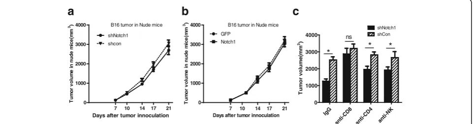
To explore whether regulation of tumor growth by Notch1 expression in melanoma cells was due to host immune status, we subcutaneously inoculated B16-shCon, B16-shNotch1, or B16-Notch1 melanoma cells into BALB/c Nude mice, which are deficient in T, B and NK cells. In contrast to immunocompetent mice, no significant differences of tumor burden were observed in BALB/c Nude mice challenged with B16-shNotch1 or B16-Notch1 cells (Fig. 2a & b). The results suggested that Notch1 might promote tumor growth in vivo via suppression of antitumor immunity.
To further determine how Notch1 affected the antitumor immunity, B16-shNotch1 or B16-shCon cells were subcuta-neously inoculated into C57BL/6 mice that were depleted of CD4+ or CD8+ T cells or NK cells and tumor growth was monitored. CD8+T cell depletion significantly compro-mised the effect of Notch1 expression on tumor growth. However, the compromised effect of CD4+ T cells or NK cells depletion was less important than CD8+ T cells in Notch1-mediated tumor growth (Fig. 2c). These results showed that regulation of tumor growth by Notch1 majorly depended on CD8+T cells in immune system.
Notch1 inhibited local antitumor immunity but not system immunity
To investigate the effect of Notch1 in tumor tissue on local anti-tumor immunity, we analyzed the immune status in the tumor microenvironment of B16 tumor-bearing mice. We found that knocking down Notch1 remarkably reduced while overexpressing Notch1
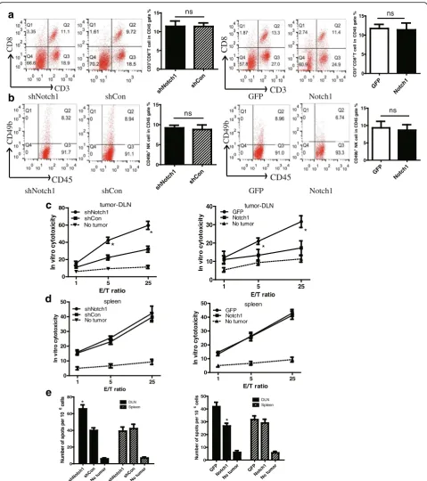
increased the percentage of CD3+CD8+ CTL and NK cells in tumor-DLNs (Fig. 3a & b). As IFN-γ is a cyto-kine crucial for T-cell activation and effector function, we then examined the IFN-γ-expressing immune cells in tumor-DLNs. Knocking down Notch1 resulted in a significant increase of the percentage of IFN-γ -expressing CD8+ T cells and IFN-γ-expressing NK cells (Fig. 3c & d). In contrast, the opposite results were observed in Notch1 overexpressing cells (Fig. 3a-d). These results suggested that Notch1 in tumor cells remarkably inhibited local antitumor immunity.
We next analyzed the spleen T lymphocytes to evaluate the system immunity. Knocking down or overexpressing Notch1 did not affect the percentage of CD3+CD8+ CTL and NK cells in splenocytes (Fig. 4a & b). Then, IFN-γ ELISPOT assay and chromium release assay were per-formed to evaluate the cytotoxicity of T cells in tumor-DLNs and spleens. In in vitro cytotoxicity assay, knocking down Notch1 significantly increased the CTL activity in tumor-DLNs but not in spleens (Fig. 4c & d). Consistent with the chromium release assay, we found that knocking down Notch1strongly increased the number of IFN-γ– producing cells in tumor-DLNs in response to B16 tumor antigen (Fig. 4e). In contrast, the opposite results were ob-served in Notch1 overexpressing cells. These data sug-gested that Notch1 in tumor tissues inhibited CTL activation in tumor-DLNs but not in spleen cells.
Notch1 expression in tumor modulated immune cell population and phenotype
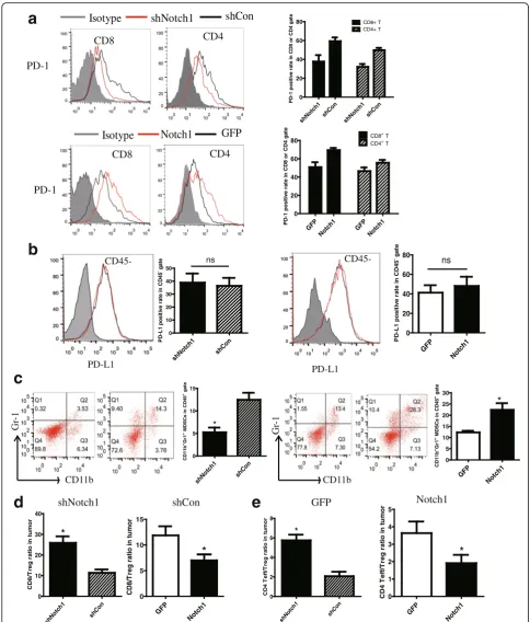
The PD-1/PD-L1 axis is a critical regulator of T cell fate and function. PD-1 has also been identified as a marker of T cell exhaustion, a hypo-functional cell state found in tumor. The expression of PD-1 on CD8+ and CD4+T cells in tumor-DLNs were analyzed by flow cytometry. Knocking down Notch1 reduced PD-1 expression while overexpressing Notch1 showed an increased PD-1 ex-pression on CD8+ and CD4+ T cells (Fig. 5a). No
0 1000 2000 3000 4000
shNotch1 shcon
7 10 14 17 21
B16 tumor in Nude mice
Days after tumor innoculation
Tu
m
o
r v
o
lu
me
i
n
n
u
d
e
mi
c
e
(mm
3)
0 1000 2000 3000 4000
GFP Notch1
7 10 14 17 21
B16 tumor in Nude mice
Days after tumor innoculation
T
u
m
o
r v
o
lum
e
in nude
m
ic
e
(m
m
3)
IgG
anti-CD
8
an
ti-CD4
anti-NK
0 1000 2000 3000 4000
shNotch1 shCon
* * *
ns
T
um
or
v
ol
um
e(
m
m
3)
a
b
c
Fig. 2Notch1 function in tumor growth depended on the immune system.aTumor growth curve of shNotch1 and shCon cells in BALB/c Nude
significant difference was observed in PD-L1 expression on CD45− melanoma cells in tumor (Fig. 5b). Tregs and MDSCs, as the immunosuppressor cells, suppress antitu-mor immunity and weaken therapeutic efficacy of im-munotherapy. To investigate the mechanism how CTLs were inhibited through Notch1 in the tumor microenvir-onment, we assessed the percentage of MDSCs and Tregs in tumor-DLNs of either Notch1 knocked down or knocked in cells. As shown in Fig. 5c-e, Notch1 in
melanoma tumor promoted more MDSCs and Tregs in-filtration in tumor microenvironment and suppressed antitumor immunity (Fig. 5c-e).
Notch1 enhanced the inhibitory effects of melanoma cells on proliferation and activation of CD8+T lymphocytes To explore the mechanism of immunosuppression induced by Notch1 in B16 cells, splenic CD8+ T cells isolated from B16 tumor-bearing mice were expanded
CD3 CD8 CD45 CD49b CD8 CD3 CD49b CD45
shNotch1 shCon GFP Notch1
CD8 CD8 CD49b shNot ch1 shCo n 0 10 20 30 40 50 60 * CD3 +CD8 +T c e ll i n C D 4 5 g a te % GF P Not ch1 0 5 10 15 20 25 * CD3 +CD8 +T c e ll i n C D 4 5 g a te % shNo tch1 shCon 0 2 4 6 8 10 12 14 * CD4 9 b
+ NK
cel l in CD 4 5 g a te % GF P Not ch1 0 2 4 6 8 10 * CD4 9 b
+ NK
c e ll i n C D45 gat e % shNotc h1 shCon 0 5 10 15 20 25 30 * IF N r e le a s in g C D 8
+ c
e ll in CD4 5 +C D8
+ ga
te % GF P Notch 1 0 2 4 6 8 10
*
IF N r e le a s in g CD8+ cel
l in C D 4 5 +CD8 + gat
e % shN otch 1 shC on 0 5 10 15 20 25 30 * IF N re leas ing N K c e ll in C D4 5 +C D 49 b
+ g
at e % GF P No tch1 0 2 4 6 8 10 12 14
*
IF N r e le a si n g NK c e ll in CD4 5 +CD4 9 b+ gat
e
%
CD49b
shNotch1 shCon GFP Notch1
shNotch1 shCon GFP Notch1
shNotch1 shCon GFP Notch1
a
b
c
d
Fig. 3Notch1 expression in melanoma cells inhibited IFN-γ-releasing CTL and NK cell infiltration in tumor . Tumor-DLNs isolated from tumor-bearing
CD8 CD45 CD49b CD49b CD45 shNotc h1 shCon No tu
mor shN
otch1shC on No tum or 0 20 40 60 80 DLN Spleen * Nu m b er o f s p o ts p e r 1 0
6 c
e
lls
GFP Not
ch1
No t um or GFP Not ch1 No tum or 0 10 20 30 40 50 DLN Spleen * Nu mb e r of s pot s pe r 1 0
6 c
e ll s 0 20 40 60 80 shNotch1 shCon No tumor
1 5 25
* * tumor-DLN E/T ratio In vi tr o c yt o to xi ci ty 0 10 20 30 40 GFP Notch1 No tumor
1 5 25
* * tumor-DLN E/T ratio In v itr o c y to tox ici ty 0 10 20 30 40 50 shNotch1 shCon No tumor
1 5 25
spleen E/T ratio In v it ro c yt o to x ici ty 0 10 20 30 40 50 GFP Notch1 No tumor
1 5 25
spleen E/T ratio In vi tr o c yt o to xi ci ty shNotch1 shCo n 0 5 10 15 ns CD3 +CD 8 +T ce ll in C D45 g a te % GFP Not ch1 0 5 10 15 ns CD 3 +C D8 +T ce ll in C D45 g a te % shNotch1 shC on 0 5 10 15 ns C D4 9b
+ NK
c e ll i n C D 4 5 g a te % GFP Notch1 0 5 10 15 ns CD 4 9 b
+ N
K c e ll in C D 4 5 g a te %
shNotch1 shCon GFP Notch1
shNotch1 shCon GFP Notch1
a
b
c
d
e
CD3 CD8 CD3Fig. 4Notch1 in tumor tissues inhibited CTL activation in tumor-DLNs but not in spleen cells. Splenocytes isolated from tumor-bearing mice were analyzed
CD11b
Gr
-1
Isotype shNotch1 shCon
CD8 CD4
CD8 CD4
Isotype
Notch1
GFP
PD-1 PD-L1 PD-1 PD-L1 CD45- CD45-shNot ch1 shC on 0 10 20 30 40
*
CD 8 /T re g ra ti o in tu m o r GFP Not ch1 0 5 10 15*
CD 8 /T re g rat io i n t u m or shNotch1 shC on 0 2 4 6 8 * CD4 T e ff/ T re g r a tio in tu m o r GFP Not ch1 0 1 2 3 4 5
*
CD 4 Te ff /T reg r a tio in t um or shNo tch1 shCo n 0 5 10 15 * CD1 1 b +Gr 1+ MD
S C s i n C D 4 5
+ ga
te GFP Not ch1 0 5 10 15 20 25 30 * CD1 1 b +Gr 1
+ MD
S C s i n C D 4 5
+ ga
te Gr -1 CD11b shN otc h1 shCon shNotc h1 shC on 0 20 40 60
80 CD8+ T
CD4+ T PD-1 p os it iv e r at e i n C D 8 o r C D 4 g at e GFP Notc h1 GF P Not ch1 0 20 40 60 80 CD8 + T CD4+ T
PD-1 p o s it iv e r a te i n C D 8 o r C D 4 g at e shN otch 1 shC on 0 10 20 30 40 50 ns PD -L 1 po si ti ve rat e i n C D 45
- g
at e GFP Notc h1 0 20 40 60 80 ns P D -L 1 p o si ti ve r a te i n CD4 5
- ga
te
a
b
c
d
e
Fig. 5Notch1 expression in tumor modulated immune cell population and phenotype. Tumor-DLNs and tumor tissue isolated from tumor-bearing
mice were analyzed for immune cell population and phenotype by flow cytometry.aFlow cytometry analysis of PD-1 expression on CD4+and CD8+ T lymphocytes in tumor-DLNs.bFlow cytometry analysis of PD-L1 expression on CD45−melanoma cells in tumor.cFlow cytometry analysis of CD11b +
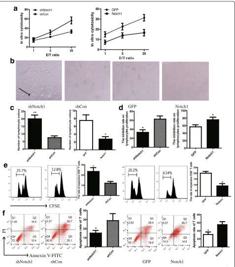
and activated with irradiated B16 cells in culture for 3 days prior to test cytotoxic function against B16-shNotch1 or B16-Notch1 cells by chromium release assays. The cytotoxicity of T cells against B16-shNotch1 was more powerful than that against B16-shCon cells (Fig. 6a). The diminished effects were observed in Notch1 overexpressing cells (Fig. 6a).
To evaluate the role of Notch1 in B16 cells on inhibit-ing T lymphocyte proliferation, CD8+T cells isolated from wild-type B16 tumor-bearing mice were co-cultured with Notch1 knocked down or knocked in B16 cells in transwell chamber for 48 h. The CD8+T cells were cultured in the upper chamber and B16 cells were in the lower chamber. According to microscopic data, knocking down Notch1 showed more lymphocyte colony formation than control group (Fig. 6b & c). In CCK8 lymphocyte proliferation assay, the proliferation rate was significantly increased in CD8+T cells which were co-cultured with B16-shNotch1 cells (Fig. 6d). The ef-fects of Notch1 on inhibiting T lymphocytes prolifera-tion was confirmed by CFSE diluprolifera-tion (Fig. 6e). Moreover, less T cells were induced apoptosis after co-cultured with B16-shNotch1 cells (Fig. 6f ). In contrast, the opposite results were observed in Notch1 overexpressing cells (Fig. 6b-f ). These results suggested that Notch1 promoted the inhibitory effects of melanoma cells on lymphocyte proliferation and activation.
The effects of Notch1 on tumor-induced immunosuppression were attributed to the upregulation of TGF-β1
To study how Notch1 affect local anti-tumor immunity in vivo and vitro, mRNA expression and supernatant secretions of TGF-β1, VEGF and IL-10 in B16 cells were measured by Q-PCR and ELISA, respectively. Notch1 knocking down remarkably reduced, while overexpres-sion of Notch1 showed significant increase in mRNA expression and supernatant secretion of TGF-β1 (Fig. 7a & b). However, no significant differences were observed in mRNA expression and supernatant secretion of IL-10 and VEGF (Fig. 7a & b). The results indicated that Notch1 upregulated tumor-derived TGF-β1 in melan-oma cells. In addition, we discovered that neutralization of TGF-β1 in the co-culture system significantly reduced the inhibitory effects of Notch1 on CD8+T lymphocyte apoptosis (Fig. 7c), proliferation (Fig. 7d) and activation (Fig. 7e) induced by melanoma cells.
To determine the secretion of immunosuppressive cytokines and IFN-γin vivo, homogenate of tumor tissue from B16 tumor-bearing mice were harvested for ELISA assay. The data showed that TGF-β1 secretion in tumor tissue was significantly reduced in B16-shNotch1 tumor, while increased in B16-Notch1 tumor in vivo (Fig. 7f ). However, there was no significant change in IL-10 and
VEGF secretions in all groups of mice (Fig. 7f ). In addition, IFN-γ secretion of homogenate supernatant in B16-shNotch1 tumor was significantly increased, sug-gesting an enhancement of antitumor immunity (Fig. 7f ). In contrast, Notch1 overexpressing tumor showed a reduction of IFN-γ secretion in homogenate super-natant. These data indicated that Notch1 in melanoma up-regulated melanoma-derived TGF-β1 and promoted tumor-induced immunosuppression in vivo.
Discussion
Malignant melanoma is a potentially fatal cancer charac-terized by rapid progression, metastasis to regional lymph nodes and distant organs, and resistance to chemotherapy and radiotherapy [14]. Melanoma has well-documented immunogenicity, which is conducive to investigating different immunotherapeutic strategies based on melanoma antigen-specific and nonspecific immunostimulation or adoptive transfer of melanoma-specific activated T cells. However, the overall outcomes of immunotherapeutic clinical studies were not satisfac-tory [15, 16]. Melanoma can produce immunosuppres-sive cytokines, such as TGF-β, VEGF and IL-10, to create an immunosuppressive microenvironment that facilitates tumor growth and blocks antitumor immune responses [17].
Among immune suppressor cells, Treg cells and MDSCs are significantly increased in hosts with ad-vanced malignancies. It is well established that Treg cells and MDSCs are essential for the control of autoimmune responses, and that their accumulations in tissues or peripheral blood of tumor-bearing mice are responsible for the suppression of anti-tumor immune effector T cell functions [18]. Cancer cells can modulate anti-tumor immune responses indirectly through the activation of Treg cells and MDSCs [19]. It has been shown that the loss of regulatory function by depletion of tumor-induced Treg cells and MDSCs might enhance immune responses, resulting in tumor reduction, while the in-creased number of Treg cells and MDSCs effectively prevented tumor destruction [20].
In the present study, Notch1 expression in melanoma cells upregulated TGF-β1 mRNA expression and pro-moted TGF-β1 secretion in tumor microenvironment, which enhanced tumor-mediated inhibition of T cell pro-liferation and activation. Furthermore, Notch1 inhibited CD8+
shNotch1 shCon GFP Notch1 shN otc h1 shCo n 0 5 10 15 20 25 ** N u m b er of l y m phocyt e col ony GFP Not ch1 0 2 4 6 8 10 * N u m b er of l y m phoc y te c o lo n y shN otc h1 shCon 0 20 40 60 80
*
T h e i n hi bi ti on r a te on l ym phocy tes pr o life ra tio n GFP Not ch1 0 20 40 60 80 100*
T he i n hi bi ti on r a te on l y m p hocyt es pr ol if er at io n CFSE Annexin V-FITC PIshNotch1 shCon GFP Notch1
0 20 40 60 80 shNotch1 shCon
1 5 25
E/T ratio In vi tr o cyt o to xi ci ty 0 10 20 30 40 GFP Notch1
1 5 25
E/T ratio In vi tr o cyt o to x ic it y shN otch 1 shCo n 0 10 20 30 * Th e r at e of expan s iv e C D 8
+ T
cel ls GFP Not ch1 0 5 10 15 20 25 * T h e ra te of expan s iv e CD8
+ T
cel ls shNot ch1 shCon 0 10 20 30 40
*
Ap o p to s is r a te of T c e lls GFP Notc h1 0 20 40 60 80 * A p opt o s is ra te of T c e llsa
b
c
e
f
d
Fig. 6Notch1 expression in melanoma cells increased tumor cell-mediated inhibition of T cell proliferation and activation.aIn vitro cytotoxicity of CD8+T
Interaction between Notch and TGF-β signaling has been identified in multiple tumors to regulate a wide variety of complicated tumor biological behaviors. For example, heightened Dll4/Notch signaling in tumor cells
can magnify TGF-β-induced pSMAD2/3 signaling and is required to sustain TGF-β-induced tumor cell growth [21]. Moreover, inhibition of Notch signaling leads to at-tenuation of both basal and TGF-β1-induced TGF-β
a
c
d
e
f
b
Fig. 7The effects of Notch1 on tumor-induced immunosuppression were attributed to the upregulation of TGF-β1.aRelative mRNA expression of
signaling in clear cell renal cell carcinoma cells, includ-ing an extensive set of genes known to be involved in migration and invasion [22]. Additionally, Notch signal-ing is required for the initiation of epithelial-mesenchymal transition and synergizes with TGF-β sig-naling pathways to promote the transition of endothelial cells to mesenchyme, as well as mesenchymal cell inva-siveness [23]. Our current findings further identified that Notch activity was a contributor to elevating melanoma-derived TGF-βsecretion, which led to the inhibition of immune cell proliferation and function, and blocked an-titumor immune responses.
TGF-β is produced by tumor cells and tumor-infiltrating immune cells within the tumor microenvir-onment. The role of TGF-β in immune suppression includes the inhibition of T cell proliferation, cytokine production and cytotoxicity [24, 25], as well as the remodeling of the tumor microenvironment and the recruitment of immune suppressive cells [26, 27]. Under some conditions, TGF-βalso inhibits antigen presenting cell function by suppressing cell maturation, inhibiting IFN-γproduction and inducing major histocompatibility complex class II down-regulation [28, 29]. Our results showed low TGF-β1 secretion and rare infiltration of Treg cells and MDSCs in tumor microenvironment after Notch1 knocking down. Therefore, we speculated that reduction of Treg cells and MDSCs in tumor sites was a possible consequence of the down-regulation of tumor-derived TGF-β1 induced by Notch1 knockdown. In addition, there were more CD8+ cytotoxic T lympho-cytes infiltration and higher IFN-γ release in tumor microenvironment, while tumor growth was significantly suppressed. This revealed a more powerful function of CD8+ cytotoxic T cells and more intense anticancer im-mune responses in vivo. The opposite effects were found in Notch1 overexpression cells. All these results strongly suggested that aberrantly elevated TGF-β1 secretion in response to abnormal activation of Notch signaling was a potential mechanism of melanoma-induced immunosuppression.
Conclusions
In conclusion, we found that Notch1 signaling in melan-oma cells facilitated tumor immune escape and pro-moted cancer progression via TGF-β1 secretion. Inhibition of Notch1 expression in melanoma cells might be considered as a potential therapeutic method for melanoma immunotherapy.
Acknowledgements
This work was supported by the following 4 Foundations: National Natural Science Foundation of China (No. 81372449); Science and Technology Planning Project of Guangdong Province, China (No.2016A020215103 and No.2014A020212537);
Science and Technology Foundation of Guangzhou, China(No.201604020009).
Authors’contributions
ZY participated in the design of the study, performed the statistical analysis and drafted the manuscript. YQ carried out the flow cytometric analysis. NL carried out cell culture and lentivirus transfection. JZ participated in animal experiments. ZC participated in animal experiments. ML carried out the Elispot assay and in vitro cytotoxicity assay. WZ carried out the CCK8 assay and apoptosis analysis. RL participated in the design of the study and revised the manuscript. SK conceived of the study and participated in its design and coordination. All authors read and approved the final manuscript.
Competing interests
The authors declare that they have no competing interests.
Publisher’s Note
Springer Nature remains neutral with regard to jurisdictional claims in published maps and institutional affiliations.
Author details
1Cancer Center, Integrated Hospital of Traditional Chinese Medicine,
Southern Medical University, No.13, Shiliugang Road, Haizhu District, Guangzhou 510315, Guangdong Province, People’s Republic of China.
2Oncology Department, Nanfang Hospital, Southern Medical University,
No.1838, North of Guangzhou Avenue, Baiyun District, Guangzhou, Guangdong Province 510515, People’s Republic of China.3Cancer Center, The First People’s Hospital of Huaihua City, Huaihua 418000, Hunan Province, People’s Republic of China.
Received: 26 October 2017 Accepted: 11 December 2017
References
1. Emens LA, Ascierto PA, Darcy PK, et al. Cancer immunotherapy:
opportunities and challenges in the rapidly evolving clinical landscape. Eur J Cancer. 2017;81:116–29.
2. Sun LX, Lin ZB, Duan XS, et al. Suppression of the production of transforming growth factor beta1, interleukin-10, and vascular endothelial growth factor in the B16F10 cells by Ganoderma lucidum polysaccharides. J Interf Cytokine Res. 2014;34(9):667–75.
3. Radtke F, MacDonald HR, Tacchini-Cottier F. Regulation of innate and adaptive immunity by notch. Nat Rev Immunol. 2013;13(6):427–37. 4. Guruharsha KG, Kankel MW, Artavanis-Tsakonas S. The notch signalling
system: recent insights into the complexity of a conserved pathway. Nat Rev Genet. 2012;13(9):654–66.
5. Golan T, Messer AR, Amitai-Lange A, et al. Interactions of melanoma cells with distal Keratinocytes trigger metastasis via notch signaling inhibition of MITF. Mol Cell. 2015;59(4):664–76.
6. Kang S, Xie J, Miao J, et al. A knockdown of Maml1 that results in melanoma cell senescence promotes an innate and adaptive immune response. Cancer Immunol Immunother. 2013;62(1):183–90.
7. Traiffort E, Ferent J. Neural stem cells and notch signaling. Med Sci (Paris). 2015;31(12):1115–25.
8. Rizzo P, Mele D, Caliceti C, et al. The role of notch in the cardiovascular system: potential adverse effects of investigational notch inhibitors. Front Oncol. 2014;4:384.
9. Qiu H, Tang X, Ma J, et al. Notch1 autoactivation via transcriptional regulation of Furin, which sustains Notch1 signaling by processing Notch1-activating proteases ADAM10 and membrane type 1 matrix
metalloproteinase. Mol Cell Biol. 2015;35(21):3622–32.
10. Liu ZJ, Xiao M, Balint K, et al. Notch1 signaling promotes primary melanoma progression by activating mitogen-activated protein kinase/
phosphatidylinositol 3-kinase-Akt pathways and up-regulating N-cadherin expression. Cancer Res. 2006;66(8):4182–90.
11. Zhang K, Wong P, Zhang L, et al. A Notch1-neuregulin1 autocrine signaling loop contributes to melanoma growth. Oncogene. 2012;31(43):4609–18. 12. Wieland E, Rodriguez-Vita J, Liebler SS, et al. Endothelial Notch1 activity
facilitates metastasis. Cancer Cell. 2017;31(3):355–67.
13. Yang Z, Qi Y, Lu C, et al. Small interfering RNA (siRNA)-mediated knockdown of Notch1 suppresses tumor growth and enhances the effect of IL-2 immunotherapy in malignant melanoma. J BUON. 2015;20(6):1553–64. 14. Nam S, Kim CW, Baek SJ, et al. The clinical features and optimal treatment
15. Rosenberg SA. Cell transfer immunotherapy for metastatic solid cancer– what clinicians need to know. Nat Rev Clin Oncol. 2011;8(10):577–85. 16. Berinstein NL. Strategies to enhance the therapeutic activity of cancer
vaccines: using melanoma as a model. Ann N Y Acad Sci. 2009;1174:107–17. 17. Zattra E, Fortina AB, Bordignon M, et al. immunosuppression and
melanocyte proliferation. Melanoma Res. 2009;19(2):63–8.
18. Lindau D, Gielen P, Kroesen M, et al. The immunosuppressive tumour network: myeloid-derived suppressor cells, regulatory T cells and natural killer T cells. Immunology. 2013;138(2):105–15.
19. Stewart TJ, Smyth MJ. Improving cancer immunotherapy by targeting tumor-induced immune suppression. Cancer Metastasis Rev. 2011;30(1):125–40. 20. RadosavljevićGD, JovanovićIP, Kanjevac TV, et al. The role of regulatory T
cells in the modulation of anti-tumor immune response. Srp Arh Celok Lek. 2013;141(3-4):262–7.
21. Ohnuki H, Jiang K, Wang D, et al. Tumor-infiltrating myeloid cells activate Dll4/notch/TGF-beta signaling to drive malignant progression. Cancer Res. 2014;74(7):2038–49.
22. Sjölund J, Boström AK, Lindgren D, et al. The notch and TGF-beta signaling pathways contribute to the aggressiveness of clear cell renal cell carcinoma. PLoS One. 2011;6(8):e23057.
23. Garside VC, Chang AC, Karsan A, et al. Co-ordinating notch, BMP, and TGF-beta signaling during heart valve development. Cell Mol Life Sci. 2013; 70(16):2899–917.
24. Hanks BA, Holtzhausen A, Evans KS, et al. Type III TGF-beta receptor downregulation generates an immunotolerant tumor microenvironment. J Clin Invest. 2013;123(9):3925–40.
25. Sisirak V, Vey N, Goutagny N, et al. Breast cancer-derived transforming growth factor-beta and tumor necrosis factor-alpha compromise interferon-alpha production by tumor-associated plasmacytoid dendritic cells. Int J Cancer. 2013;133(3):771–8.
26. Li MO, Flavell RA. TGF-beta: a master of all T cell trades. Cell. 2008; 134(3):392–404.
27. Kawano M, Itonaga I, Iwasaki T, et al. Anti-TGF-beta antibody combined with dendritic cells produce antitumor effects in osteosarcoma. Clin Orthop Relat Res. 2012;470(8):2288–94.
28. Ansa-Addo EA, Zhang Y, Yang Y, et al. Membrane-organizing protein moesin controls Treg differentiation and antitumor immunity via TGF-beta signaling. J Clin Invest. 2017;127(4):1321–37.
29. Van der Jeught K, Joe PT, Bialkowski L, et al. Intratumoral administration of mRNA encoding a fusokine consisting of IFN-beta and the ectodomain of the TGF-beta receptor II potentiates antitumor immunity. Oncotarget. 2014; 5(20):10100–13.
• We accept pre-submission inquiries
• Our selector tool helps you to find the most relevant journal • We provide round the clock customer support
• Convenient online submission • Thorough peer review
• Inclusion in PubMed and all major indexing services • Maximum visibility for your research
Submit your manuscript at www.biomedcentral.com/submit