Original Article
*Corresponding author: Department of Biochemistry, Faculty of Medicine, Iran University of Medical Sciences,
The Effect of Incubation Time on the Activity and Stability
of Factor VIII during the Preparation Process
Kourosh Kabir
1Ph.D., Hassan Hosseini
2Ph.D., Mehdy Jahed Zargar
3M.Sc., Zeynab Mandeh
2M.Sc., Fatemeh Amrollahi
2M.Sc.
Navid Farahmandian
4M.Sc., Elham Bahreini
4*Ph.D.
1Social Determinants of Health Research Center, Alborz University of Medical Sciences, Karaj, Iran.
2Blood Transfusion Researcher Center, Institute for Higher Education and Research in Transfusion Medicine,
Tehran, Iran.
3Department of Hematology, Faculty of Paramedical, Alborz University of Medical Sciences, Karaj, Iran.
4Department of Biochemistry, Faculty of Medicine, Iran University of Medical Sciences, Tehran, Iran.
A B S T R A C T
Article history
Received 25 May 2019 Accepted 17 Jul 2019 Available online 31 Aug 2019
Key words
Blood Transfusion Factor VIII Hemophilia Incubation time
Background and Aims: Hemophilia is a rare autoimmune disorder caused by autoantibodies directed in the majority of the cases against clotting factor VIII (FVIII). FVIII is extracted from human plasma or engineered from mammalian cell cultures using recombinant DNA technology. In Iran, most of the used FVIII is prepared from human plasma in Iranian Blood Transfusion Organization. It was seen important to estimate its stability and activity from bleeding time until product preparation.
Material and Methods: In the analytical study, 60 healthy male donors (20-50 years old), 15 donors from each blood group, were selected after obtaining informed consent. Donors' blood was collected in QUADRI-PACKs and centrifuged after keeping at 24°C for 2 hours. The separated plasma was divided into three groups and incubated in the lab (22°C) for 0, 90, and 180 minutes, respectively. Then, samples were stored at -20°C for one month. Afterward, the
plasma was thawed, and FVIII activity was assayed.
Results: The activity of FVIII significantly (p<0.05) reduced by delay in freezing; after the time of 0 min: 134.84%±42, after 90 min: 126.88%±38, after 180 min: 120.22%±34. At all incubation times, the highest and the lowest FVIII activity were observed in A and O blood groups, respectively (p<0.05). FVIII activity was increased along with increasing age up to 35-40, but it decreased in subjects of 40-50 years old. These experiments confirmed that the longer the delay in freezing fresh frozen plasma, the greater the decrease in FVIII stability.
Conclusions:. According to the results of this study, the best blood donors for FVIII product are those with blood group A in the age range of 40-35 years.
Introduction
FVIII is a plasma metal ion-dependent protein
that its deficiency associates with hemophilia
A [1]. The gene for FVIII is located on the X
chromosome (Xq28). It is synthesized as a
multi-domain, a single-chain molecule with a
molecular mass of approximately 300 kDa.
FVIII functions as a cofactor for the serine
protease, factor IXa, in the anionic
phospho-lipid surface-dependent conversion of factor X
to Xa [2].
The basic treatment to prevent bleeding
in people with hemophilia A is factor
replacement therapy. This is the infusion of
FVIII to control bleeding. It comes from two
sources: human plasma or DNA technology
called recombinant FVIII. Fresh frozen plasma
(FFP) was the first manner of treatment for
hemophilia A, but it contained only low
amounts of FVII, and large volumes of FFP
were needed to inject to stop bleeding
episodes. In the mid-1960s, FFP was allowed
to thaw in the cold. The precipitated plasma,
with more FVIII in a smaller volume, could be
stored in frozen form as "cryoprecipitate" [3, 4].
By the late 1960s, FVIII was separated from
pooled plasma by developing methods
and lyophilized in packaged bottles in an
accurate dose. This lets the patients with
hemophilia A to home treatment [3]. By the
early 1980s, it was found that deadly
blood-borne viruses, including hepatitis viruses and
human immunodeficiency viruses, could be
potentially transmitted by human blood and its
derived products. Viral inactivation methods
and methods used to screen viruses in blood
donation greatly improved the safety of
plasma-derived products; yet there were still
concerns about this [5]. In 1984, recombinant
human FVIII was produced by gene cloning
method [6]. Although the risk of pathogen
transmission may be minimized by
recombinant factor VIII, some study reported
that the patients' immune system would be
more stimulated by recombinant form [7].
Plasma-derived FVIII concentrates have von
willebrand factor (VWF), which would mask
the epitope sites on the FVIII molecule or
would prevent FVIII endocytosis by dendritic
cells [7-9].
Although recombinant FVIII concentrates are
readily accessible, in a developing country
such as Iran, cryoprecipitate is still an
important plasma product to provide a
concentrated form of factor VIII. Because of
the low half-life of the enzyme which is about
8-12 hours, it is important to optimize the
steps of cryoprecipitate production [10].
Among the most critical factors affecting
yield are storage time of whole blood and
procedures for freezing, thawing, and
reconstitution [11]. The most variable factor
that may affect the activity of FVIII is the time
and temperature between donation and the
start of the freezing process before providing
cryoprecipitate. Also, it was hypothesized that
the activity of the FVIII might be different
among different blood groups. Considering
three different incubation times before FFP
preparation, the activity and stability of FVIII
were investigated among different blood
groups.
Materials and Methods
This study was conducted in Alborz Blood
Transfusion Organization. Sixty male blood
donors (age: 30-50 years), fifteen from each
blood groups, were selected following health
examinations. The study was approved by
the Ethics Committee of Alborz Blood
Transfusion Organization. The aim of the
study and methodology were explained to all
the participants, and they signed written
informed consent. People with any kind of
medication and high blood pressure and those
who had problems with blood clotting were
prevented from entering into the study. Also,
the partial thromboplastin time test performed
for all participants who did not have the
above-mentioned problems to ensure they had
not any problem in the intrinsic pathway of
blood coagulation. The acceptable partial
thromboplastin time test was in the range of
35-40 sec. The blood group of the participants
was determined by HiPer® Blood Grouping kit.
Blood collection
Sixty units of whole blood (fifteen units from
each blood groups A, B, AB, O) were
collected (450±45 mL) from random donors in
QUADRI-PACK and conserved with
citrate-phosphate-dextrose adenine. QUADRI-PACK
is a storage set used for red blood cells that
divides the original red blood cells unit into
four total bags. Immediately after blood
collection, blood units were incubated at
20-24°C for 2 hours. Then, the whole blood was
centrifuged 3600g for 9 min. For each unit, the
plasma was separated from precipitated cells
into one bag. After weighing the separated
plasma, it was divided into three equal
volumes, weighed again. After sealing each of
the three bags, one bag was immediately
stored at -20°C, and the second and third bags
were stored at -20°C after 90 and 180 min,
respectively, for one month.
Measurement of FVIII activity
FFPs were thawed in a water bath at 37°C
before measurement of FVIII activity. The
activity of FVIII was measured in duplicate
using one-stage clotting assay with reagents
from Diagnostica Stago and STA instrument.
FVIII activity was expressed as a percentage
of the reference plasma, which had an assigned
value of 100% [12].
Statistical analysis
SPSS statistics software was used to perform
only statistical operations. FVIII activities
were expressed as the mean±standard
deviation for the three times of plasma
preparation. The normality of the data was
determined. ANOVA and Tukey’s test
(pair-wise comparisons) were used to determine
differences between individual groups. P
values less than 0.05 were considered
statistically significant.
Results
After each FFP preparation, it was divided into
three equal volumes as mentioned before
section. One of three volumes was immediate
transmitted into -20°C. The second and third
volumes were stored at -20°C after 90 min. and
180 min. staying in lab temperature (22°C),
respectively. The immediately time was
considered as the first time, and 90 min. and
180 min. were considered as the second and
third times, respectively. The FVIII activities
(mean±SD) with the 95% confidence interval in
three lab incubation times were as 134.84%±42,
126.88%±38 for the first, second, and third
times respectively. 120.22%±34. Hence, FVIII
activity was decreased significantly (p<0.05) by
delay in FFP freezing after preparation.
Comparison of FVIII activity in blood
groups
The mean FVIII activity (%) was examined
for the four ABO blood groups (Table 1).
One-way ANOVA analysis showed significant
differences among blood groups (p<0.001) and
also among defined times for each group
(P<0.001) in FVIII activity. Subjects with blood
group A had a significantly higher FVIII
activity than others (p<0.05) as followed by B,
AB, and O blood groups. Although, FVIII
activity in B blood group was significantly
higher than AB and O blood groups (p<0.05),
the difference among AB and O blood groups
did not show any statistical significance
(p>0.05). All blood groups showed a significant
decrease in the FVIII activity with increasing
delay in FFP freezing after preparation.
Comparison of FVIII activity between
different age groups
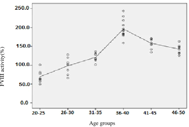
Figure 1 shows the level of FVIII activity in
different age groups of each ABO group. The
correlations between FVIII activity and age
were checked using regression analysis. Our
results revealed a linear and positive correlation
(r=0.9, p<0.001) between FVIII activity and age
up to 35-40 years; however, a negative
correlation was observed (r= -0.54, p<0.05) by
increasing age (Fig. 2).
Table1. Comparison of FVIII activity among four ABO blood groups during three examination times
Blood group Activity (%) 1th time Activity (%) 2nd time Activity (%) 3rd time P-value
A 149.92±38 142.57±42 135.14±39 p<0.05
AB 128.11±46 118.96±45 109.41±41 p<0.05
B 138.10±44 131±42 128.49±41 p<0.05
O 123.26 ±34 115±40 107.86±42 p<0.05
p-value p<0.001 p<0.001 p<0.001
Values are presented as means ± SD.
Discussion
FVIII is one of the clotting factors processed at
the Blood Transfusion Center for hemophilia
A patients. Although concentrated and
lyophilized FVIII and its recombinant form
have been available for many years,
cryoprecipitate prepared from FFP have, to
date, been used in some countries such as Iran.
To optimize its production, it is essential to
know the effect of various factors such as
delay times on the FVIII activity during its
preparation. The half-life of FVIII is short and
about 8-12 hours.
Fig. 1. Comparison of FVIII activity among different age groups. BG=Blood groups
Fig. 2. Correlations between FVIII activity and age groups
Like most of the proteins, it is more stable at
low temperature and gradually loses its
activity out of the refrigerator and rapidly in
high temperature. In this study, the initial
incubation time (2 h) and temperature
(22-24°C) after blood donation and before
centrifuge of whole blood were considered
according to the routine program performed in
Age groups
F
VIII ac
ti
v
it
y
(%
)
Age groups
Age groups Age groups
Age groups
Blood Transfusion Center in Alborz province.
The results of the study showed that 90 and
180 min delay in FFP freezing significantly
decreases FVIII activity in compassion to
immediate FFP freezing. Carlebjörk et al.
reported that in plasma FVIII was stable for at
least two h at room temperature [13]. Smith et
al. showed that the percentage of FVIII
activity significantly decreases by holding
plasma at 1-6°C for 2, 8, 15 hrs, respectively,
before freezing [14]. Swärd-Nilsson et al.
found that storage at room temperature for six
h causes a small but statistically significant
decrease in FVIII. They concluded that for an
optimal yield of FVIII, freezing should start
within four h after plasma donation. Our
results demonstrated that FFP freezing should
be done within two h after plasma donation
[15].
VWF is the specific carrier of factor VIII in
plasma and protects it from proteolytic
degradation, prolonging its half-life in
circulation and efficiently localizing it at the
site of vascular injury. According to the
previous studies, there is a close relationship
between plasma levels of Von Willebrand and
factor VIII [16]. It is a large multimeric
glycoprotein that its plasma levels differ
among people. The variability in its plasma
levels depends on some factors such as age,
race, ABO and Lewis blood groups,
epinephrine, inflammatory mediators, and
endocrine hormones [16, 17]. Some studies
reported that group O and group AB subjects
have the lowest and the highest plasma Von
Willebrand levels, respectively [17, 18]. Other
studies, based on genotype found that
genotype OO individuals have the lowest
plasma VWF levels and heterozygous
individuals for the O allele (genotypes AO,
BO) possess significantly lower plasma VWF
levels than those not carrying an O allele
(genotypes AA, AB, BB) [16, 19]. According
to our results, the order of increase in FVIII
activity was from blood group O with the
lowest, then AB, B and with highest in blood
group A. Song et al. found FVIII activity as
the lowest in subjects with blood group O and
the highest in those with either B or AB. They
described the variations in results likely due to
intrinsic genetic variability and environmental
factors [20]. Also, the results of FVIII activity
and its relationship with blood group partially
accord with the results of VWF. As Smith et
al. suggested, the transport and chaperoning
function of VWF for FVIII may be responsible
for the association between ABO and FVIII
activity [21].
In this study, the relationship between FVIII
activity and age groups was also evaluated
among the blood groups. Wang et al. reported
that FVIII and VWF levels show significant
and positive relationships with age [22].
Cohen et al. revealed a linear increase in VWF
and FVIII with increasing age [23]. Our results
identified that FVIII activity increased along
with increasing age up to 35-40, but it
decreases in subjects of 40-50 years old. The
reduction in FVIII activity was not statistically
important in the subjects who were over 40
years old. Since the age defined for blood
donation is up to 50 years, the changes in
FVIII were evaluated up to middle age. Little
is known about the mechanisms that control
the changes in VWF and FVIII levels
with age. Aging is characterized by the
accumulation of damage and other harmful
changes, leading to decrease in some protein
activity, although an increase in their gene
expression may be observed [24]. In spite of
these interpretations, the evaluation of such
changes in FVIII activity with increase in age
need more molecular and biochemical studies.
Conclusion
Given that FVIII is one of unstable blood
clotting factors and as a quality index for FFP
and cryoprecipitate, it is necessary to optimize
FFP production conditions to prevent a decrease
in FVIII activity. According to the results of
this study, the best time of plasma freezing is
immediately after FFP production, and the best
blood group and age for FVIII product are
blood group A and the age group 35-40.
Conflict of Interest
The authors have no conflict of interest.
Acknowledgments
This work was supported by Alborz Blood Transfusion Organization. Thanks to the members of Blood donation section of Karaj for their contribution in improving this study.
Reference
[1]. Wakabayashi H, Freas J, Zhou Q, Fay PJ. Residues 110–126 in the A1 domain of factor VIII contain a Ca2+ binding site required for cofactor activity. J Biol Chem. 2004; 279(13): 12677-2684.
[2]. Fay PJ. Activation of factor VIII and mechanisms of cofactor action. Blood Rev. 2004; 18(1): 1-15.
[3]. Lusher JM, Milestones in hemophilia and concepts in future clinical trial design. Semin Hematol. 2006; 43(1): 83-7.
[4]. Fijnvandraat K, Peters M, Ten Cate JW.
Inter‐individual variation in half‐life of infused
recombinant factor VIII is related to pre‐infusion
von Willebrand factor antigen levels. Br J Haematol. 1995; 91(2): 474-76.
[5]. Franchini M, Mannucci PM. Past, present and future of hemophilia: a narrative review. Orphanet J Rare Dis. 2012; 7(1): 24-29.
[6]. Lynch CM, Israel DI, Kaufman RJ, Miller AD. Sequences in the coding region of clotting factor VIII act as dominant inhibitors of RNA accumulation and protein production. Human Gene Therapy 1993; 4(3): 259-72.
[7]. Lusher JM, Arkin S, Abildgaard CF, Schwartz RS. Recombinant factor VIII for the treatment of previously untreated patients with hemophilia A-safety, efficacy, and development of inhibitors. New Eng J Med. 1993; 328(7): 453-59.
[8]. Dasgupta S, Repessé Y, Bayry J, Navarrete AM, Wootla B, Delignat S, et al. VWF protects FVIII from endocytosis by dendritic cells and subsequent presentation to immune effectors.
Blood 2007; 109(2): 610-12.
[9]. Scharrer I, Bray G, Neutzling O. Incidence of inhibitors in haemophilia A patientsa review of recent studies of recombinant and plasma-derived factor VIII concentrates. Haemophilia 1999; 5: 145-54.
[10]. Mannucci PM, Chediak J, Hanna W, Byrnes J, Ledford M, Ewenstein BM, et al. Treatment of von Willebrand disease with a high-purity factor VIII/von Willebrand factor concentrate: a prospective, multicenter study. Blood 2002; 99(2): 450-56.
[11]. Pool JG, Shannon AE. Production of high-potency concentrates of antihemophilic globulin in a closed-bag system: assay in vitro and in vivo. New Eng J Med. 1965; 273(27): 1443-447. [12]. Rodgers SE, Duncan EM, Barbulescu DM,
Quinn DM, Lloyd JV. In vitro kinetics of factor VIII activity in patients with mild haemophilia
A and a discrepancy between one‐stage and
two‐stage factor VIII assay results. Br J
Haematol. 2007; 136(1): 138-45.
[13]. Carlebjörk G, Oswaldsson U, Rosén S. A simple and accurate microplate assay for the determination of factor VIII activity. Thrombos Res. 1987; 47(1): 5-14.
[14]. Smith JF, Ness PM, Moroff G, Luban NL. Retention of coagulation factors in plasma frozen after extended holding at 1-6 C. Vox Sanguinis. 2000; 78(1): 28-30.
[15]. Swärd‐Nilsson AM, Persson PO, Johnson U,
Lethagen S. Factors influencing factor VIII activity in frozen plasma. Vox sanguinis 2006;
90(1): 33-9.
[16]. Franchini M, Capra F, Targher G, Montagnana M, Lippi G. Relationship between ABO blood group and von Willebrand factor levels: from biology to clinical implications. Thrombos J. 2007; 5(1): 14.
[17]. Gallinaro L, Cattini MG, Sztukowska M, Padrini R, Sartorello F, Pontara E, et al. A shorter von Willebrand factor survival in O blood group subjects explains how ABO determinants influence plasma von Willebrand factor. Blood 2008; 111(7): 3540-545.
[18]. Gill JC, Endres-Brooks J, Bauer PJ, Marks WJ, Montgomery RR. The effect of ABO blood group on the diagnosis of von Willebrand disease. Blood 1987; 69(6): 1691-695.
[19]. O'donnell J, Laffan M. The relationship
between ABO histo‐blood group, factor VIII and
von Willebrand factor. Transf Med. 2001; 11(4): 343-51.
[20]. Song J, Chen F, Campos M, Bolgiano D, Houck K, Chambless LE, et al. Quantitative influence of abo blood groups on factor viii and its ratio to von willebrand factor, novel
observations from an aric study of 11,673 subjects. PloS one 2015; 10(8): e0132626. [21]. Smith NL, Chen MH, Dehghan A, Strachan
DP, Basu S, Soranzo N, et al. Novel associations of multiple genetic loci with plasma levels of factor VII, factor VIII, and von Willebrand factor. Circulation 2010; 121(12): 1382-392. [22]. Wang Z, Dou M, Du X, Ma L, Sun P, Cao H,
et al. Influences of ABO blood group, age and gender on plasma coagulation factor VIII, fibrinogen, von Willebrand factor and Adamts13 levels in a Chinese population. Peer J. 2017; 5: 3156.
[23]. Cohen W, Castelli C, Alessi MC, Aillaud MF, Bouvet S, Saut N, et al. ABO blood group and von Willebrand factor levels partially explained the incomplete penetrance of congenital thrombophilia. Arterioscleros, Thrombos, Vascul Biol. 2012; 32(8): 2021-2028.
[24]. Gonskikh Y, Polacek N. Alterations of the translation apparatus during aging and stress response. Mech Ageing Dev. 2017; 168(5): 30-6.