R E S E A R C H A R T I C L E
Open Access
Telomere protein Rap1 is a charge resistant
scaffolding protein in chromosomal
bouquet formation
Hanna Amelina, Shaan Subramaniam, Vera Moiseeva, Christine Anne Armstrong, Siân Rosanna Pearson
and Kazunori Tomita
*Abstract
Background:Chromosomes reorganize in early meiotic prophase to form the so-called telomere bouquet. In fission
yeast, telomeres localize to the nuclear periphery via interaction of the telomeric protein Rap1 with the membrane protein Bqt4. During meiotic prophase, the meiotic proteins Bqt1-2 bind Rap1 and tether to the spindle pole body to form the bouquet. Although it is known that this polarized chromosomal arrangement plays a crucial role in meiotic progression, the molecular mechanisms of telomere bouquet regulation are poorly understood.
Results:Here, we detected high levels of Rap1 phospho-modification throughout meiotic prophase, and identified
a maximum of 35 phosphorylation sites. Concomitant phosphomimetic mutation of the modification sites suggests that Rap1 hyper-phosphorylation does not directly regulate telomere bouquet formation or dissociation. Despite the negative charge conferred by its highly phosphorylated state, Rap1 maintains interactions with its binding partners. Interestingly, mutations that change the charge of negatively charged residues within the Bqt1-2 binding site of Rap1 abolished the affinity to the Bqt1-2 complex, suggesting that the intrinsic negative charge of Rap1 is crucial for telomere bouquet formation.
Conclusions:Whereas Rap1 hyper-phosphorylation observed in meiotic prophase does not have an apparent role
in bouquet formation, the intrinsic negative charge of Rap1 is important for forming interactions with its binding partners. Thus, Rap1 is able to retain bouquet formation under heavily phosphorylated status.
Keywords:Telomere, Shelterin complex,Schizosaccharomyces pombe,Phosphorylation, Meiosis
Background
Telomeres are specialized nucleoprotein structures that form the natural ends of linear chromosomes. While telomeres are mostly known for their essential function in chromosome maintenance, they also play an important role in meiotic progression [1]. During meiotic prophase, the position of chromosomes within the nucleus is dra-matically reorganized and telomeres cluster within a lim-ited area of the nuclear envelope to form the so-called telomere bouquet [2, 3]. This conserved reorganization of chromosomes during meiotic prophase has been observed in most eukaryotic organisms and is shown to promote homolog pairing and meiotic recombination [4]. Recent
studies suggest that the bouquet also plays a crucial role in meiotic spindle formation [5].
In fission yeast Schizosaccharomyces pombe, the telo-mere bouquet is observed throughout meiotic prophase. This period is also known as the‘horsetail nucleus’stage, during which the nucleus elongates and oscillates back and forth between the cell poles, following the spindle pole body or SPB (the yeast equivalent of the centro-some) driven by cytoplasmic microtubules [6]. Bouquet formation is achieved by expression of a pair of meiosis-specific proteins, Bqt1 and Bqt2, which bridge the telo-meric proteins Taz1 and Rap1 to the SPB component Sad1 [7]. To ensure telomere attachment to the SPB, telo-meres must be tethered to the nuclear envelope via the interaction between Rap1 and the inner nuclear mem-brane complex Bqt3 and Bqt4 [8]. Disruption of any of * Correspondence:k.tomita@ucl.ac.uk
Chromosome Maintenance Group, UCL Cancer Institute, University College London, London WC1E 6DD, UK
these components leads to failure of telomere clustering and defective spore formation in meiosis [7–11]. Sporula-tion defects in the bouquet mutants occur mainly due to impaired spindle formation and partly due to compro-mised meiotic centromere assembly, followed by chromo-some segregation defects [5, 12]. Recent studies suggest that recruitment of a single telomere tract or centromere to the SPB is sufficient to confer functional spindles [13, 14]. Hence, the telomere bouquet does not only func-tion in alignment of chromosomes but is also crucial for the recruitment of a chromosome to the SPB to create a functional meiotic spindle.
Although the bouquet composition and its function are becoming better understood, the molecular mecha-nisms that govern dissociation of telomeres from the SPB remain elusive. Interestingly, in fission yeast, polar-ized bouquet configuration is maintained until the end of meiotic prophase, and upon entry into the first mei-otic division, telomeres dissociate from the SPB in a con-certed manner, dubbed‘telomere fireworks’[5]. Another interesting observation is that Bqt1 and Bqt2 do not localize to telomeres at the onset of the first meiotic div-ision [7]. Moreover, Rap1 is highly phosphorylated in mi-totic cells [15]. We therefore hypothesized that disruption of the interaction between Rap1 and the Bqt1-2 complex, potentially by means of post-translational modifications of one or both interacting partners, may be responsible for telomere dissociation from the SPB. In this report, we in-vestigated whether Rap1 is involved in the termination of the telomere bouquet. Comprehensive phosphoproteomic analysis of the meiotic Rap1 protein revealed that it is pro-gressively phosphorylated throughout meiotic prophase and the number of phosphosites peaks after completion of the bouquet stage. Surprisingly, this massive phosphoryl-ation of Rap1 is dispensable for telomere bouquet dissoci-ation, as indicated by our live microscopy analysis and protein interaction studies of the phospho-mutants. Our study illuminates that Rap1 is a protein resistant to nega-tive charge and functions as a ‘scaffolding’ protein in the telomere bouquet.
Results
Rap1 is hyper-phosphorylated in meiosis
To investigate the stability of the Rap1 protein through-out meiosis, a homozygous diploid (h−/h−)
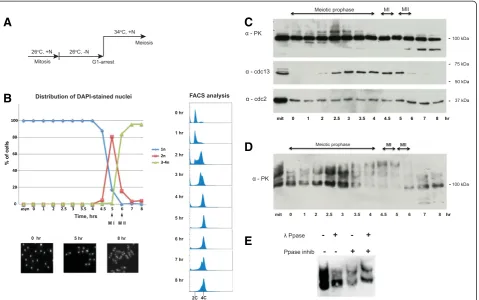
temperature-sensitive pat1-114 strain carrying a mat-Pc cassette was utilized to synchronize meiosis [16, 17]. Meiosis was induced after nitrogen starvation, followed by a temperature shift from permissive (26°C) to restrictive conditions (34°C) (Fig. 1a). Progression of meiosis was monitored by assessing the number of nuclei and DNA content per cell from fractions collected at 30-minute or 1-hour intervals during the synchronization proced-ure (Fig. 1b). In order to assess Rap1 protein stability
during meiosis, Rap1 was endogenously tagged with PK (V5) epitope peptide and detected by V5 anti-bodies. Western blotting analysis of synchronous cul-ture extracts showed that Rap1 protein is rather stably expressed during meiosis, although lower molecular weight, potentially truncated forms of Rap1, were ob-served at the end of meiosis (Fig. 1c, top panel).
Interestingly, a number of distinctly shifted bands of Rap1 were detected during meiotic prophase. Similar shifted bands of Rap1 have also been recently reported [18]. To determine if Rap1 is phosphorylated during meiosis, cell extracts were further analyzed using Phos-tag™SDS-PAGE [19]. Phos-tag™gel analysis revealed that the Rap1 protein is highly phosphorylated during mei-osis. Strikingly, the maximum level of phosphorylation was observed at 4.5–5 hr, when almost none of the fast-migrating forms of Rap1 were detected (Fig. 1d). Phos-phatase treatment confirmed that the shifted bands ob-served at 4.5–5 hr represented phosphorylated forms of Rap1 (Fig. 1e). Thus, our data indicates that Rap1 phos-phorylation accumulates as meiotic prophase progresses, and Rap1 becomes hyper-phosphorylated at the onset of meiosis I, when the bouquet stage ends [5].
Mass spectrometry analysis of Rap1 reveals an increasing number of phosphosites detected upon completion of the bouquet stage
Hyper-phosphorylation of Rap1 in meiosis is dispensable for telomere bouquet formation and dissociation
Since we observed that Rap1 phosphorylation peaks at meiosis I, we speculated that the resulting highly nega-tive charge of Rap1 is responsible for the change in its affinity to the Bqt1-2 complex. In order to mimic hyper-phosphorylated Rap1, all validated phosphorylation sites from S-212 to S-562 were substituted with negatively charged glutamate residues (rap1-32E) (Fig. 3a). To moni-tor telomeres and the SPB through meiosis, endogenous Taz1 and Sid4 were tagged with YFP and mCherry, respect-ively. To our surprise, the phosphomimeticrap1-32E mu-tants did not exhibit any detectable meiotic defects and their telomeres clustered and dissociated from the SPB in a timely manner very similar to that of the wild-type (Fig. 3b). Accordingly, rap1-32E mutants exhibited no sporulation defects (Fig. 3c). The corresponding non-phosphorylatable mutant form of Rap1 (rap1-32A) also did not cause defects in meiotic progression and telomere bouquet behaviour (Fig. 3a,b,c). Western blot analysis from meiotic cell
extracts confirmed that the mutant forms of Rap1 were stably expressed, and the phospho-modification of Rap1-32A was significantly reduced (Additional file 2). Finally, our yeast two-hybrid assay confirmed that the Bqt1/2 bind-ing domain of Rap1 falls within 216–388 amino acids, and introduced cluster mutations did not affect its interaction with the Bqt1-2 complex (Fig. 3d).
Suspecting that some phospho-modifications might remain unidentified in our study, five additional serine and threonine residues (S-317, T-321, S-322, T-328 and S-364), along with 12 detected phosphosites within and adjacent to the Bqt1/2 binding domain, were all substituted to glutamate (rap1-17E) or alanine (rap1-17A) (Fig. 3a). However, these mutations also did not cause any defects in meiosis (Fig. 3b,c). Thus, we conclude that accumulation of negative charge at the Bqt1-2 binding domain of Rap1 does not affect its ability to form the bouquet.
Becauserap1-32Aandrap1-32Ebear mutations within the binding domain of the telomerase negative regulator Poz1 [21], we checked whether telomere length regulation
C
A
E
D
B
Fig. 1Rap1 is hyper-phosphorylated in meiosis.aSchematic diagram of meiotic culture synchronization using homozygous diploid cells carrying the temperature-sensitivepat1-114mutation and themat-Pccassette.bDistribution graph of the number of nuclei in meiocytes through meiosis (top left) and images of DAPI-stained cells from indicated fractions (bottom left). FACS analysis shows DNA duplication from 2C to 4C (right).
was impaired in the rap1 phospho-mutants. Since C-terminus tagging of Rap1 impaired telomere length homeostasis (Fig. 3e), the PK epitope tag was fused to the N-terminus. Although phosphomimetic forms of the Rap1 protein (Rap1-32E and 17E) migrate slower than wild-type Rap1, none of the cluster mutations affected protein stability (Fig. 3f). The strains carrying mutant Rap1 main-tained their telomere length comparable to that of wild-type (Fig. 3e). Accordingly, all mutants retained their ability to interact with Poz1 by the yeast two-hybrid assay (Fig. 3g). Additionally, both32Aand32Emutant forms of Rap1 retained the ability to interact with Bqt4 (Fig. 3h). Indeed, telomere localization to the nuclear periphery in interphase was not impaired in rap1-32A and 32E mu-tants (Additional file 3). Thus, hyper-phosphorylation of Rap1 observed in meiosis does not appear to have a role in telomere bouquet regulation. Furthermore, our muta-genesis analysis suggests that Rap1 is able to resist high negative charge changes without affecting its function in meiosis or telomere length homeostasis.
Intrinsic negative charge of Bqt1/2 binding domain of Rap1 is crucial for telomere bouquet formation
Rap1 protein is negatively charged, and the Bqt1/2 binding region is particularly rich in hydrophobic and negatively charged amino acid residues. Some of these negatively charged residues (D-335, D-337, D-338 and E-342) are well-conserved among fission yeast species (Fig. 4a). Importantly, mutation analysis indicated that Rap1-DD337AA (D337A and D338A mutations) no longer interacts with the Bqt1-2 complex, but retains
its ability to interact with Bqt4 and Poz1 in yeast two-hybrid assay (Fig. 3d,g,h).
To study the function of Rap1-DD337AA, endogenous rap1 was mutated and fused to YFP. Accordingly, rap1-DD337AAmutants were defective in sporulation (Figs. 3c and 4b). Live cell imaging of the mutant showed that Rap1-DD337AA localized to telomeres, as determined by co-localization to Taz1 (Fig. 4c), but did not cluster at the SPB in meiotic prophase (Fig. 4d). Furthermore, in many cases the SPB was destabilized and detached from the nucleus and, as a consequence, aberrant chromo-some segregation was observed (Fig. 4d). These meiotic phenotypes are characteristic of rap1Δ mutants as well as the bouquet-defective mutants [5]. However, Rap1-DD337AA was stably expressed and telomere length of
the rap1-DD337AA mutant was the same as that of
wild-type (Fig. 3e,f). Additionally, telomeres of the mutant cells were retained at the nuclear periphery in interphase (Additional file 3). Thus, rap1-DD337AA is a meiosis-specific loss-of-function mutation, and negatively charged aspartates at positions 337 and 338 are crucial for bouquet formation.
Discussion
In this study we have shown that the level of phosphor-ylation of Rap1 gradually increases during the course of meiotic prophase, peaking in meiosis I. The SPB is known to recruit a number of kinases and phosphatases that modify its subunits, and these modifications play a critical role in regulating mitotic commitment and meiotic progression [22–25]. Since telomere heterochromatin comes into close contact with the meiotic SPB during
chromosomal bouquet configuration, the telomere-bound proteins are likely to be exposed to these kinases. Additionally, DNA damage checkpoint kinases are acti-vated during meiotic recombination [26]. In fact, some phosphorylations originate from telomere-associated ki-nases as indicated by reduced shifted bands of Rap1 at 4–4.5 hr in the absence of Taz1 (Additional file 4). Never-theless, our study suggests that the hyper-phosphorylation of Rap1 observed during meiosis is not directly involved in the regulation of the bouquet. Rap1 can withstand sig-nificant charge changes that do not affect interactions with its binding partners and its function in meiosis. Hence, rather than a functional regulatory protein, meiotic Rap1 appears to be a‘scaffolding’protein, that is targeted by multiple kinases. Interestingly, while mutating 32
phosphorylation sites did not alter Rap1 function, muta-tion of only two highly conserved residues (D-337 and D-338) disrupted its ability to bind to the Bqt1-2 complex, causing pronounced defects in telomere clustering and chromosome segregation. In contrast, the binding part-ners Bqt1 and Bqt2 are positively charged, suggesting that Rap1 binds to the Bqt1-2 complex through hydrogen bonding interactions. Thus, we speculate that negatively charged residues at Rap1’s interaction surfaces are evolu-tionarily conserved in order to retain affinity under shifts in charge occurring throughout meiotic prophase.
Rap1 is also highly phosphorylated in the mitotic cell cycle, particularly in M-phase. Among the ation sites reported for mitotic Rap1, five phosphoryl-ation sites (S-213, T-378, S-422, S-456 and S-513) were
B
C
A
G
H
D
F
E
shown to have an inhibitory effect on Rap1-Bqt4 inter-action, which was demonstrated by the phosphomimetic rap1-5D/5E mutants [15]. In mitosis, Cdc2 phosphory-lates three of these sites in order to temporarily release telomeres from the nuclear envelope. This mechanism assists faithful chromosome segregation in anaphase. However,rap1-5Dandrap1-5Emutants do not have any sporulation defects [15], which suggests that the bouquet is intact. This is surprising since the Rap1-Bqt4 inter-action is required for telomere clustering in meiosis [8],
and raises the possibility that telomeres remain associ-ated with Bqt4 and the SPB via different mechanisms. Notably, four residues of Rap1 including Cdc2 targets were also found to be phosphorylated throughout mei-otic prophase in our study (except for S-456, detected only at 4.5 hr) (Additional file 1). Unlike the Rap1-5E mutant protein, our phosphomimetic cluster mutant Rap1-32E, which includes 5E mutation (Fig. 3a), was able to interact with Bqt4. Thus, we predict that pre-meiotic hyper-phosphorylation of Rap1 (or particular
A
B
C
D
phosphorylations among them) counteracts Cdc2 kinase action to preserve affinity to Bqt4, and thereby maintain telomere localization to the nuclear membrane and bou-quet configuration.
Conclusions
Rap1 hyper-phosphorylation observed during meiotic prophase does not have a direct role in telomere bou-quet regulation. Rap1 uses its negatively charged amino acid residues to bind the Bqt1-2 complex. Therefore the interaction is not affected by changes in net charge caused through progressive hyper-phosphorylation.
Methods
Yeast genetics and plasmids
The genotypes of the strains used for this study are listed in Additional file 5. All media and supplements were pur-chased from Formedium™(Hunstanton, UK). Fission yeast was grown at 32°C in standard rich media (YES) unless in-dicated. Epitope tag insertion at the C-terminus was de-scribed previously [27]. A plasmid for N-taggedrap1was constructed by cloning the rap1 gene, including 1,360 bases of the upstream and 1,500 bases of the downstream regions, with a primer set including aKpnI site. The start codon of rap1+ was replaced with a single V5 (PK) se-quence. A kanMX6 cassette was inserted 700 bases up-stream of the gene. The resulting plasmid pRap1a-nPK was digested withKpnI and replaced theura4+cassette at the rap1 gene locus in rap1Δ cells. The transformants were backcrossed with a wild-type strain and cultured for 2 weeks before analysis of telomere length. To generate rap1mutants, cluster-mutatedrap1gBlocks were synthe-sized (Integrated DNA Technologies, Coralville, IA, USA) and replaced wild-typerap1+in the pRap1a-nPK plasmid and the yeast two-hybrid pGAD and pGBK plasmids.
Yeast two-hybrid assays
The assay was conducted according to the Matchmaker Gold Yeast Two-Hybrid System manual (Clontech, Mountain View, CA, USA). Expression vectors for the GAL binding domain (BD) fused proteins and the GAL activation domain (AD) fused proteins were generated by subcloning of the indicated cDNAs into pGBK and pGAD, respectively. Expression of BD and AD fused proteins were confirmed by Western blotting using anti-myc and anti-HA antibodies, respectively. To express Bqt1 and Bqt2 together in the yeast two- hybrid plas-mids,bqt2+cDNA was inserted at the start codon of the bqt1+
cDNA sequence; the resulting plasmid was named pGBK-Bqt2-1.
pat1-114 synchronization
Cells were first cultured in YE media at 26°C overnight until late stationery phase, then transferred to EMM
media containing a nitrogen source and incubated for 24 hr until mid-log phase (OD = 0.5–0.7). The cells were next transferred to EMM media without a nitrogen source (EMM-N) by filtering, and cells were incubated for another 15–16 hr to arrest cells at G1 phase. To in-activate the pat1 kinase gene and induce meiosis, the temperature was shifted up to 34°C, cultures were sup-plemented with one-fifth volume of EMM media pre-warmed to 34°C, and meiotic fractions were collected at the required time point.
Western blotting and Phos-tag gel
Whole-cell protein extracts prepared using a trichloroacetic acid method were separated by SDS-PAGE using 10% acrylamide gels. Western blotting was performed with anti-V5 peptide (Bio-Rad, Hercules, CA, USA), anti-Cdc2 (Santa Cruz Biotechnology, Dallas, TX, USA) and anti-Cdc13 (Santa Cruz Biotechnology) antibodies following a stand-ard protocol. For detection of phosphorylated Rap1 forms, 7.5% acrylamide gels were supplemented with 25 μM PhosTag ligand (AAL-107; NARD Institute, Amagasaki, Japan) and 50μM MnCl2, according to the protocol [19].
A Phos-tag gel was treated with 1 mM EDTA prior to transfer. Two technical replicates of the Western blotting image in Fig. 1c are shown in Additional file 6.
Telomere Southern blotting
Southern blotting was performed as described previously [27]. Equal amounts of EcoRI-digested DNA fragments were separated on a 1% agarose gel and subjected to Southern blotting with a telomere probe.
Preparation of cell extracts and immunoprecipitation for mass spectrometry
Six litres of synchronous meiotic cells expressing Rap1-3xPK were harvested at 3.5 hr (early prophase) or 4.5 hr (late prophase) following the pat1-114 synchronization protocol as described above. Rap1-3xPK was immuno-precipitated from native cell extracts in RIPA buffer (50 mM Tris–HCl, pH 8; 150 mM NaCl; 1% NP-40; 0.5% sodium deoxycholate, and 0.1% SDS), supple-mented with 1X PhosSTOP (Roche, Basel, Switzerland); 1× cOmplete EDTA-free protease inhibitor (Roche); 1 mM PMSF, 1 mM DTT, 0.1 ng/mL MG132 (Sigma-Aldrich, St Louis, MO); and 10 U/mL TURBO DNase (Ambion, Life Technologies, Carlsbad, CA, USA), using Dynabeads M-270 epoxy (Life Technologies) pre-coated with anti-V5 peptide antibodies.
Phosphatase treatment of native cell extracts
Native whole-cell extracts were prepared using modi-fied HB buffer (50 mM HEPES-KOH, pH 7.5; 140 mM NaCl, 0.1% NP-40; 1 mM MnCl2). Then 20μl of lysate
phosphatase inhibitors; both phosphatase and inhibi-tors; or water as a control. The samples were incubated at 30°C for 1 hour before being separated on SDS-PAGE gel.
Further details about microscopy and mass spectrom-etry methods can be found in Additional file 7. A list of phosphopeptides detected by mass spectrometry is shown in Additional file 8.
Open access of data
The mass spectrometry proteomics data have been deposited to the ProteomeXchange consortium [1] via the PRIDE partner repository with the dataset identifier PXD001841.
Additional files
Additional file 1:List of Rap1 phosphorylation sites identified in the study.
Additional file 2:Western blot images of TCA-extracted Rap1 proteins from meiotic cells. Indicated cells were induced to undergo meiosis using thepat1-114mutation, and were collected from the 4.5 hr fraction.Cell extracts were separated in regular gradient (left) and Phos-tag SDS-PAGE (right), and subjected to Western blot using anti-PK(V5) antibody. Unlike wild-type meiotic Rap1 that shows multiple bands, Rap1-32A exhibits a single band in the Phos-tag gel, suggesting the phospho-modifications were diminished in this mutant form.
Additional file 3:Localization of telomeres in mitotic interphase.(A) Projected images of cells in mitotic interphase. Taz1-YFP, Sid4-mCherry and Bqt3-Cerulean were used to visualize telomeres, spindle pole body and the membrane, respectively. White and grey arrows indicate examples of telomeres localized at the nuclear periphery and away from the nuclear envelope, respectively. Scale bar equals 5μm. (B) Percentages of telomeres localized at the nuclear periphery in interphase cells. Cells containing one nucleus and one Sid4-mCherry focus (not dissociated) were selected as mitotic interphase cells. Telomeres were scored as localized at the nuclear periphery when the distance between the brightest Taz1-YFP focus and Bqt3-Cerulean-labeled nuclear membrane was less than 0.4μm using SoftWoRks (Applied Precision; GE Healthcare, Chalfont St Giles, UK). (C) Middle z-section image of the cells in mitotic interphase. Example of telomeres detached from the nuclear envelope (left) and telomeres localized at the nuclear periphery (right). Scale bar equals 5μm.
Additional file 4:Reduced phosphorylation of Rap1 intaz1Δ. Separation of phosphorylated Rap1-3xPK on a Phos-tag gel fromtaz1Δ synchronized meiotic cell fractions at indicated time. Anti-Cdc2 (CDK) and anti-Cdc13 (Cyclin B) antibodies were used as a loading control and meiosis synchronicity marker, respectively. Note that Taz1 is required for Rap1 localization at telomeres.
Additional file 5:Fission yeast strain list.
Additional file 6:Technical triplicates of Phos-tag gel related to Fig. 1d.
Additional file 7:Mass spectrometry and microscopy methods. Additional file 8:Mass spectrometry data for identified phosphopeptides.
Abbreviations
AD:Activation domain; BD: Binding domain; SPB: Spindle pole body.
Competing interests
The authors declare that they have no competing interests.
’
HA carried out most of the experiments. HA and KT designed the study and wrote manuscript. SS performed yeast two-hybrid analysis. SRP assisted with yeast two-hybrid assays. VM assisted with live cell imaging. CAA supported yeast genetics analysis. HA, KT, SS and VM generated strains and plasmids. All authors read and approved the final manuscript.
Acknowledgments
We thank the Proteomics and Metabolomics Core Facility at the Institute for Cancer Research (ICR) for mass spectrometry analysis of the samples and technical advice on the sample preparation. We thank Junko Kanoh (Osaka University, Japan) for critical reading before submission. This work is supported mainly by the European Research Council (281722-HRMCB) and partly by Cancer Research UK (C36439/A12097) and the Cancer Research UK - UCL Centre.
Received: 5 March 2015 Accepted: 4 June 2015
References
1. Jain D, Cooper JP. Telomeric strategies: means to an end. Annu Rev Genet. 2010;44:243–69.
2. Harper L, Golubovskaya I, Cande WZ. A bouquet of chromosomes. J Cell Sci. 2004;117:4025–32.
3. Scherthan H. A bouquet makes ends meet. Nat Rev Mol Cell Biol. 2001;2:621–7.
4. Niwa O, Shimanuki M, Miki F. Telomere-led bouquet formation facilitates homologous chromosome pairing and restricts ectopic interaction in fission yeast meiosis. EMBO J. 2000;19:3831–40.
5. Tomita K, Cooper JP. The telomere bouquet controls the meiotic spindle. Cell. 2007;130:113–26.
6. Chikashige Y, Ding DQ, Funabiki H, Haraguchi T, Mashiko S, Yanagida M, et al. Telomere-led premeiotic chromosome movement in fission yeast. Science. 1994;264:270–3.
7. Chikashige Y, Tsutsumi C, Yamane M, Okamasa K, Haraguchi T, Hiraoka Y. Meiotic proteins bqt1 and bqt2 tether telomeres to form the bouquet arrangement of chromosomes. Cell. 2006;125:59–69.
8. Chikashige Y, Yamane M, Okamasa K, Tsutsumi C, Kojidani T, Sato M, et al. Membrane proteins Bqt3 and−4 anchor telomeres to the nuclear envelope to ensure chromosomal bouquet formation. J Cell Biol. 2009;187:413–27. 9. Chikashige Y, Hiraoka Y. Telomere binding of the Rap1 protein is required
for meiosis in fission yeast. Curr Biol. 2001;11:1618–23.
10. Cooper JP, Watanabe Y, Nurse P. Fission yeast Taz1 protein is required for meiotic telomere clustering and recombination. Nature. 1998;392:828–31. 11. Kanoh J, Ishikawa F. spRap1 and spRif1, recruited to telomeres by Taz1, are
essential for telomere function in fission yeast. Curr Biol. 2001;11:1624–30. 12. Klutstein M, Fennell A, Fernandez-Alvarez A, Cooper JP. The telomere bouquet
regulates meiotic centromere assembly. Nat Cell Biol. 2015;17:458–69. 13. Fennell A, Fernandez-Alvarez A, Tomita K, Cooper JP. Telomeres and
centromeres have interchangeable roles in promoting meiotic spindle formation. J Cell Biol. 2015;208:415–28.
14. Tomita K, Bez C, Fennell A, Cooper JP. A single internal telomere tract ensures meiotic spindle formation. EMBO Rep. 2013;14:252–60. 15. Fujita I, Nishihara Y, Tanaka M, Tsujii H, Chikashige Y, Watanabe Y, et al.
Telomere-nuclear envelope dissociation promoted by Rap1 phosphorylation ensures faithful chromosome segregation. Curr Biol. 2012;22:1932–7. 16. Chikashige Y, Kurokawa R, Haraguchi T, Hiraoka Y. Meiosis induced by
inactivation of Pat1 kinase proceeds with aberrant nuclear positioning of centromeres in the fission yeast Schizosaccharomyces pombe. Genes Cells. 2004;9:671–84.
17. Bahler J, Schuchert P, Grimm C, Kohli J. Synchronized meiosis and recombination in fission yeast: observations with pat1-114 diploid cells. Curr Genet. 1991;19:445–51.
18. Kanoh J. Release of chromosomes from the nuclear envelope: a universal mechanism for eukaryotic mitosis? Nucleus. 2013;4:100–4.
19. Kinoshita E, Kinoshita-Kikuta E, Takiyama K, Koike T. Phosphate-binding tag, a new tool to visualize phosphorylated proteins. Mol Cell Proteomics. 2006;5:749–57.
21. Miyoshi T, Kanoh J, Saito M, Ishikawa F. Fission yeast Pot1-Tpp1 protects telomeres and regulates telomere length. Science. 2008;320:1341–4. 22. Funaya C, Samarasinghe S, Pruggnaller S, Ohta M, Connolly Y, Muller J, et al.
Transient structure associated with the spindle pole body directs meiotic microtubule reorganization in S. pombe. Curr Biol. 2012;22:562–74. 23. Kim S, Meyer R, Chuong H, Dawson DS. Dual mechanisms prevent premature chromosome segregation during meiosis. Genes Dev. 2013;27:2139–46.
24. Ohta M, Sato M, Yamamoto M. Spindle pole body components are reorganized during fission yeast meiosis. Mol Biol Cell. 2012;23:1799–811. 25. Grallert A, Boke E, Hagting A, Hodgson B, Connolly Y, Griffiths JR, et al.
A PP1-PP2A phosphatase relay controls mitotic progression. Nature. 2015;517:94–8.
26. Shimada M, Nabeshima K, Tougan T, Nojima H. The meiotic recombination checkpoint is regulated by checkpoint rad + genes in fission yeast. EMBO J. 2002;21:2807–18.
27. Armstrong CA, Pearson SR, Amelina H, Moiseeva V, Tomita K. Telomerase activation after recruitment in fission yeast. Curr Biol. 2014;24:2006–11.
Submit your next manuscript to BioMed Central and take full advantage of:
• Convenient online submission
• Thorough peer review
• No space constraints or color figure charges
• Immediate publication on acceptance
• Inclusion in PubMed, CAS, Scopus and Google Scholar
• Research which is freely available for redistribution