www.ijper.org
Application of 3D QSAR and Docking Studies in
Optimization of Perylene diimides as Anti Cancer
Agent
Catna Nagaraj Hemalatha1, Vijey Aanandhi Muthkumar*2
1Department of Pharmaceutical Chemistry, School of Pharmaceutical Sciences, Vels Institute of Science, Technology and Advanced
Studies, VISTAS, Pallavaram, Chennai, Tamil Nadu, INDIA.
2Department of Pharmaceutical Chemistry and Analysis, School of Pharmaceutical Sciences, Vels Institute of Science, Technology and
Advanced Studies, VISTAS, Pallavaram, Chennai, Tamil Nadu, INDIA.
ABSTRACT
Introduction: Telomerase is an enzyme which binds to telomeres and increases its
length which leads to extension of lifespan of cells. These enzymes are expressed at detectable levels in cancer cells which makes an attractive target for cancer therapy. The G Quadruplex ligands which bind to telomerase with respect to duplex genomic DNA is of special importance. The Perylene di imides are selected, designed and QSAR study has been done, finally from the QSAR results’ docking has been done by G4LDB database. To compare and to narrow down the Docking results from G4LDB database, we have chosen AutoDock tool by selecting a target Telomerase protein (PDB ID: 4B18) to analyse the binding affinity of the protein with respect to the Perylene diimides. The best scored compounds will be efficient for designing new molecules as well as for the anti-cancer therapy. Material: G4LDB Database, AutoDock 4.2, Discovery Studio Visualizer 4.1. Methods: The study was to investigate and compare the results from G4LDB database with the AutoDock results for anti cancer activity of Perylene di imides. The results are visualized by Discovery Studio 4.1 Visualizer. Compound 20 and compound 48 shows best ligand interaction with the selected targets from G4LDB database. From the AutoDock results the compounds are docked with the specific Telomerase protein 4B18 and Compound 11 shows good binding energy when compared with the PIPER. Results: Compounds 11, 20 and 48 showed good biological activity also possessing best binding affinity with the target. Discussion: From the QSAR and Docking studies (G4LDB and AutoDock), 3 compounds (11, 20 and 48) showed good biological activity possessing a strong correlation coefficient, endorses that Perylene derivatives are having strong affinity with the targets. Docking has been done from the results of QSAR study, targeting Telomerase protein to study the binding affinity with the target. Conclusions: From the results, the best compounds will be efficient to inhibit telomerase enzyme and these compounds can be used to design new molecules which will be effective for anti-cancer therapy.
Key words: Perylene Derivatives, QSAR Plus, G-Quadruplex Ligand Database, Docking.
DOI: 10.5530/ijper.52.4.77 Correspondence:
Dr. M. Vijey Aanandhi Department of Pharmaceuti-cal Chemistry and Analysis, School of Pharmaceutical Sciences, Vels Institute of Science, Technology and Advanced Studies, VISTAS, Pallavaram , Chennai.Tamil Nadu, INDIA.
Phone: +919840959519 E-mail: hodpchemistry@ velsuniv.ac.in
INTRODUCTION
Telomerase enzyme and its related proteins are having high expression in most type of tumour cells and it has been touted as
most efficient and potent for cancer therapy.
The compounds which interact or inhibit
these proteins are Telomerase Inhibitors.
The Guanine rich Quadruplex DNA (G4)
Submission Date: 09-08-2017;
Revision Date: 05-12-2017;
Accepted Date: 17-05-2018
which is present in telomeric DNA stabi-lizes and affects the gene promoters, in
turn disrupts the biological process. Per -ylene di imides are a promising class of G-Quadruplex Ligands which stabilize the G-Quadruplex structure, by stacking on
study we demonstrated the structural activity relation-ship of Observed and predicted Biological activity and it
has been done in QSARPlus Module in VLifeMolecu
-lar Design Suite and performed docking study by using
G4LDB Database (Guanine Rich Ligand Database).
This QSAR module facilitates evaluation of descriptors and generates QSAR equation for predicting
biologi-cal activity of new molecules.1-2 Based upon the study,
new compounds are designed as the results shows the presence of steric descriptors in benzene ring with positive
co efficient indicated the importance of steric interac -tions, bulky group can substitute and the presence of
electrostatic descriptors with positive coefficient near to the bay region, while negative co efficient near to the
imide indicated that electro negative groups should be substitutes on imide ring and the appearance of
descrip-tors H-Bond acceptor with a positive coefficient suggest that an increased activity of the compound. These are significant in developing the novel perylene di imide derivatives.3 Further from the QSAR results, Docking
has been done in G4LDB Database, is a unique and comprehensive database which compiles dataset of 28 G-Quadruplex Complex structures/targets are included
in the database which was obtained from Protein Data Bank (PDB).4 In this study, to compare the results of Docking, we had done docking in AutoDock 4.2 to narrow down the results. From the QSAR and Dock -ing results of G4LDB 9 compounds have been chosen
for the study. The target selected for this study was a Telomerase protein (4B18:PDB ID) and by using this
as target, docked the nine compounds to get to know
the binding energy, ligand efficiency and the number of hydrogen bonds interacted with the target.5
MATERIALS AND METHODS
Molecular Docking was performed by the AutoDock 4.2 Tools. It helps to understand the binding properties in a steady state environment. In this study we used
4B18-a telomerase protein increasesthe binding affinity
for importin proteins and promotes nuclear import of
hTERT.
AutoDock calculations are performed in several steps: i)
Preparation of coordinate files using AutoDock Tools, ii) Pre-calculation of atomic affinities using Auto Grid,
iii) Docking of Ligands using AutoDock, and iv) Analysis of results using AutoDock Tools, Discovery Studio
4.1Visualizer to visualize the interaction studies.6 preparation of ligands for docking
Perylene di imide ligands were taken. The compounds/ ligands are the results of QSAR and G4LDB Database.
Accelrys Draw 4.2 was used to draw the structures and to convert 2D to 3D. All ligands are converted to PDB Format by using Discovery studio Visualizer which was used for coordinate files, which includes atomic partial charges and atom types. Torsion angles were calculated to assign the fixable and non-bonded rotations of the molecule.
Preparation of Protein (4B18)
High resolution crystallographic structures were
retrieved from RCSB Protein Data Bank (www.rcsb.org).
To understand the biological activity of a drug molecule as prototype therapeutic agent, the knowledge of binding
selectivity towards protein environment is every essential.
Docking study was performed to understand and
correlate their biological efficacy towards the selected binding domain of the protein. We used the AutoDock 4.2 MGL tools version 1.5.6 software packages for the
molecular docking experiment and Discovery Studio
Visualizer to visualize the results. Polar Hydrogen atoms were added to the protein. The deletion of the both
water molecules and the inorganic charges were done
to avoid error. Gasteiger Charge of the macromolecule was added. Ligand docking was carried out applying the
Lamarckian Genetic Algorithm (LGA) implemented
in recognize the binding site. The grid size and spacing was set to recognize the binding site. The lowest binding
energy conformers were selected out of different
conformers. Other miscellaneous parameters were
assigned to the default values obtained from AutoDock
4.2 program.
Molecular Docking Analysis was done by the output file of the docking that was generated after the study. The
binding energy, Inhibition Constant, and the number
of hydrogen bonds were considered for the analysis.
Compound 11, 20 and 48 showed potent binding inter-actions with the protein 4B18 and compared with the
standard PIPER. The visualization of the 3 best com
-pounds are visualized by Discovery Studio Visualizer
and the ligand interactions with the target, the distance between the ligand interactions are marked and the results are showed in Figure a – p for the compound
– 11, 20, 48 and PIPER. The protein 4B18 was modelled by using PROCHECK, open source server and the Ramachandra plot for the protein was studied.
The best compounds are compared with the standard
RESULTS AND CONCLUSION
QSAR study has been carried out by V Life MDS Software- QSAR Plus Module to predict and compare
the biological activity of standard and newly designed
compounds. QSAR has been done by developing variable regression methods. The compounds are
divided into training and test set compounds by manual
selection, random and sphere exclusion methods.7 The
models are developed and based on regression values; we selected eight equations to design new compounds
of perylene di imides. Out of 497 compounds, 59
compounds possess better biological activity when
compared with the standard compounds.
These 59 selected compounds from QSAR study has
been chosen for docking study. Docking has been done by G-Quadruplex Ligand Database (G4LDB).8,9 This
is an online database which was having in built tools
and performed by Open Babel 2.3.0 to predict the binding affinity with the targets. The targets (1LIH, 1NZM,
3CE5, 3SC8 and 2HRI) for the docking are selected based upon the literature survey and the selected
compounds are docked. The results are visualized by Discovery studio Visualizer 4.1 Visualizer and the
results of QSAR and G4LDB docked are tabulated
in Table 1 and 2. From the results 9 compounds are
selected and the results of these compounds are visualized
using Discovery Studio 4.1 Visualizer. To narrow down
the results, 2 best compounds are selected, and the
compounds are Compound 20 and Compound 48. The
hydrogen bond interactions and binding free energy levels, pKi values are compared with the standard
PIPER compound.10,11
From the results of G4LDB Database, the 9
com-pounds are docked with the specific protein (PDB ID: 4B18), a telomerase protein. Docking has been done by Auto Dock 4.2. Finally, from the AutoDock results,
Compound 11 shows good binding energy when
compared with the standard PIPER compound. The
study states as from the G4LDB Database 2 compounds
posses good binding affinity, and from the Auto Dock
Compound 11 possess good binding energy. Based on
the results from QSAR, G4LDB and AutoDock results compounds 11, 20 and 48 may have potent anti-cancer activity and from these compounds scaffolds we can
design schemes for synthesis. In future studies we can
use these scaffolds for experimental designing work for
in vitro as well for invivo methods.
The best potent compounds are studied to know the interactions with the nearby aminoacids and the ligand
interactions are visualized by online PROCHECK
server12,13 and the figures for the compounds are
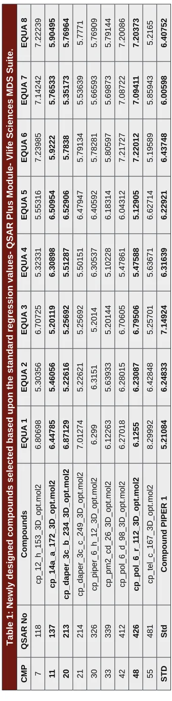
Table 1: Newly designed compounds selected based upon the standard regression values- QSAR Plus Module- Vlife Sciences MDS Suite.
CMP QSAR No Compounds EQUA 1 EQUA 2 EQUA 3 EQUA 4 EQUA 5 EQUA 6 EQUA 7 EQUA 8 7 11 8 cp_12_h_153_3D_opt.mol2 6.80698 5.30356 6.70725 5.32331 5.55316 7.23985 7.14242 7.22239 11 137 cp_14a_a_172_3D_opt.mol2 6.44785 5.46056 5.201 19 6.30898 6.50954 5.9222 5.76533 5.90495 20 213 cp_daper_3c_b_234_3D_opt.mol2 6.87129 5.22616 5.25692 5.51287 6.52906 5.7838 5.35173 5.76964 21 214 cp_daper_3c_c_249_3D_opt.mol2 7.01274 5.22621 5.25692 5.50151 6.47947 5.79134 5.53639 5.7771 30 326 cp_piper_6_h_12_3D_opt.mol2 6.299 6.3151 5.2014 6.30537 6.40592 5.78281 5.66593 5.76909 33 339 cp_pm2_cd_26_3D_opt.mol2 6.12263 5.63933 5.20144 5.10228 6.18314 5.80597 5.69873 5.79144 42 412 cp_pol_6_d_98_3D_opt.mol2 6.27018 6.28015 6.70605 5.47861 6.04312 7.21727 7.08722 7.20086 48 426 cp_pol_6_r_1 12_3D_opt.mol2 6.1255 6.23087 6.79506 5.47588 5.12905 7.22012 7.0941 1 7.20373 55 481 cp_tel_c_167_3D_opt.mol2 8.29992 6.42848 5.25701 5.63671 6.62714 5.19589 5.85943 5.2165 STD Std
Compound PIPER 1
Table 2: pKi values – G-Quadruplex Ligand Database.
S. No Compounds 1L1H 1NZM 3CE5 3SC8 2HRI
1 CMP_11 7.33 12.4 8.34 6.5 4.26
2 CMP_20 5.01 12.41 8.57 7.21 6.01
7 CMP_48 8.66 12.1 8.54 6.65 6.6
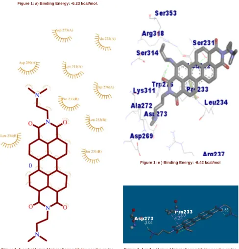
Figure 1: a) Binding Energy: -6.23 kcal/mol.
Figure 1: b and c) Ligand Interactions with the nearby amino acids.
Figure 1: d) Hydrogen Bond Acceptor and Donor Representation of Protein with the Ligand.
Figure 1: e ) Binding Energy: -6.42 kcal/mol
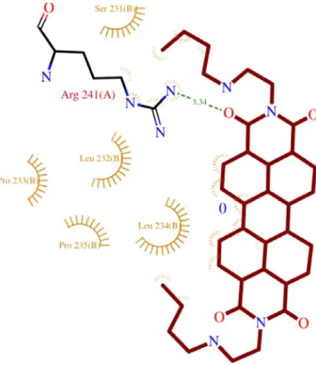
Figure 1: f and g ) Ligand Interactions with the nearby amino acids.
Figure 1: h) Hydrogen Bond Acceptor and Donor Representation of Protein with the Ligand.
Figure 1: i) Binding Energy: -6.59 kcal/mol.
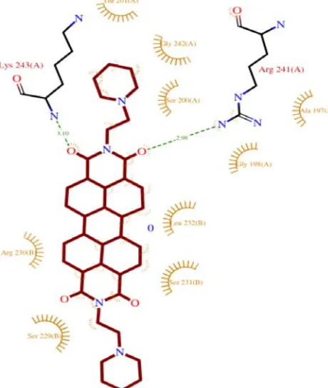
Figure 1: j) Ligand Interactions with the nearby amino acids.
Figure 1: k) Ligand Interactions with the nearby amino acids.
Figure 1: m) Binding Energy: -5.59 kcal/mol.
Figure 1: p) Hydrogen Bond Acceptor and Donor Representation of Protein with the Ligand.
Figure 1: q) Ramachandra Plot for the protein 4B18. Figure 1: n) Ligand Interactions with the nearby amino acids.
Figure 1: o) Ligand Interactions with the nearby amino acids.
shown respectively in Figure c(Compound 11), Figure g(compound 48), Figure k (Compound 48) and for
Standard PIPER, Figure o. The Ramachandra plot for the protein was shown in Figure q.
DISCUSSION
Telomerase which is responsible for maintaining the
length of the Telomere and scietific evidences proves
it exhibits in tumor cells, and we chosen G-Quadruplex
their biological activity and descriptors are studied.
From the QSAR results, we analysed as the perylene di imide derivatives are possessing the biological activity with the presenceof steric descriptors in benzene ring
with positive coefficients which indicates the importance
of steric interactions, also buly groups can substitute and the presence of electrostatic descriptors with positive
coefficeint near to the bay region, while negative coef
-ficients near to the imide ring indicates that electro
negative groups should be substitutes on imide ring and the appearance of descriptors H- bond acceptor with
a positive coefficent suggest that an increased activity of the compound. These are significant in developing the novel perylene di imide derivates. Based upon these results, we have selected 59 compounds from 491 newly designed compounds, where the selection was based on the biological activity (r2) ranging from 5 to 9 from all
the 8 equations for the synthesis.
From the results of QSAR, Docking has been done by
G4LDB Database and Autodock and the PDB ID for
G4LDB database are 1LIH, 1NZM, 3CE5, 3SC8 and
2HRI and for AutoDock 4B18 were chosen as target and the binding energy, inhibition constant values of
the 9 compounds are tabulated in Table 3. Consolidating
the results of Docking, we selected best three compounds (Compound 11, 20 and 48) which were having better
binding affinity and binding energy when compared with the standard PIPER compound. Hence these
compounds can be used as a Scaffold to design more compounds of perylene di imides as well by using these
scaffold, we can synthesize more compounds. These
compounds may possess more potent and inhibitory activity for the telomerase enzyme which tends to
apop-tosis. Molecular modelling study helps to open future de nova modelling of new compound to treat cancer.
Table 3: Molecular Docking Study of Perylene ligands with the protein 4B18 using AutoDock 4.2 Soft-ware.
S No Compound Confor mation
Binding Energy
Ligand
Efficiency
Inhibition Constant/ Units
No of Hydrogen Bonds
Hydrogen Contacts
Biological Activity
1. Compound
_7 4th -5.33 -0.12 122.99µM 1 7th:0:N38 5.87
2. Compound
_11 1st -6.23 -0.14 26.99 µM 1 4B18:A:GLN223:OE1 5.66
3. Compound
_20 2nd -6.42 -0.16 19.79 µM 1 11th:0:O18 5.59
4. Compound
_21 4th -3.16 -0.07 4.84 mM 1 4B18:B:LYS236:N 5.30
5. Compound
_30 2nd -5.75 -0.12 60.84 µM NIL 20th: 0:O35 5.35
6. Compound
_33 4th -1.62 -0.04 64.72 mM NIL 4B18:A:TRP276:HE1 5.94
7. Compound
_42 4th -2.89 -0.06 7.63 mM 1 21st: 0:O35 4.96
8. Compound
_48 10th -6.59 -0.04 67.8 mM 1 4B18:B:SER229:OG 4.84
9. Compound
_55 9th -3.15 0.07 4.94 mM 2 NIL 5.75
10. PIPER 4th -5.59 -0.12 80.55 µM 2 NIL 6.24
ACKNOWLEDGEMENT
The authors are thankful to Vels University (VISTAS)
and its management for providing research facilities and
encouragement. The author is obliged to DBT -
Government of India (BT/Bio-care/03/10047/2013-14)
for providing financial assistance to carry out the research work.
CONFLICT OF INTEREST
The authors declare that they have no competing
interests.
ABBREVIATIONS
PIPER: PIPER
[N,N’-bis-(2-(1-piperidino)ethyl)-3,4,9,10-perylene tetracarboxylic acid diimide], G4LDB:
Guanine Rich Ligand Database, QSAR: Quantitative
Structure Activity Relationship, VLife Science MDS- V Life Sciences Molecular Design Suite, PDB- Protein Data Bank, MGL- Molecular Graphics Laboratory, RCSB- Research Collaboratory for Structural Bioinformatics.
REFERENCES
1. Quantitative Structure Activity Relationship (QSAR) VLife Sciences Technologies Pvt Ltd. - VLife Product Documentation- Tutorial QSAR. - Obtaining 3D QSAR models by kNN – MFA method using VLife MDS. 2. Quantitative Structure Activity Relationship (QSAR) VLife Sciences
Technologies Pvt Ltd. - VLife Product Documentation- Tutorial
QSAR.-Obtaining conventional QSAR model by Partial Least Squares Regression Method using VLife MDS.
3. Hemalatha CN, Aanandhi MV.”G-Quadruplex Ligands as Stabilizer targeting Telomerase as Anti-Cancer Agents”. Asian Journal of Pharmaceutical and Clinical Research. 2017;10:50-3.
4. Hemalatha CN, Aanandhi MV. 3D QSAR and Docking study of Perylene-
di imides analogues as potent apoptosis inducer and efficacious anticancer
agent. Indian Drugs, Dec 2017 Issue, In Press. 2017.
5. Blackburn EH. Telomeres and telomerase: Their mechanisms of action and the effects of altering their functions. FEBS Lett. 2005;579(4):859-62. 6. Sanner MF. Python: A Programming Language for Software Integration and
Development. J Mol Graphics Mod. 1999;17(1):57-61.
7. Jain SV, Ghate M, Bhadoriya KS, Bari SB, Chaudhari A, Borse JS. 2D, 3D-QSAR and docking studies of 1, 2, 3-thiadiazole thioacetanilides analogues as potent HIV-1 non-nucleoside reverse transcriptase inhibitors. Organic and Medicinal Chemistry Letters. 2012;2(1):22. DOI: 10.1186/2191-2858-2-22.
8. Franceschin M, Rossetti L, D’Ambrosio A, Schirripa S, Bianco A, Ortaggi G, et al. Natural and synthetic G-quadruplex interactive berberine derivatives. Bioorg Med Chem Lett. 2006;16(6):1707-11.
9. Li Q, Xiang JF, Yang QF, Sun HX, Guan AJ, Tang YL. G4LDB: A database for discovering and studying G-quadruplex ligands. Nucleic Acids Research. 2012;41(D1):D1115-23.
10. Salvati E. Telomerase damage induced by the G-Quadruplex ligand RHPS4 has an anti tumor effect. Journal of clinical investigation. 2007;117(11):3236-47. 11. Rossetti L, Franceschin M, Schirripa S, Bianco A, Ortaggi G, Savino M.
Selective interactions of peylene derivatives having different side chains with inter- and intramolecular G-Quadruplex DNA structures. A correlation with telomerase inhibition. Bioorganic and Muedicinal Chemistry Letters. 2005;15(2):413-20.
12. Laskowski RA, Mac Arthur MW, Moss DS, Thornton JM. PROCHECK - a program to check the stereochemical quality of protein structures. J App Cryst. 1993;26(2):283-91.
13. Laskowski RA, Rullmannn JA, Mac Arthur MW, Kaptein R, Thornton JM. AQUA and PROCHECK-NMR: Programs for checking the quality of protein structures solved by NMR. J Biomol NMR. 1996;8(4):477-86. [PubMed id: 9008363]
• From QSAR 59 compounds are selected for Docking study. Docking has been done by G-Quadruplex Ligand Database (G4LDB). This is an online database which was having in built tools and performed by Open Babel 2.3.0 to predict the binding affinity with the targets. The targets (1LIH, 1NZM, 3CE5, 3SC8 and 2HRI) for the docking are selected based upon the literature survey and the selected compounds are docked. The results are visualized by Discovery studio Visualizer 4.1 Visualizer. From the results 9 com-pounds are selected and the results of these comcom-pounds are visualized using Discovery Studio 4.1 Visual-izer. To narrow down the results, 2 best compounds are selected and the compounds are Compound 20 and Compound 48. The hydrogen bond interactions and binding free energy levels, pKi values are com-pared with the standard PIPER compound.
• From the results of G4LDB Database, the 9 compounds are docked with the specific protein (PDB ID: 4B18), a telomerase protein. Docking has been done by Auto Dock 4.2. Finally from the AutoDock results, Compound 11 shows good binding energy when compared with the standard PIPER compound. The study states as from the G4LDB Database 2 compounds posses good binding affinity, and from the Auto Dock Compound 11 possess good binding energy.
• The best potent compounds are studied to know the interactions with the nearby amino acids and the ligand interactions are visualized by online PROCHECK server.
• From the insilico studies, Compound 11, 20 and 48 has been showing best binding energy with respect to the target telomerase enzyme.
Cite this article: Hemalatha CN and Muthkumar VA. Application of 3D QSAR and Docking Studies in Optimization of Perylene diimides as Anti Cancer Agent. Indian J of Pharmaceutical Education and Research. 2018;52(4):666-75.
PICTORIAL ABSTRACT
Ms. C.N.Hemalatha, M.Pharm., (Ph.D) is a Research Scholar carrying out Pharmaceutical Chemistry research at School of Pharmaceutical Sciences, Vels Institute of Science, Technology and Advanced Studies, VISTAS, Chennai. About Authors